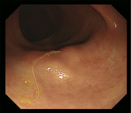
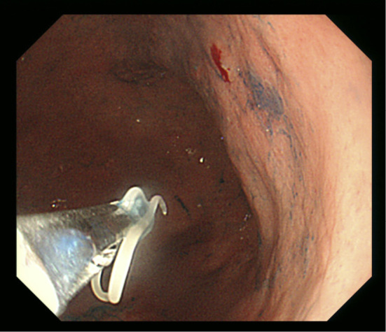
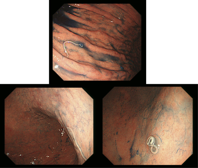
A 71-year-old man with no notable medical history consumed raw mackerel as sushi. Two days later, he incidentally underwent esophagogastroduodenoscopy (EGD) and colonoscopy on the same day. EGD revealed three Anisakis larvae penetrating the gastric mucosa (Picture 1), and colonoscopy revealed an Anisakis larva penetrating the mucosa of the transverse colon (Picture 2). These larvae were removed with biopsy forceps (Picture 3). We did not conduct a blood test or computed tomography due to his lack of symptoms before and during endoscopy. With the widespread use of screening endoscopy, reports of asymptomatic anisakiasis are increasing (1,2). However, to our knowledge, no cases of the simultaneous endoscopic diagnosis of gastric and colonic anisakiasis have been reported. In the present case, a laxative might have carried the gastric Anisakis larvae to the colon. We should make a careful diagnosis when we encounter Anisakis larvae in such a case.
Picture 1.
Picture 2.

Picture 3.

The authors state that they have no Conflict of Interest (COI).
References
- 1. Tamai Y, Kobayashi K. Asymptomatic colonic anisakiasis. Intern Med 54: 675, 2015. [DOI] [PubMed] [Google Scholar]
- 2. Angelo Z, Giuseppina B, Daniela B, et al. . Asymptomatic Anisakis and erosive lesions in the colon. Ann Gastroenterol 31: 246, 2018. [DOI] [PMC free article] [PubMed] [Google Scholar]


