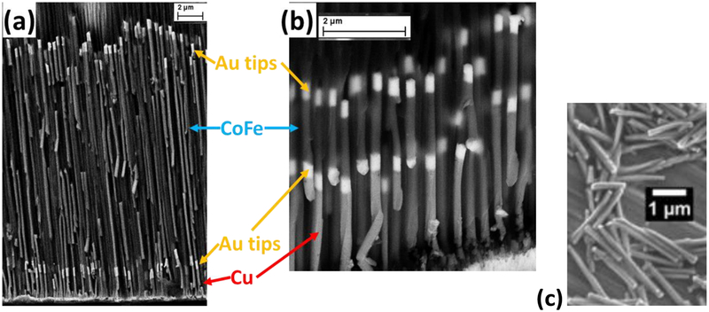
Fig. 2.
Cross-sectional SEM images of Co35Fe65nanowires 16 μm (a) and 2 μm (b) aligned in an AAO template. The Cu layers at the bottom were dissolved by 1 M FeNO3 before dissolving the AAO template. Note, some of the NWs were broken when the AAO template was cleaved to make these cross-sectional images. (c) Released Au-tipped Ni nanowires.

