Abstract
We investigated whether Tbx1, the gene for 22q11.2 deletion syndrome (22q11.2DS) and Foxi3, both required for segmentation of the pharyngeal apparatus (PA) to individual arches, genetically interact. We found that all Tbx1+/-;Foxi3+/- double heterozygous mouse embryos had thymus and parathyroid gland defects, similar to those in 22q11.2DS patients. We then examined Tbx1 and Foxi3 heterozygous, null as well as conditional Tbx1Cre and Sox172A-iCre/+ null mutant embryos. While Tbx1Cre/+;Foxi3f/f embryos had absent thymus and parathyroid glands, Foxi3-/- and Sox172A-iCre/+;Foxi3f/f endoderm conditional mutant embryos had in addition, interrupted aortic arch type B and retroesophageal origin of the right subclavian artery, which are all features of 22q11.2DS. Tbx1Cre/+;Foxi3f/f embryos had failed invagination of the third pharyngeal pouch with greatly reduced Gcm2 and Foxn1 expression, thereby explaining the absence of thymus and parathyroid glands. Immunofluorescence on tissue sections with E-cadherin and ZO-1 antibodies in wildtype mouse embryos at E8.5-E10.5, revealed that multilayers of epithelial cells form where cells are invaginating as a normal process. We noted that excessive multilayers formed in Foxi3-/-, Sox172A-iCre/+;Foxi3f/f as well as Tbx1 null mutant embryos where invagination should have occurred. Several genes expressed in the PA epithelia were downregulated in both Tbx1 and Foxi3 null mutant embryos including Notch pathway genes Jag1, Hes1, and Hey1, suggesting that they may, along with other genes, act downstream to explain the observed genetic interaction. We found Alcam and Fibronectin extracellular matrix proteins were reduced in expression in Foxi3 null but not Tbx1 null embryos, suggesting that some, but not all of the downstream mechanisms are shared.
Author summary
The mechanisms required for segmentation of the pharyngeal apparatus (PA) to individual arches are not precisely delineated in mammalian species. Using mouse models, we found that two transcription factor genes, Tbx1, the gene for 22q11.2 deletion syndrome and Foxi3, genetically interact in the third pharyngeal pouch endoderm during thymus and parathyroid gland development. When examining wildtype embryos, we found that each arch is surrounded by epithelial cells derived from the endoderm and ectoderm that undergo dynamic processes during PA segmentation. Invagination co-occurs with formation of multilayers of epithelia that become juxtaposed. The cells are then reorganized to form a dual-layer of tightly intercalated cells at the mature pouch-cleft junction. In Tbx1 and Foxi3 null mutant embryos, these processes are disrupted. Further, the endoderm cells form extensive multilayers in the region where cells should normally invaginate. When examining genes that may act downstream of Tbx1 and Foxi3 we found several including Notch pathway genes Jag1, Hes1, and Hey1 are downregulated in both mutant embryos. Together, we show that Tbx1 and Foxi3 are important for regulating PA segmentation through cellular and genetic mechanisms that may be critical in 22q11.2 deletion syndrome patients.
Introduction
The pharyngeal apparatus (PA) is an evolutionarily conserved structure that forms early in vertebrate embryos. The PA develops as a series of bulges, termed arches, found on the lateral surface of the head region of the embryo. During mammalian development, five pairs of pharyngeal arches numbered PA1, PA2, PA3, PA4, and PA6 (the fifth PA is transient) form subsequently, over time, from the rostral to caudal part of the head region of the embryo [1]. The process involved in the formation of each arch is referred to as pharyngeal segmentation. Each arch contributes to different craniofacial muscles, nerves and skeletal structures. PA1 contributes to the skull, incus and malleus of the middle ear, jaw, nerves and muscles of mastication. PA2, contributes to the skull, stapes in the middle ear, facial muscles, jaw, and upper neck skeletal structures. In addition to skeletal structures, muscles and nerves, PA3 is required to form the thymus and parathyroid glands. In mouse embryos, the thymus and parathyroid glands are derived only from PA3, but in humans the inferior parathyroid gland is derived from PA3 whereas PA4 contributes to the superior parathyroid gland. PA4 and PA6 contribute to the aortic arch and arterial branches [2, 3].
Each pharyngeal arch is surrounded by endoderm and ectoderm derived epithelial cells forming pharyngeal pouches and clefts, respectively. Mesoderm and neural crest derived mesenchyme cells occupy the center of each arch [1, 4]. The epithelia are needed to invaginate and promote segmentation to individual arches. The pharyngeal endoderm (PE), which is the focus of this study, receives and sends signals from the mesenchyme to initiate morphogenesis and invaginate [5]. Once each arch segments, proper patterning is also required to form derivative structures. The PE sends distinct signals in each arch to promote normal patterning [6–9]. Abnormal PA segmentation or patterning during development will cause defects within the later structures derived from the PA and leads to human birth defect disorders.
One particular gene important for PA segmentation is Tbx1, encoding a T-box transcription factor implicated in 22q11.2 deletion syndrome (DiGeorge syndrome [MIM# 188400]; velo-cardio-facial syndrome [MIM# 192430]). Tbx1-/- homozygous null mutant mouse embryos die at birth with hypoplastic and intermittent missing craniofacial muscles [10], cleft palate, absent thymus and parathyroid glands, as well as a persistent truncus arteriosus (PTA) with a ventricular septal defect (VSD) [11–13]. The first arch forms in Tbx1-/- embryos but the distal PA fails to become segmented, thereby explaining, in part, why the PA derived structures are malformed [11–13]. Tbx1 is expressed in the mesoderm of the head region in early mouse embryos and then throughout the endoderm, mesoderm, and distal ectoderm of the PA, while each arch forms, and becomes reduced at mouse embryonic day (E)10.5 [12, 14, 15]. Tissue specific inactivation of Tbx1 has been performed using the Cre-loxP system [4, 16–19]. It was found that Tbx1 is required in all three tissues for development of the derivative organs affected in the null mutant embryos [17, 19–21]. Since the PE is critically important for segmentation of the distal PA [22, 23], it is important to understand the genes and processes that might act downstream.
Another gene that has been shown to be important for normal PA segmentation is Foxi3, which encodes a Forkhead box (Fox) transcription factor. Foxi3 is expressed in the ectoderm in the head region early in embryonic development and is then expressed in the epithelia of the PA from around the same stages as when Tbx1 is expressed [24]. Foxi3 is also important for epithelial cell differentiation within the epidermis [25]. Heterozygous mutations were discovered in Foxi3 in several hairless dog breeds with hair follicle and teeth defects [26]. Global Foxi3-/- null mutant mouse embryos fail to form endodermal pouches and this results in failed PA segmentation leading to severe defects in the skull, jaw, and ears [27–29]. It has been shown that Foxi3 may have a cell non-autonomous effect on craniofacial neural crest cell survival because these cells undergo apoptosis in the mutant embryos at E10.0 [27].
In this study, we tested whether there is a genetic interaction between Tbx1 and Foxi3 during mouse embryonic development. We discovered that these two factors interact at minimum, in the third pharyngeal pouch endoderm, needed to form the thymus and parathyroid glands. Further, we found that inactivation of Foxi3 results in cardiovascular anomalies. We characterized the process of pharyngeal segmentation and found that global inactivation of Tbx1 and Foxi3 both result in failure of the epithelia to properly invaginate along with an expansion of multilayers of PE cells leading to failed segmentation of the distal PA. We identified some shared downstream genes that were reduced in expression in either null mutant and suggest that they may share some similar molecular mechanisms.
Results
Tbx1 acts upstream of Foxi3 in embryogenesis
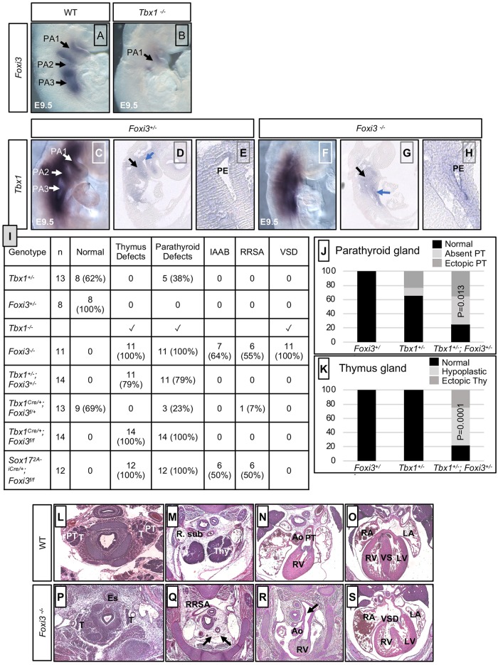
Since loss of Foxi3 or Tbx1 disrupt the segmentation of the PA, we tested whether Foxi3 might act upstream or downstream of Tbx1. Foxi3 is normally expressed throughout the epithelia of the PA, while Tbx1 is more broadly expressed (Fig 1A and 1C). Whole mount in situ hybridization (WMISH) using a Foxi3 antisense mRNA probe on Tbx1-/- mouse embryos and wildtype (WT) littermate controls at E9.5, revealed that Foxi3 expression was reduced in PA1 and absent in the unsegmented distal pharyngeal apparatus in Tbx1 null mutant embryos (Fig 1A and 1B). To determine if Tbx1 expression is affected in Foxi3-/- embryos, WMISH followed by tissue sectioning using a Tbx1 probe on Foxi3+/- control littermate and Foxi3-/- mouse embryos was performed and we found that the Tbx1 expression pattern was maintained in the pharyngeal mesoderm and endoderm despite the lack of segmentation of the distal PA (Fig 1C–1H). This indicates that either Foxi3 acts downstream of Tbx1 in the same genetic pathway and/or that the cells expressing Foxi3 were lost in the Tbx1 null mutant embryos.
Fig 1. Tbx1 and Foxi3 genetically interact for thymus and parathyroid gland development.
(A-B) Whole mount in situ hybridization (WMISH) using an antisense Foxi3 probe on wildtype (WT; A) and Tbx1-/- mouse embryos at E9.5 (B); n = 3 for each genotype. Individual pharyngeal arches, PA1, PA2 and PA3 are indicated and the arrows point to the corresponding arch. (C-H) WMISH using an antisense Tbx1 probe on whole mount and sagittal sections of control (Foxi3+/-; C-E) and Foxi3-/- (F-H) mouse embryos at E9.5 (n = 3 for each genotype). Black arrow indicates the core mesoderm and blue arrow indicates the endoderm. Zoomed in image of the pharyngeal pouch (E, H) next to the blue arrow (D, G), to demonstrate that Tbx1 is still expressed in the control and mutant embryos, respectively. Pharyngeal endoderm is indicated as PE. (I) Table summarizing defects found in each embryo with the genotype listed in the first column on the left. The second column indicates the number of embryos. The rest of the columns indicate the number and percent (in parentheses) with various defects as determined by histological analysis at E15.5. Absent thymus and parathyroid glands were observed in all Tbx1-/-, Foxi3-/- and Sox172A-iCre/+;Foxi3f/f embryos. Abbreviations: interrupted aortic arch type B (IAAB), retro-esophageal right subclavian artery (RRSA) and ventricular septal defect (VSD). (J-K) Bar graphs summarizing parathyroid (J) and thymus (K) defects found in Tbx1+/-, Foxi3+/-, and Tbx1+/-;Foxi3+/- embryos versus controls. Fisher’s exact two tailed test was used to determine significance between defects observed in Tbx1+/- and Tbx1+/-;Foxi3+/- embryos. Defects observed in Tbx1+/-;Foxi3+/- embryos include ectopic or absent parathyroid glands as well as ectopic and hypoplastic or absent thymus glands. (L-S) Histological sections of control (WT) embryos (L-O) and Foxi3-/- embryos (P-S) at E15.5. Foxi3-/- embryo with absent thymus glands and RRSA is shown (Q); IAAB is also present (R) as well as a VSD (S). Abbreviations: right subclavian artery (R. sub), thymus (Thy), aorta (Ao), pulmonary trunk (PT), right atrium (RA), left atrium (LA), right ventricle (RV), left ventricle (LV), ventricular septum (VS). More examples of defects that occurred in other mutant embryos are shown in S1 and S4 Figs.
Inactivation of Foxi3 leads to thymus, parathyroid and aortic arch defects
It has been previously shown that Foxi3-/- embryos have absent jaw bones, abnormal mandible, deformed maxilla bones, absent jugal (bony arch of zygoma, cheek bone) and smaller palatines, misshapen Meckel’s cartilage, and absent ears [27, 29]. At E9.5, segmentation of the PA to individual arches did not occur in Foxi3-/- embryos (Fig 1F and 1G), which is consistent with previous findings [27]. There is little known about its role in formation of later embryonic structures from the distal PA derived from PA3-6. We found that at E15.5, Foxi3-/- embryos had absent thymus and parathyroid glands (100%; n = 11), interrupted aortic arch type B (IAAB, 63%; n = 7), ventricular septal defect (VSD; 100%; n = 11) and retro-esophageal right subclavian artery (RRSA, 55%; n = 6) as listed in Fig 1I and shown in Fig 1L–1S. Some had both a RRSA and IAAB (18%; n = 2), while the remaining had either RRSA or IAAB (81%; n = 9; Fig 1I and 1P–1S).
Tbx1 and Foxi3 genetically interact in the pharyngeal apparatus
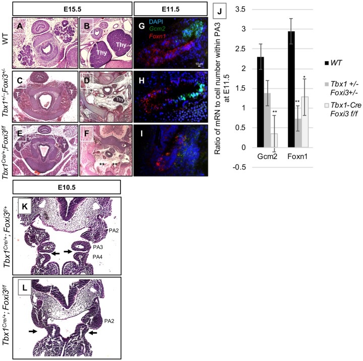
Based upon the similarities in the distal PA derived defects, and that Foxi3 was reduced in expression in Tbx1-/- embryos, we tested whether there could be a genetic interaction between the two genes. We first tested whether single heterozygous Foxi3+/- [27] or Tbx1+/- [11] embryos had defects. At E15.5, Fox3+/- embryos were normal (n = 8) and Tbx1+/- embryos had a normal thymus gland and had ectopic or absent parathyroid glands in 38% of the embryos (n = 5; Fig 1I–1K; S1A–S1H Fig). Normally, the parathyroid glands should be found adjacent to the thyroid glands, where the two carotid arteries are present nearby in the same section. When ectopic in Tbx1+/- or Tbx1+/-;Foxi3+/- embryos, parathyroid glands were found in a more caudal position in the embryo that is separate from the thyroid glands (Fig 2C; S1C and S1 Fig) versus controls (Fig 2A; S1A Fig). When ectopic in Tbx1+/-;Foxi3+/- embryos, thymus glands were more rostrally located than normal and were present at the same level of the embryos as the carotid arteries (Fig 2D; S1F Fig), as compared to control embryos, where the thymus glands were located at the branchpoint between the innominate and right carotid artery (Fig 2B; S1B and S1D Fig). Hypoplastic thymus glands were smaller in size than normal glands (Fig 2D; S1F Fig). At E15.5, all double heterozygous Tbx1+/-;Foxi3+/- embryos had either a hypoplastic and/or ectopic thymus and parathyroid glands (n = 14) and this increase is statistically significant (Fig 1J and 1K). More than half of double heterozygous embryos had both a hypoplastic thymus and ectopic parathyroid glands in comparison to WT controls (57%; n = 8/14; Figs 1J, 1K and 2A–2D; S1A–S1H Fig).
Fig 2. Thymus and parathyroid gland defects in Tbx1+/-;Foxi3+/- and Tbx1Cre/+;Foxi3f/f embryos.
(A-F) Transverse histology sections stained with H&E of WT (A-B), Tbx1+/-;Foxi3+/- (C-D) and Tbx1Cre/+;Foxi3f/f (E-F) embryos at E15.5. Abbreviations: thyroid (T) and thymus (Thy). Asterisks in A indicate parathyroid glands. Asterisks in F indicate absent thymus glands. Numbers include: WT, n = 16; Tbx1+/-;Foxi3+/-, n = 13; Tbx1Cre/+;Foxi3f/f, n = 14. Normal parathyroid glands are located adjacent to thyroid glands (A) as compared to when they are not present adjacent to the thyroid glands in mutant embryos (C, E). Thymus glands are located at the branchpoint between the innominate and right carotid artery (B) but are more rostrally located and smaller (D) or absent (E) in mutant embryos. (G-I) RNAscope in situ hybridization with mRNA probes for Gcm2 (green) and Foxn1 (red) probes on sagittal sections in WT (G), Tbx1+/-;Foxi3+/- (H) and Tbx1Cre/+;Foxi3f/f (I) embryos at E11.5. Gcm2 marks parathyroid gland precursor cells and Foxn1 marks thymus gland precursor cells; n = 3 for each genotype. (J) Quantification of RNAscope experiments. Number of mRNA signal dots was quantified in proportion to number of cells present in each tissue section. P-values were determined using the t-test. Two stars indicate a P-value <0.005 and one star indicates a P-value <0.05. (K-L) H&E coronal histology sections of Tbx1Cre/+;Foxi3f/+ (K), and Tbx1Cre/+;Foxi3f/f (L) embryos at E10.5. Arrows indicate the third pharyngeal pouch and morphology defects. Third pouch is absent in L. Tbx1Cre/+;Foxi3f/+, n = 6 and Tbx1Cre/+;Foxi3f/f, n = 8.
Expression of Gcm2 (glial cells missing homolog 2) and Foxn1 (Forkhead box protein N1) mark the parathyroid-fated and thymus-fated domains in the pharyngeal endoderm of PA3, respectively [30–32]. We performed RNAscope in situ hybridization on tissue sections with probes for Gcm2 and Foxn1 at E11.5 when both of these genes are expressed. In Tbx1+/-;Foxi3+/- embryos, expression of both markers was slightly reduced in intensity in PA3 in comparison to WT littermate controls (Fig 2G and 2H). When expression was quantified in comparison to WT embryos, both genes were reduced in expression in the PA3 derivative region in the double heterozygous embryos, but only reduction of Foxn1 was statistically significant (Fig 2J). The presence of some expression of these two genes is consistent with the occurrence of milder thymus and parathyroid gland defects (hypoplastic thymus and/or ectopic parathyroid) in these embryos as compared to null mutant embryos (Fig 1I; S1A–S1H Fig; Fig 2A–2D). Histology sections of Tbx1+/-;Foxi3+/- embryos at E10.5 were examined to see if there were defects in PA3. We noted a slightly narrowing of the space between PA3 and PA4 in the double heterozygous embryos, but no other malformations were detected (S1I–S1M Fig). Again, the phenotype at E15.5 is relatively mild in comparison to either null mutant, in which the organs were completely absent. To gain more insights into the basis of the observed defects, we examined where Tbx1 and Foxi3 are co-expressed.
Tbx1 and Foxi3 are co-expressed in the pharyngeal pouches and clefts
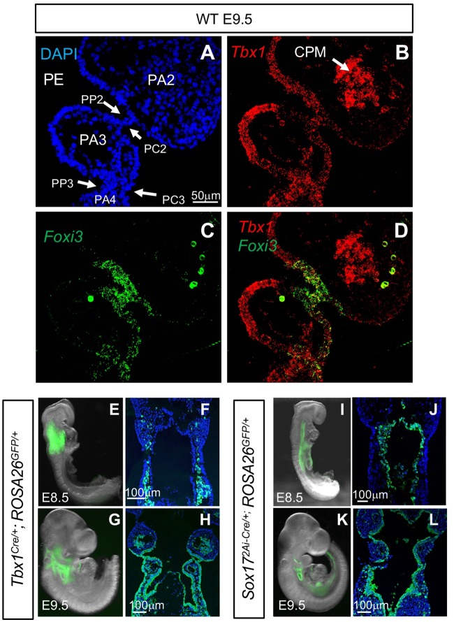
The PA develops from E8.5-E10.5, in which PA3 forms by E9.5. In situ hybridization using RNAscope probes was performed on coronal tissue sections from WT embryos to determine if there is overlap between Foxi3 and Tbx1 mRNA expression at E9.5 (Fig 3A–3D). There was strong expression of Tbx1 in the pharyngeal endoderm and cardiopharyngeal mesoderm (Fig 3B), as has been previously reported [33, 34]. Tbx1 was also expressed in the pharyngeal endoderm and ectoderm of the distal PA (Fig 3B) as has been previously reported [33]. Foxi3 expression was localized exclusively to the pharyngeal pouches and clefts as published in the past [35]. Expression of Foxi3 was particularly strong in the junction between the pharyngeal pouch and cleft that lies between PA2 and PA3 (Fig 3C). Expression of Foxi3 was also detected where invagination of the epithelia is taking place to form the separation between PA3 and PA4 (Fig 3C). Co-expression of Tbx1 and Foxi3 in the same cells was detected in the second and third pharyngeal pouch and cleft and where invagination was taking place (Fig 3D). The third pharyngeal pouch is where co-expression of both genes occurred and this is the same region where the thymus and parathyroid glands will form. This provides supporting evidence that there could be a genetic interaction between the two genes. We then decided to inactivate both alleles of Foxi3 using the Tbx1Cre mouse line [36], where we would expect more obvious developmental defects at these early stages than when we inactivate one allele.
Fig 3. Foxi3 and Tbx1 expression in embryonic pharyngeal pouches and Tbx1Cre and Sox172A-iCre lineages.
(A-D) RNAscope in situ hybridization with mRNA probes for Foxi3 and Tbx1 on coronal sections from a WT embryo at E9.5. Tbx1 expression is shown in red and Foxi3 expression is shown in green. Abbreviations: Pharyngeal endoderm (PE); pharyngeal pouch 2 (PP2); pharyngeal cleft 2 (PC2); pharyngeal pouch 3 (PP3); pharyngeal cleft 3 (PC3); and cardiopharyngeal mesoderm (CPM). Total of n = 3 embryos. (E-H) Lineage tracing of Tbx1Cre/+;Rosa26GFPf/+ embryos. GFP expression is visible in whole mount embryos and the corresponding coronal sections at E8.5 (E-F) and at E9.5 (G-H); n = 3 for each genotype and stage. (I-L) Lineage tracing of Sox172A-iCre/+;Rosa26GFPf/+ embryos. GFP expression is visible in whole mount embryos and the corresponding coronal sections at E8.5 (I-J) and E9.5 (K-L); n = 4 for each genotype and stage.
Conditional inactivation of Foxi3 results in defects in the distal pharyngeal apparatus
To establish the role of Foxi3 within the Tbx1 lineage to explain the basis of PA3 derived defects in double heterozygous embryos, we inactivated it using Tbx1Cre/+ knockin mice [36]. For this, Tbx1Cre/+ mice were crossed with a Foxi3 floxed allele (Foxi3f/+) and the double heterozygous mice were crossed with Foxi3f/f mice to inactivate both alleles of Foxi3. We also crossed the Tbx1Cre/+ mice with a Rosa26GFP/GFP allele to detect the Tbx1 lineage using the GFP reporter. GFP fluorescence was observed in the pharyngeal mesoderm at E8.5 (Fig 3E and 3F) and in both the pharyngeal mesoderm plus epithelia of the PA at E9.5 (Fig 3G and 3H). The Tbx1Cre/+ is a knock-in allele for Tbx1 and it is heterozygous for Tbx1 [36]. GFP fluorescence was not detected in the epithelia at E8.5. This was unexpected because Tbx1 mRNA expression occurs in the epithelia at this stage [37]. Thus, there is a difference in timing of Tbx1 expression and detection of GFP fluorescence, which marks recombination of loxP sites and translation of sufficient GFP to be visualized. This timing difference may explain why Tbx1Cre/+;Foxi3f/+ embryos at E15.5 did not exhibit a more severe phenotype than what occurred in Tbx1+/- embryos, as compared to that in Tbx1+/-;Foxi3+/- embryos (Fig 1I).
At E15.5, Tbx1Cre/+;Foxi3f/f mutant embryos were compared to Tbx1Cre/+;Foxi3f/+ controls (n = 13) to determine if there were any PA derived defects (Fig 1I). Reduction of Foxi3 expression in the pouches, but not the pharyngeal clefts, within the PA in Tbx1Cre/+;Foxi3f/f embryos was observed in S2 Fig. The Tbx1Cre/+;Foxi3f/f embryos had absent thymus and parathyroid glands (100% n = 14; Fig 2E and 2F), similar to what occurred in Foxi3-/- embryos (Fig 1I). To determine if Foxn1 (thymus) and Gcm2 (parathyroid) expression [32] was affected, we performed RNAscope in situ hybridization with probes for these genes and found that the expression of both genes in the PA3 region were significantly reduced in conditional null versus wildtype controls (Fig 2G, 2I and 2J). We noted that there was no separation between PA3 and PA4 at stage E10.5 (Fig 2L) as compared to the presence of a separation in Tbx1Cre/+;Foxi3f/+ controls (Fig 2K). Reduction in expression of Foxn1 and Gcm2 as well as the presence of morphology defects at E10.5 might explain why the thymus and parathyroid glands did not form. Despite having absent thymus and parathyroid glands, these mutant embryos did not have cardiovascular or aortic arch defects (Fig 1I and S3A–S3F Fig). India ink injections confirmed presence of aortic arch arteries 3, 4 and 6, in WT and Tbx1Cre/+;Foxi3f/f embryos (S3G and S3H Fig). While expression of Foxi3 was significantly reduced as determined by WMISH (S3A–S3D Fig) in conditional mutant embryos, we also tested Tbx1Cre/+;Foxi3f/- embryos to further inactivate Foxi3 and found that the embryos lacked the thymus and parathyroid glands but similar to above, they had no intracardiac or aortic arch defects (n = 8; S3I–S3L Fig). Therefore, there is an interaction between Tbx1 and Foxi3 in the third pharyngeal pouch endoderm. We did not detect PA4 derived defects in the Tbx1Cre/+;Foxi3f/- embryos at E15.5. There are two different possibilities to explain basis for the lack of aortic arch or branching anomalies derived from PA4. One possibility is that there is no genetic interaction in the fourth pharyngeal pouch. Another possibility could be due to timing of Tbx1 gene expression and delayed timing of Cre activity using the Tbx1Cre allele, within the PE (Fig 3E–3H), although its less likely an issue in Tbx1Cre/+;Foxi3f/- embryos. We then next decided to inactivate Foxi3 in the PE to understand its tissue specific function.
Foxi3 is expressed in the epithelia in the PA (Figs 1A, 3C and 3D and [24]). To determine the role of Foxi3 within the PE, we performed tissue specific inactivation of Foxi3 using the Sox172A-iCre/+ allele [38]. We first confirmed that the PE population (and endothelial cells) is marked by GFP expression using Sox172A-iCre/+;ROSA26GFP/+ embryos (Fig 3I–3L). Foxi3 is not expressed in endothelial cells. Recombination occurred prior to E8.5 as indicated by robust green fluorescence that was present at E8.5 (Fig 3I–3L). Foxi3 expression was reduced in these conditional mutants as detected by WMISH (S2E–S2H Fig). We found that Sox172A-iCre/+;Foxi3f/f embryos had failed segmentation of the distal PA as compared to Sox172A-iCre/+;Foxi3f/+ controls (S4A and S4C Fig). The PA was not as severely affected as in the Foxi3-/- null mutant embryos (S4B Fig), as PA2 was present, albeit hypoplastic (S4C Fig). At E15.5, all Sox172A-iCre/+;Foxi3f/f embryos had absent thymus and parathyroid glands (100%; n = 12; Fig 1I). Cardiovascular defects occurred in 66% of embryos that included IAAB (50% n = 6), and/or RRSA (50% n = 6; Fig 1I and S4D–S4F Fig). As compared to Foxi3-/- mutant embryos that always had an aortic arch defect plus a VSD, Sox172A-iCre/+;Foxi3f/f mutant embryos had an IAAB but had normal septation of the ventricles (Fig 1I). Several mutant embryos had both an RRSA and IAAB (33% n = 3), while 33% (n = 3) had just an RRSA and 33% (n = 3) had an IAAB, whereas a few had normal aortic arches and normal branching (33% n = 3; Fig 1I). India ink injections showed that the 4th aortic arch artery was absent in Sox172A-iCre/+;Foxi3f/f embryos at E10.5, similar to what we observed in Foxi3-/- embryos (S4G–S4L Fig). Absence of the 4th aortic arch arteries would lead to the observed cardiovascular defects that occurred in the embryos. Based upon this data, Foxi3 expression is required in the PE for classic 22q11.2DS phenotypes. Further, Foxi3 in the ectoderm may have an additional role in the septation of the ventricles.
Loss of Foxi3 in the Tbx1 expressing lineage disrupts segmentation between PA3-4
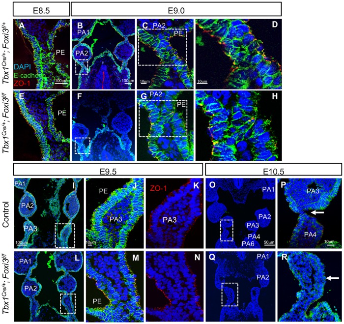
In the Tbx1Cre/+;Foxi3f/f mutant embryos, only PA3 derivative structures were affected, being the thymus and parathyroid glands. As indicated above, the pouch and cleft between PA3 and PA4 did not form and was missing at E10.5, when segmentation was complete (Fig 2K and 2L). E-cadherin is a cell-cell adhesion protein forming adherens junctions that bind cells tightly to each other and it marks epithelial cells (Reviewed in [39]). ZO-1 (Zona occluden-1) forms permeable barriers in adherens junctions and is a marker for the presence of apical/basal polarity among epithelial cells, with expression specifically on the apical side of the cell facing a lumen [40]. Immunofluorescence using antibodies to E-cadherin and ZO-1 was performed on WT, Tbx1Cre/+;Foxi3f/+ and Tbx1Cre/+;Foxi3f/f embryos at E9.5, to visualize the structure of the epithelial cell population. At E8.5 and E9.0 there was no noticeable difference between the mutant and control embryos (Fig 4A–4H). But, at E9.5, invagination of the partially stratified pharyngeal endoderm and ectoderm between PA3 and PA4 did not take place (Fig 4L–4N) as compared to controls (Fig 4I–4K). The PE of PA3 maintained its epithelial identity and cell polarity as marked by expression of E-cadherin (Fig 4I, 4J, 4L and 4M) and ZO-1 (Fig 4K and 4N). It is possible that instead of failure to invaginate, there was a delay. However, at E10.5, the third pouch and cleft were absent in the conditional mutant embryos (Fig 4Q and 4R) in comparison to controls (Fig 4O and 4P), so that there was no distinction between PA3 and PA4 as shown by H&E staining (Fig 2K and 2L). Thus, the genetic interaction between the two genes is at minimum, in PA3.
Fig 4. Inactivation of Foxi3 within the Tbx1Cre/+ lineage disrupts PA3 morphogenesis.
(A-R) DAPI (blue), E-cadherin (green), and ZO-1 (red) antibodies were used for immunofluorescence on coronal sections from embryos at E8.5-E9.0 (n = 3, each), E9.5 (n = 5) and E10.5 (n = 4) in Tbx1Cre/+;Foxi3f/+ (A-D, O-P) or WT (I-K) controls and in Tbx1Cre/+;Foxi3f/f (E-H, L-N, Q-R) mutant embryos. White boxes indicate the location of the higher magnification images. White arrows in E10.5 images (P, R) indicate 3rd pharyngeal pouch in control and absent 3rd pharyngeal pouch in mutant embryos.
Process of pharyngeal segmentation in wildtype and mutant embryos
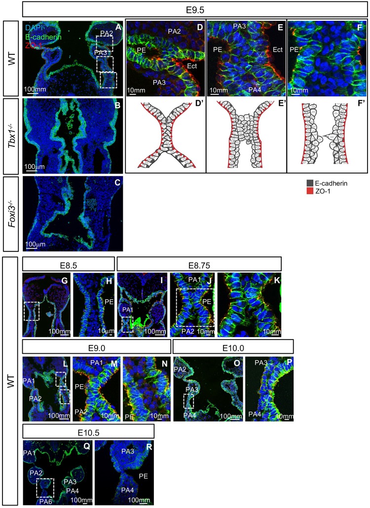
Immunofluorescence using antibodies to E-cadherin and ZO-1 was performed to detect differences between WT, Tbx1 and Foxi3 null mutant embryos at E9.5. While individual arches were present in the PA in WT embryos, segmentation of the distal PA, PA2-6, did not occur in Tbx1 or Foxi3 null mutant embryos at this stage (Fig 5A–5C). In the Tbx1-/- and Foxi3-/- embryos, there was a multilayered stratified epithelium, possibly in areas where cells would begin to invaginate, which appeared thicker than in WT embryos (Fig 5A–5C).
Fig 5. PA segmentation in wildtype, Tbx1 and Foxi3 mutant embryos.
(A-F) To visualize epithelial cells within the PA, DAPI (blue), E-cadherin (green), and ZO-1 (red) antibodies were used for immunofluorescence on WT (A), Tbx1-/- (B), and Foxi3-/- (C) coronal sections at E9.5 (WT, n = 5; Tbx1-/-, n = 4 and Foxi3-/-, n = 5). White boxes (A) indicate where the region was magnified (D-F). (D) Second pouch and cleft where segmentation is complete and epithelial cells formed a dual intercalated layer. (E) Third pouch and clefts are forming at E9.5. The epithelium is stratified. (F) Location of future PA4-PA6, where invagination is initiated, as can be observed by cell projections from the epithelia. PE indicates the pharyngeal endoderm and Ect. indicates the ectoderm for each section. (D’-F’) Cartoon illustrating the segmentation process from images in D-F. (G-R). DAPI (blue), E-cadherin (green), and ZO-1 (red) antibodies were used for immunofluorescence to visualize epithelial cells within the PA in coronal sections from WT E8.5 (G-H), E8.75 (I-K), E9.0 (L-M), E10.0 (O-P), E10.5 (Q-R) embryos. White boxes indicate the location of higher magnification images. PE indicates pharyngeal endoderm; n = 4 for each stage.
To examine whether multilayers of epithelia are present as a normal part of epithelial cell dynamics, we carefully examined the PA segments in WT embryos at E9.5 (Fig 5A and 5D–5F). During normal segmentation of the distal PA, the endoderm and ectoderm invaginate toward each other to form a pouch and cleft, which becomes juxtaposed thereby providing a physical separation of the rostral and caudal arch. This process occurs dynamically in a rostral to caudal manner over time, such that the process of segmentation of different arches can be observed at one stage.
The junction of the pouch and cleft between PA2 and PA3 was completely formed by E9.5 and consisted of a tightly organized intercalated dual layer of cells, likely one layer of endoderm and one of ectoderm, in which ZO-1 is expressed at the outer face of each layer, on their apical surface (Fig 5D and 5D’). To determine the process of segmentation, we examined the region where the junction of the pouch and cleft between PA3 and PA4 was forming (Fig 5E and 5E’). We noted that there were multiple layers of a partially stratified epithelium where the pharyngeal epithelia became juxtaposed to each other (Fig 5E and 5E’). The outer cells expressed ZO-1 on their apical surface, but the inner cells did not (Fig 5E and 5F). It was as though a zippering process was beginning in the central region such that ZO-1 negative cells rostrally and caudally were being pushed out. More caudally, epithelial cells on either side of the mesenchyme appeared to be extending processes towards each other at the point where the cells were beginning to invaginate to form the next segment (Fig 5F and 5F’). Thus, the process of segmentation involves cell movement and repositioning as well as communication of the endoderm and ectoderm in order to form mature pouch-cleft junctions.
Additional stages of E8.5, E8.75, E9.0, E10.0 and E10.5 were examined to further characterize epithelial cell dynamics and ascertain whether there were fundamental differences at different stages in WT embryos (Fig 5G–5R). The first transition of pouch morphogenesis began at E8.5 when the endoderm and ectoderm initiate the process of invagination to eventually separate PA1 from PA2 (Fig 5G and 5H). We did not observe multilayers of epithelial cells at E8.5 (Fig 5G and 5H). As invagination was completing between PA1 and PA2, at E8.75 (Fig 5I), the pharyngeal endoderm and ectoderm, consisting of two to a few layers, became loosely juxtaposed (Fig 5J and 5K). This is similar to the pouch-cleft formation for PA3-4 at E9.5 (Fig 5E and 5E’). At E9.0 (Fig 5L), the junction between first pouch and cleft that separated PA1 and PA2 formed a tight intercalated dual cell layer (Fig 5M) that appears similar to that observed in Fig 5D and 5D’. At E9.0, the second pouch and cleft between PA2-PA3, consisting of a few layers of epithelium, became juxtaposed and intercalated (Fig 5N). At E9.0 and E9.5, there were two to three layers of endoderm cells in the caudal PA, as compared to less layers in the rostral PA, where the mature pouch-cleft junction occurred. At E10.0, the pouches and clefts formed a pouch-cleft junction that was almost mature in between PA3-4 (Fig 5O and 5P). At E10.5, PA formation was complete with the presence of dual layer mature pouch-cleft junctions between each arch (Fig 5Q and 5R).
Global Tbx1 inactivation results in excessive layers of endoderm cells
As indicated in Fig 5B, segmentation of the distal PA in Tbx1-/- embryos did not occur and additional layers of epithelia were present at E9.5, possibly where the cells would begin to invaginate. However, proliferation assays at E8.5 and E9.5 revealed no significant changes in cell proliferation in null mutant embryos as compared to controls (S5A–S5G Fig) as has been previously reported at E9.5 [18]. ZO-1 expression was normal at E8.5 (S5H and S5J Fig) and E9.5 (S5I and S5K Fig), and the apical side of only the outer facing cells expressed ZO-1 (S5H–S5K Fig). The PA in Tbx1-/- embryos was shorter in length and cell number quantification revealed that there were significantly more epithelial cells within the shortened PA at E9.5, but not at E8.5 (S5L Fig). This suggests that more cells were packed into a smaller PA at E9.5. We then performed endoderm specific inactivation to determine whether this process was cell type autonomous. Inactivation of Tbx1 in the PE using the Sox172A-iCre/+ allele results in a normal first and hypoplastic second arch, as compared to an absent second arch in global null mutant embryos. This is similar to the situation with endoderm inactivation of Foxi3 (S4C Fig). Invagination of the PE did not take place and this resulted in failed segmentation of the distal arches [18], as we confirmed (S5M and S5N Fig). Similar to what was observed in the global Tbx1 null mutant embryos, E-cadherin and ZO-1 expression was normal in the conditional mutant embryos as compared to the Sox172A-iCre/+;Tbx1f/+ littermates at E9.5 and E10.5 (S5M and S5P Fig). As compared to the global null mutant, we did not observe excessive multilayers in the conditional mutant embryos at E9.5 (S5M and S5N Fig). We did observe multilayers of PE cells in the distal PA at E10.5 as compared to controls (S5O and S5P Fig), suggesting some differences between the conditional mutant embryos compared to null mutant embryos.
Excessive multilayered epithelium occurs in the PA of Foxi3 mutant embryos
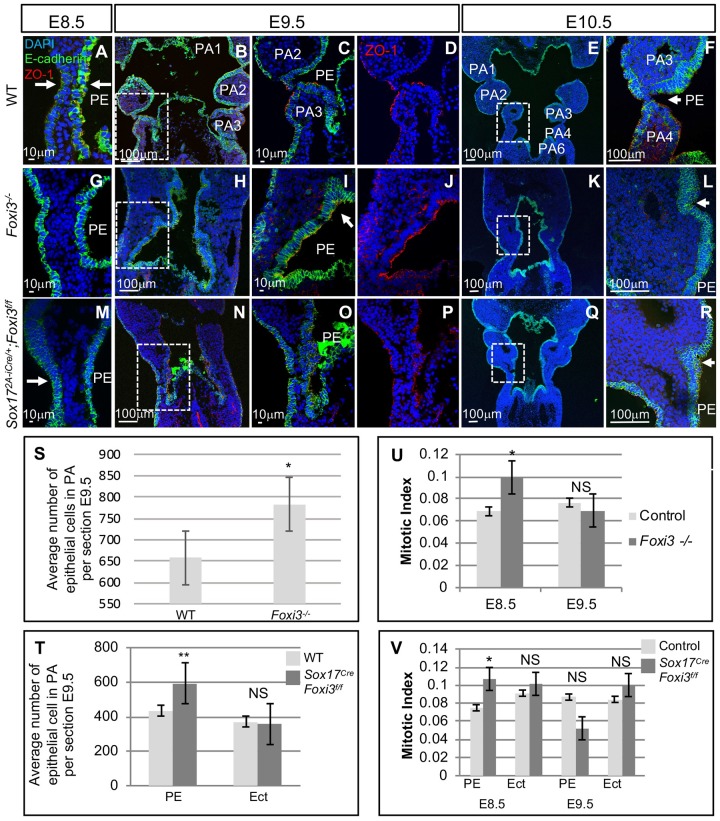
We next examined WT, Foxi3-/- and Sox172A-iCre/+;Foxi3f/f embryos at E8.5-E10.5 to understand if the defects observed in Foxi3 null mutant embryos occurred in a tissue specific manner (Fig 6). Invagination defects began at E8.5 in both Foxi3-/- and Sox172A-iCre/+;Foxi3f/f embryos (Fig 6A, 6G and 6M). At E9.5, regions with excessive stratified multilayers of endoderm cells, especially at the points where the cells would be turning inwards to invaginate, were found in Foxi3-/- (Fig 6H–6J) and Sox172A-iCre/+;Foxi3f/f embryos (Fig 6N–6P) versus WT controls (Fig 6B–6D). All endoderm cells in WT, Foxi3-/- and Sox172A-iCre/+;Foxi3f/f embryos, expressed E-cadherin and the outermost cells expressed ZO-1 on the apical side, indicating that cells did not lose epithelial identity or polarity (Fig 6A–6R). At E9.5 and E10.5, the epithelial cells in Foxi3-/- embryos began to invaginate but never advanced (Fig 6K and 6L) as compared to WT embryos (Fig 6E and 6F). At E10.5, the epithelia partially invaginated in the Sox172A-iCre/+;Foxi3f/f embryos, that seemed more complete for the ectoderm than endoderm (Fig 6Q and 6R). Overall, this data indicates a tissue autonomous role of Foxi3 in the endoderm during segmentation of the PA.
Fig 6. Foxi3-/- and Sox17-2A-iCre/+;Foxi3f/f mutant embryos have defects in PA segmentation.
(A-R) DAPI (blue), E-cadherin (green), and ZO-1 (red) mark epithelial cells on coronal sections from WT (A-F), Foxi3-/- (G-L) and Sox172A-iCre/+;Foxi3f/f (M-R) embryos from E8.5-E10.5 (n = 4, each). White boxes indicate the location of higher magnification images. White arrows indicate where invagination occurs in the PE and ectoderm. (S-T) Quantification of epithelial cells within the PA at E9.5 in WT controls and mutant embryos. Error bars represent standard error. One asterisk indicates P-value <0.05; two asterisks indicate P-value <0.005 (n = 4, each). (U-V) Quantification of proliferation assays includes counting E-cadherin positive epithelial cells within the PA and calculation of the mitotic index, which is the ratio of pH3 positive cells within the E-cadherin positive epithelial cells. Error bars represent standard error, and asterisks indicate P-value <0.05. Pharyngeal endoderm (PE) and ectoderm (Ect) were quantified separately in conditional mutant embryos. At E8.5 and E9.5 a total of n = 6 and n = 4, were examined, respectively, for control and mutant embryos. Controls include WT, Foxi3+/-, and Sox172A-iCre/+;Foxi3f/+ littermates.
Cell proliferation is increased in Foxi3 mutant embryos at E8.5 but not at E9.5
E-cadherin positive epithelial cells within the PA in WT versus Foxi3-/- and WT embryos were quantified and there was a 45% (P-value <0.03) increase of epithelial cell number within the PA of Foxi3 null mutants as compared to controls (S6 Fig) This may be correlated with the additional cell layers observed in the mutant embryos. There was a 40% increase in the number of endodermal cells (P-value <0.006), but no increase (P-value <0.4) was detected of ectoderm cells in the PA of Sox172A-iCre/+;Foxi3f/f mutant embryos versus controls, at E9.5 (Fig 6T). We then decided to test whether cell proliferation was increased at E8.5 and E9.5. For this, a pH3 antibody was used to mark proliferating cells on serial sections on mutant versus control embryos at E8.5 and E9.5 (Fig 6U and 6V; S6 Fig). When calculating the ratio between proliferating cells versus total E-cadherin positive cells, there was a significant increase at E8.5 in the endoderm of Foxi3-/- and Sox172A-iCre/+;Foxi3f/f embryos versus controls (Fig 6U and 6V; S6 Fig). At E9.5 there was no difference in proliferation between mutant and control embryos (Fig 6U and 6V; S6 Fig). This indicates that increases of cell proliferation at E8.5 can partially explain why there is an increase of layers of epithelial cells within the PA at E9.5 but not at E10.5.
Notch-pathway gene expression is reduced in Foxi3 and Tbx1 null embryos
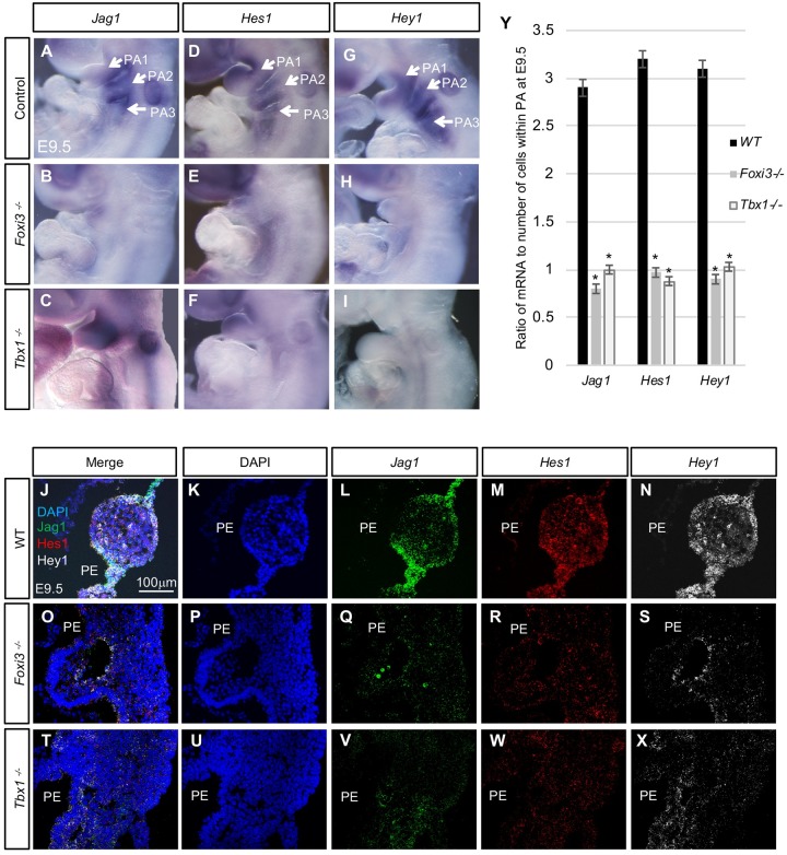
Notch signaling is critical for many aspects of embryonic development such as for skeletal development [41, 42] and cardiovascular development [21, 43, 44]. Notch signaling might have a possible role in thymus gland development [21, 45]. It has also been shown that Notch pathway genes, Jagged1 (Jag1) and Hes1 act downstream of Tbx1 during embryogenesis [21, 46, 47]. We therefore wanted to determine if Jag1, Hey1, and Hes1 may be regulated by Tbx1 and Foxi3 during PA formation. To test this, we performed WMISH and RNAscope experiments on WT, Tbx1-/- and Foxi3-/- mutant embryos at E9.5. By WMISH, Jag1, Hes1 and Hey1 expression in the pharyngeal pouch-cleft regions in WT embryos was reduced in both Foxi3 and Tbx1 null mutant embryos (Fig 7A–7I). Three-color RNAscope assays were performed on tissue sections from WT, Tbx1-/- and Foxi3-/- mutant embryos at E9.5, to examine expression level changes of Jag1, Hes1 and Hey1 (Fig 7J–7X). As in the WMISH experiments, Jag1 expression was localized to the pouch-cleft junctions, Hes1 was expressed at a low level throughout the PA in WT embryos and Hey1 was expressed more broadly in the PA (Fig 7J–7X). Expression was quantified and we found that levels of all three genes were significantly reduced in null mutant embryos (Fig 7Y). We also examined expression patterns of additional genes that are expressed in the PE.
Fig 7. Expression of Notch pathway genes Jag1, Hes1, and Hey1 were downregulated in Foxi3 and Tbx1 null mutant embryos.
(A-I) WMISH with antisense Jag1, Hes1, and Hey1 mRNA probes on whole mount embryos at E9.5. Jag1 antisense probe on WT (A), Foxi3-/- (B) and Tbx1-/- mutant embryos (C); n = 3 of each genotype. Hes1 probe on WT (D), Foxi3-/- (E) and Tbx1-/- (F) mutant embryos; n = 2–3 for each genotype. Hey1 probe on WT (G), Foxi3-/- (H) and Tbx1-/- (I) mutant embryos; n = 2, each. (J-X) RNAscope in situ hybridization with mRNA probes for Jag1, Hes1, and Hey1 on coronal sections of WT (J-N), Foxi3-/- (O-S), and Tbx1-/- (T-X) embryos at E9.5; n = 3, each genotype. Merged channels are shown (J, O, and T); DAPI is shown in blue color (K, P, and U); Jag1 mRNA expression is in green (L, Q, and V); Hes1 mRNA expression is in red (M, R, and W); and Hey1 expression is in white (N, S and X). The sections correspond to the same rostral-caudal location within in each embryo. (Y) Quantification of RNAscope experiments on WT versus Tbx1-/- and Foxi3-/- embryos at E9.5. Nuclei and mRNA were quantified for each section and probe. The total number of mRNA signal dots was divided by the total number of cells for each replicate. Graph represents the average ratio. Asterisks indicate P-values < 0.05.
Isl1 (Islet1) is expressed in the second heart field mesoderm and endoderm, among other tissues during early embryogenesis. Based upon WMISH, there was not a dramatic difference in expression in WT, Foxi3-/- and Tbx1-/- embryos at E9.5 (S7A–S7C Fig). FGF signaling has been shown to act genetically downstream of Tbx1 [48, 49] and Foxi3 [27]. We previously found that Fgf3 is reduced in expression when Tbx1 is inactivated [50]. Using WMISH, we also found that Fgf3 expression was reduced in Foxi3-/- embryos at E9.5 (S7D and S7E Fig). Expression in the otic vesicle was gone because the structure doesn’t form in Foxi3 null mutant embryos. Expression of another PE specific transcription factor gene, Pax8 (Paired box 8) was reduced within the PA in both Tbx1 and Foxi3 null mutants at E9.5 (S7F–S7H Fig). Pax8 is important for thyroid gland development and acts downstream of Foxi3 [51]. Pax9 is a transcription factor that is normally expressed within the PE and marks the pouches during embryogenesis [27]. We also confirmed that Pax9 mRNA expression is reduced but not absent in the PA in Tbx1 mutant embryos [52] in comparison to WT controls using WMISH and RNAscope (S7I–S7M Fig). Other studies reported that Pax9 expression was mis-regulated in Foxi3-/- embryos [27]. Our data indicated that Pax9 expression was reduced in Foxi3-/- embryos, although this does not rule out that it was also mis-regulated (S7J and S7M Fig). All together this shows that there are genes that act downstream of both Tbx1 and Foxi3.
Extracellular matrix proteins in Tbx1 and Foxi3 null mutant mouse embryos
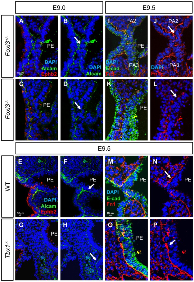
In addition to observing expression of known genes important for pharyngeal segmentation, we also investigated expression of activated leukocyte cell adhesion molecule (Alcam; also called CD166, Neurolin, or DM-GRASP), Ephrinb2, and Fibronectin (Fn1) in Foxi3 and Tbx1 null mutant embryos. This is because these extracellular proteins have roles in epithelial cell function and endodermal pouch formation in zebrafish [53] [54–57]. In Foxi3-/- embryos, Alcam expression was reduced in the pharyngeal endoderm (Fig 8C and 8D) as compared to heterozygous controls (Fig 8A and 8B). In contrast, Alcam protein expression appeared unchanged in Tbx1-/- embryos (Fig 8G and 8H) in comparison to WT controls (Fig 8E and 8F). In zebrafish, ephrinb2 is required to prevent epithelial cells from rearranging once PE segmentation is complete [58]. We did not observe a change of Ephrinb2 expression in epithelial cells in Foxi3+/- and WT controls as well as Foxi3-/- or Tbx1-/- mutant embryos (Fig 8A, 8C, 8E and 8G). Fibronectin protein is an extracellular matrix protein that is present outside the basal surface of the epithelia closest to the adjacent mesenchyme and is also expressed in the mesenchyme [56, 57]. Fibronectin expression was absent or expression was spotty in cells adjacent to the endoderm and ectoderm in Foxi3-/- mutant (Fig 8K and 8L) versus Foxi3+/- control embryos (Fig 8I and 8J). Fibronectin expression in Tbx1-/- mutant embryos was increased (Fig 8O and 8P) as compared to control littermates (Fig 8M and 8N), which is consistent with previous findings [59]. This indicates some differences between Tbx1 and Foxi3 functions. We note that some of the changes in patterns of expression could be due to morphological defects in the null mutant embryos.
Fig 8. Alcam and Fibronectin expression is reduced in epithelial cells within the PA at E9.5 in Foxi3-/-, but not in Tbx1-/- embryos.
(A-H) DAPI (blue), Alcam (green) and Ephrin b2 (red) antibodies were used to examine coronal sections in embryos at E9.0. Alcam localizes to the basal side of epithelium and Ephrin b2 localizes to the apical side. Sections from Foxi3+/- control embryos are shown with all three fluorescence color channels (A) and only DAPI and Alcam (B). Sections from Foxi3-/- embryos are shown with all three channels (C) and only DAPI and Alcam (D). Sections from WT littermates of Tbx1-/- embryos are shown with all three channels (E) and only DAPI and Alcam (F). Tbx1-/- embryos are shown with all three channels (G) and only DAPI and Alcam (H). White arrows indicate where Alcam expression is reduced in Foxi3-/- mutant embryos, but is present in Tbx1-/- embryos in comparison to control embryos. (I-P); DAPI (blue), E-cadherin (E-cad, green) and Fibronectin (Fn1, red) antibodies mark epithelial cells and the intracellular matrix on coronal sections at E9.5. (I-J) Sections from Foxi3+/- control embryos shown with all three fluorescent channels (I) and only the red and blue channels (J). (K-L) Sections from Foxi3-/- embryos showing all three channels (K) and only the red and blue channels (L). White arrows indicate where expression is spotty and inconsistent in Foxi3-/- mutants. (M-P) Sections from WT (M-N) and Tbx1-/- embryos (O-P) showing all three channels (M and O) and only the red and blue channels (N and P). White arrow in N and P indicates where Fibronectin expression is increased in Tbx1-/- mutants. n = 3 for each genotype in each experiment.
Discussion
In this report, we found that there is a genetic interaction between Tbx1 and Foxi3 in the formation of the thymus and parathyroid glands from the third pharyngeal pouch in mammals. Inactivation of Foxi3 in the Tbx1 domain resulted in absent thymus and parathyroid glands and inactivation in the endoderm resulted additionally in aortic arch defects that are similar as is observed in patients with 22q11.2DS. Expression of Jag1, Hey1 Fgf3, Pax8 and Pax9 was reduced in both Tbx1 and Foxi3 null mutant embryos, suggesting some shared downstream genes. We investigated the cellular mechanisms by which pharyngeal pouch-cleft junctions form in the process of pharyngeal segmentation. We found that the epithelial cells invaginate and form temporary multilayers in which cells become juxtaposed, repositioned and tightly intercalated to form junctions between the endoderm and ectoderm. Global inactivation of both genes resulted in failed invagination and excessive multilayers of endoderm cells. We identified autonomous and non-autonomous functions in this process. Together, this study adds new genetic, molecular and cellular insights into the process of pharyngeal segmentation in mammals.
Epithelial cells undergo dynamic transitions in the vertebrate PA
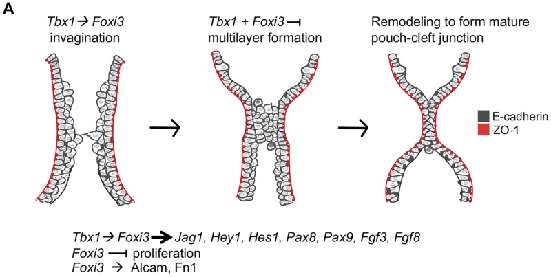
During vertebrate embryonic development, the segmentation of the distal PA is needed to create individual arches that later form derivative structures including the thymus and parathyroid glands [23, 60]. We revisited the process of pharyngeal segmentation to better understand the functions of Tbx1 and Foxi3. Our data indicates that there are a few major epithelial transitions required for morphogenesis, as shown in the model in Fig 9. In the first transition, invagination of the endoderm and ectoderm takes place. Next, a few layers of a partially stratified epithelium forms in the region where invagination occurs starting at E8.75-E9.0. The internal layers of epithelial cells in the partially stratified epithelium do not express the cell polarity protein, ZO-1. Interestingly, it appears as if endoderm and ectoderm cells extend processes towards each other as illustrated in Fig 9. In the final transition, as invagination is completed and the endoderm and ectoderm meet, the multilayers of loosely organized cells form a tightly organized dual intercalated pouch-cleft junction, in which ZO-1 is expressed on the apical surfaces. It can be hypothesized that a zippering process initiates in the center of the forming junction of the epithelia, in which cells become reorganized. E-cadherin expression remains throughout the process indicating that the cells retain at least some of their epithelial properties.
Fig 9. Summary cartoon of Tbx1 and Foxi3 functions in PA segmentation.
Cartoon of epithelial cells during PA segmentation for each arch. Tbx1 and Foxi3 are co-expressed where segmentation to individual pharyngeal arches occurs. When segmentation of the distal PA begins at E8.5, invagination of E-cadherin expressing cells (dark gray) is initiated and a partially stratified multilayer of epithelium forms by E8.75-E9.0, near the point where invagination continues. Tbx1 acts upstream of Foxi3 to promote proper invagination as indicated. The outermost cell layer maintains apical/basal polarity as depicted by ZO-1 expression (red), while the inner layers do not. The epithelial cells begin to project towards each other as shown. Next, a multilayer is juxtaposed where invagination has advanced, and a zippering process ensues from the center of the region, where cells are reorganized. Finally, a dual-layer of intercalated epithelial cells are formed at the mature pouch-cleft junction and both arches are separated. Foxi3 appears to inhibit excess proliferation of PE cells early, while Tbx1 doesn’t alter proliferation. In Tbx1 null mutant embryos more cells are present in the shortened PA. Inactivation of Tbx1 or Foxi3 results in the appearance of excessive layers of endoderm cells, in particular, where invagination is initiating. Therefore, these genes might both promote invagination and restrict excessive multilayers during PA segmentation. We found that Tbx1 and Foxi3 may act in the same pathway upstream of some Notch pathway genes, Pax8 and Pax9, as well as Fgf3. Some of these genes might be required pharyngeal segmentation. It is previously known that Tbx1 and Foxi3 act upstream of Fgf8. While loss of Foxi3 resulted in reduction of Alcam and Fibronectin expression in the extracellular matrix, global loss of Tbx1 did not have the same role. Thus, this data explains, in part, the basis of the genetic interaction between the two genes.
A similar process has been described for branching morphogenesis to form pancreatic ducts from the distal foregut endoderm [61]. In pancreatic organogenesis, a single layer of polarized foregut endoderm will invaginate into the mesenchyme to form branches and ducts composed of differentiated cells. In this process, a single layer of polarized epithelial cells is dynamically transformed to a multilayered epithelium, followed by a second transition back to a monolayer of polarized epithelial cells in the newly formed duct [61, 62]. As for the pharyngeal endoderm, the pancreatic endoderm cells express E-cadherin during this process and the row of cells on the apical side expresses ZO-1 [61, 62].
A few years ago, dynamic transitions of the pharyngeal endoderm were described in zebrafish [54]. During pharyngeal pouch formation, there is a two-step transition to form a temporary stratified epithelium from two layers of cells, which revert back to two opposing layers when pouch formation is complete [54]. In zebrafish and in mouse embryos, E-cadherin is expressed throughout the segmentation process. Apical/basal polarity is lost as detected by lack of ZO-1 expression in internal epithelial cells in zebrafish [54, 63] as we found in mouse embryos. In zebrafish, this process is regulated by signals from the ectoderm and mesoderm to the PE, and is in part, non-autonomous. Specifically, in zebrafish it was found that non-canonical Wnt (Wnt11r) signaling emanating from the mesoderm is required to regulate the process of segmentation [54]. Independently, Wnt4a signals from the ectoderm upstream of the extracellular matrix protein, Alcama (Alcam in mammals) and E-cadherin within cells, are needed to transition from multilayers to two cell layers [54].
Tbx1 functions to promote invagination and restrict excessive multilayer formation in the PE
Tbx1 is expressed in the mesoderm as well as the epithelia. In zebrafish, mesodermal tbx1 regulates expression of wnt11r (non-canonical Wnt) and fgf8a (Fibroblast growth factor 8a) morphogens that signal to the PE to promote pouch formation [20]. Similarly, in mice, inactivation of Tbx1 in the mesoderm results failed segmentation of the distal PA [34]. Thus, the data in zebrafish and mouse is consistent for non-autonomous roles of mesodermal Tbx1 in pharyngeal segmentation. It was previously found that Tbx1 has autonomous roles in the PE for segmentation of the distal PA using the Sox172A-iCre/+ allele [18]. We also found that invagination failed when Tbx1 was inactivated using this allele. In Tbx1 null mutant embryos, excessive multilayers formed by E9.5. We did not observe excessive multilayers when Tbx1 was inactivated in the endoderm at E9.5, although there were some additional layers by E10.5. We speculate that excessive multilayer formation may be partially suppressed in endodermal conditional mutant embryos because Tbx1 is still expressed in the mesoderm.
Distinct roles of Foxi3 in epithelial cell dynamics
In mammals and zebrafish, there are three Foxi class genes, Foxi1, Foxi2 and Foxi3. In zebrafish, foxi1 has a similar expression pattern and function to that of Foxi3 [51]. Recently, it was found that inactivation of foxi1 resulted in a failure to transition from a multilayered epithelium to a simple dual layered pharyngeal pouch and excessive multilayers formed [64]. This is similar to our findings for Foxi3 function in mammals, however we found that Foxi3 is also required in the invagination process. Further, in zebrafish, foxi1 appears to have its major role in the ectoderm to signal non-autonomously to the PE [64]. In foxi1 null mutant zebrafish, Alcama expression was normal. In contrast, in the mouse, loss of Foxi3 in the PE resulted in failed segmentation along with reduced Alcam expression. It is possible that ectodermal Foxi3 might have important signaling roles, but these would be independent to PE functions. Nevertheless, it appears that there are some differences in the function of these homologs in different vertebrates. There are also some differences in zebrafish in regards to tbx1 function. In zebrafish, the tbx1 gene does not have an autonomous role in the PE in pouch formation [20]. In contrast, Tbx1 has autonomous roles in the PE in the mouse.
Differences in Alcam expression levels in Foxi3 and Tbx1 null mutant embryos, implicate some mechanistic differences in gene function on the extracellular milieu. Eph-ephrin signaling in adjacent cells is important for cell migration. In zebrafish, EphB2 and EphB3 are required to maintain E-cadherin expression during budding morphogenesis of the endoderm from the foregut [62]. During pharyngeal pouch morphogenesis in zebrafish, EphrinB signaling is required to increase E-cadherin expression in the second transition of pharyngeal segmentation [58]. In foxi1 mutant zebrafish, expression of EphrinB2a was not changed [64]. Ephrin2a is expressed in a similar pattern to ZO-1. As in zebrafish, we found that Ephrin b2 was not reduced in Foxi3 mutant mouse embryos, and therefore is not directly implicated downstream of Foxi3.
In zebrafish, foxi1 in the pharyngeal ectoderm initiates wnt4a signaling and that this is required for the second transition of the endoderm to form a final mature segment [64]. In endoderm specific Foxi3 conditional mutant mouse embryos, the ectoderm was able to invaginate properly, but segmentation failed. It is possible that endodermal cells might not have been able to properly respond to signals emanating from the ectoderm. Our data also indicates that invagination of the ectoderm is not dependent on invagination of the endoderm. Rather, the ectoderm can initiate invagination independently. This is consistent with studies performed in shark and chick embryos where the endoderm remains separate from the ectoderm throughout epithelial cell invagination [23]. Further work will need to be done to understand the role of extracellular matrix proteins and signaling on epithelial cell dynamics in mouse models.
Shared genetic mechanisms downstream of Tbx1 and Foxi3
The FGF signaling pathway was previously found to be disrupted in the pharyngeal epithelia of both Tbx1 and Foxi3 null mutant embryos. Fgf8 [49] is expressed in the pharyngeal epithelia as well as the mesoderm and is required for early zebrafish [65] and mouse embryogenesis [66–68]. Inactivation of Fgf8 in the pharyngeal epithelia in mouse embryos resulted in similar phenotypic defects in the distal pharyngeal apparatus as the Tbx1 null mutant embryos [69]. Previous published work shows that Fgf8 expression is reduced in the pharyngeal pouches in Tbx1 null mutant mouse embryos and the two genes, Tbx1 and Fgf8 genetically interact [48]. Both fgf3 and fgf8 are required for segmentation of the distal PA in zebrafish [7]. Relevant to this report, Fgf8 expression was reduced Foxi3 null mutant mouse embryos and addition of exogenous fgf3 partially rescued defects in foxi1 morphants [27]. As expected, in this report, we found that Fgf3 is reduced in expression in Foxi3 mutant embryos. This suggests that Tbx1 and Foxi3 might act in the same genetic pathway as Fgf3 and Fgf8 as well as other FGF ligand genes. We found Pax8 and Pax9 expression was also reduced in the PE in Foxi3 and Tbx1 null mutant embryos, and it is possible that in particular, Pax9 is critical for PA segmentation [70]. In addition, we found genes in the Notch pathway reduced in expression in both Tbx1 and Foxi3 null mutant embryos as well.
The Notch pathway has many diverse roles in embryogenesis by regulating Notch effectors of the Hey/Her/Hes class of transcription factors. We previously noted that Jagged1 (Jag1), encoding a cell surface Notch ligand, was reduced in expression the pharyngeal arch epithelia in Tbx1 null mutant embryos [46]. In another report, expression of Hes1, encoding a Notch downstream effector, was reduced in expression in Tbx1 null mutant embryos and further, Hes1 null mutant embryos had similar thymus and aortic arch artery defects as Tbx1 null mutant embryos [21]. More recently, it was reported that Notch pathway genes were altered downstream of failed pharyngeal segmentation in mouse embryos due to inactivation of the transcription co-activator, Eya1 in mouse models [71]. In that study, Jag1, Hes1 and Hey1 expression was altered or reduced in the pharyngeal epithelia [71].
In this report, we found that Jag1, Hes1 and Hey1 were reduced in expression in Tbx1 and Foxi3 null mutant mouse embryos. Tbx1 is still expressed in the abnormal pharyngeal epithelia in Foxi3 null embryos, suggesting that perhaps the loss of expression that is observed might be due to downregulation of expression of Notch pathway genes and other genes described above. It is possible that some of the defects that were observed could be due to reduction of Notch signaling. Although our data support a possible role, it is not known if Notch pathway genes are required for segmentation of the PA. Therefore, more studies are needed to be done in the future to test a possible role for Notch signaling in this process.
Translational insights
Patients with 22q11.2DS have defects within structures derived from the PA including craniofacial dysmorphism, T-cell deficiencies or dysfunction, hypocalcemia, as well as aortic arch and cardiac outflow tract defects [72]. TBX1 is the major candidate gene for these defects, and it is required for PA segmentation [17, 18, 20]. Based on results presented in this report, we suggest that Tbx1 may act upstream of Foxi3 in this process. One question is whether individuals might be identified that have mutations in FOXI3. There has been one report of a patient with a deletion of one allele of FOXI3 that had severe ear defects, mild craniofacial defects, and missing arteries derived from PA1 and PA2 [73]. These symptoms are due to defects of structures derived from the PA but are different from those typically observed in patients with 22q11.2DS.
The phenotypic expression of 22q11.2DS varies extensively, implicating the existence of genetic or environmental modifiers. It would be interesting to determine whether DNA sequence variants in FOXI3 or other downstream genes, such as FGF pathway genes, PAX9, or Notch pathway genes, might act as potential modifiers of phenotype in individuals with 22q11.2DS. Analysis of sequence from a large cohort of individuals with 22q11.2DS will be required to test this possibility.
Materials and methods
Ethics statement
All experiments using mice were carried out according to regulatory standards defined by the National Institutes of Health and the Institute for Animal Studies, Albert Einstein College of Medicine (https://www.einstein.yu.edu/administration/animal-studies/), IACUC protocol # 2016–0507.
Mouse mutant alleles
The following mouse mutant alleles used in this study have been previously described: Foxi3f/f (flox = f), Foxi3+/- [27], Tbx1Cre/+ [36] Sox172A-iCre/+ [38], Tbx1+/- [11] and ROSA26GFPf/+ (RCE: loxP) [74]. Foxi3-/- embryos were generated by inter-crossing Foxi3+/- mice. Double Tbx1 and Foxi3 heterozygous embryos were generated by inter-crossing Tbx1+/- and Foxi3+/- mice. Tbx1Cre/+;Foxi3f/f and Sox172A-iCre/+;Foxi3f/f embryos were generated by crossing male Tbx1Cre/+;Foxi3f/+ or Sox172A-iCre/+;Foxi3f/+ mice with Foxi3f/f females. Foxi3+/-, Tbx1+/-, Tbx1Cre/+;Foxi3f/+, Sox172A-iCre/+;Foxi3f/+, Sox172A-iCre/+;Tbx1f/+ and wildtype littermates were used as controls for the experiments, as indicated.
The Foxi3+/-, Sox172A-iCre/+, and Tbx1Cre/+ mice were backcrossed 10 generations to a Swiss Webster background from a mixed C57Bl/6, Swiss Webster background. The PCR strategies for mouse genotyping have been described in the original reports and are available upon request.
Mouse embryo heart histology and phenotypic analysis
Mouse embryos were isolated in phosphate-buffered saline (PBS) and fixed overnight in 10% neutral buffered formalin (Sigma Corp.). Following fixation, the embryos were dehydrated through a graded ethanol series, embedded in paraffin and sectioned at 10 μm. All histological sections were stained with hematoxylin and eosin (H&E) using standard protocols in the Einstein Histopathology Core Facility (http://www.einstein.yu.edu/histopathology/page.aspx). A total of 80 embryos, including controls, at E15.5 were obtained from more than 10 independent crosses and analyzed morphologically using light microscopy. Fisher’s exact test was used to determine if parathyroid and thymus gland defects were significant in Tbx1+/-; Foxi3+/- compared to Tbx1+/- embryos.
RNAscope in situ hybridization and quantification
RNAscope in situ hybridization with non-radioactive mRNA probes was performed as previously described [75]. Tissue was fixed in 4% paraformaldehyde (PFA) for 24 hours at 4°C and then cryopreserved in 30% sucrose in PBS overnight at 4°C. Embryos were embedded in OCT and cryosectioned at 10 μm thickness. RNAscope probes for Tbx1, Foxi3, Pax9, Hey1, Hes1, Jag1, Foxn1, and Gcm2 were generated by Advanced Cell Diagnostics. Quantification was performed using Volocity Software (Perkin Elmer Corporation) where each nuclei and mRNA signal dot were counted. Each probe was calculated separately. The ratios of cell number to number of signal dots was calculated for each embryo (n = 3). P-values were determined using the Student’s t-test.
Whole mount in situ hybridization
Whole-mount RNA in situ hybridization with non-radioactive probes was performed as previously described [76, 77], using PCR-based probes for Foxi3 [24], Tbx1 [78], Jag1 [79], and Hey1 [80]. The probe for Hes1 was generated from a cDNA plasmid clone [21, 81]. Following the whole mount RNA in situ hybridization protocol, the embryos were fixed in 4% PFA and then dehydrated through a series of graded ethanol steps, embedded in paraffin, and sectioned at 10 μm thickness. Minimum of 2–4 embryos from 2–3 independent litters were analyzed for each experiment.
Immunofluorescence on embryo sections
Embryos were collected at various stages: E8.5 (7–10 somite pairs), E9.0 (15–19 somite pairs), E9.5 (20–23 somite pairs) E10.0 (24–29 somite pairs), and E10.5 (30–33 somite pairs). Fixation was carried out in 4% PFA in PBS at 4°C for two hours. After fixation, tissue was washed in PBS and then cryoprotected in 30% sucrose in PBS overnight at 4°C. Embryos were embedded in OCT and cryosectioned at 10 μm. After fixation, frozen sections were obtained as described and then permeabilized in 0.5% Triton X-100 for 5 min. Blocking was performed with 5% goat serum in PBS/0.1% Triton X-100 (PBT) for 1 hour. Primary antibody was diluted in blocking solution and incubated for 1 hour. Primary antibodies used included: E-cadherin (BD Transduction laboratories 610181, 1:200 mouse), ZO-1 (Invitrogen 61–7300, 1:200 rabbit), Fibronectin (ab2413, 1:100 rabbit), Alcam (R&D BAM6561, 1:50 mouse), Ephrin b2 (ab150411, 1:200 rabbit) and GFP (ab6673 1:500 goat). Proliferation of cells was assessed by immunofluorescence using antibody anti-phospho Histone H3 (Ser10), which is a mitosis marker (06–570 Millipore). Sections were washed in PBT and incubated with a secondary antibody for 1 hour. Secondary antibodies used were Alexa Fluor 488 goat a-mouse IgG (Invitrogen A32723) at 1:500 and Alexa Fluor 568 donkey a-rabbit IgG (Invitrogen A11019) at 1:500. Slides were mounted in hard-set mounting medium with DAPI (Vector Labs H-1500). Images were then captured using a Zeiss Axio Observer microscope with an apotome.
Cell number and proliferation quantification on tissue sections
To count epithelial cell number, we obtained 10 μm serial coronal sections of control, Tbx1-/-, Foxi3-/-, and Sox172A-iCre/+;Foxi3f/f embryos, which were collected and stained with an antibody for E-cadherin. To ensure that the cell quantification was accurate, we counted E-cadherin positive cells in the PA in every other section throughout each embryo. We did not count epithelial cells that were not part of the PA. When counting cells, we matched the embryos by stage using somite counts, and we matched the sections by position within the embryo. We also ensured that for each pair of control and mutant embryos, we counted the same number of sections (10–12 per embryo). We counted all phosphoH3 positive epithelial cells in each section and calculated the ratio of proliferating cells within the pharyngeal epithelium. For Tbx1-/- mutant embryos, since the PA is shorter in comparison to WT littermates, we also calculated the size of the PA using ImageJ. We then counted the E-cadherin positive cells of the PA, marking the epithelium, and divided this number by the size of the PA. The mean and standard error of the average cell counts for controls and mutant embryos were determined and they were compared using the t-test. Representations of the complete PA region from at least 3–6 embryos per genotype from at least 3 independent litters were used in each assay.
Supporting information
(A-F) Transverse histology sections stained with H&E of Foxi3+/- (A-B), Tbx1+/- (C-D) and Foxi3+/-;Tbx1+/- (E-F) embryos at E15.5. Abbreviations: parathyroid (PT), thyroid (T) and thymus (Thy). (G-H) Tables summarizing parathyroid (G) and thymus (H) defects observed. Numbers of glands observed was indicated (two per embryo). Foxi3+/-, n = 8; Tbx1+/-, n = 13 and Foxi3+/-;Tbx1+/-, n = 14 embryos at E15.5. Normally parathyroid glands are adjacent to thyroid glands (A). Parathyroid glands were scored as being ectopic when they were more caudally located than the thyroid glands in mutant embryos (C, E). Thymus glands were noted as hypoplastic that were smaller in size (F) than normal (B, D). Ectopic thymus glands were scored as such that were more rostrally located in comparison to control embryos (F). (I-M) Coronal histology sections stained with H&E of WT (I-J) and Tbx1+/-;Foxi3+/- (K-M) embryos at E10.5. Black arrows indicate the third pharyngeal pouch (I-M). Related to Figs 1 and 2.
(TIF)
(A-H) WMISH using antisense Foxi3 mRNA probe on Tbx1Cre/+;Foxi3f/+ control (A-B), Tbx1Cre/+;Foxi3f/f conditional mutant embryos (C-D), Sox172A-iCre/+;Foxi3f/+ control (E-F) and Sox172A-iCre/+;Foxi3f/f conditional mutant embryos (G-H) with corresponding coronal sections. Black arrows indicate where Foxi3 mRNA expression is reduced within conditional mutant embryos. Related to Figs 1–4 and 6.
(TIF)
(A-F) Transverse histology sections stained with H&E of WT control (A-C) and Tbx1Cre/+;Foxi3f/f mutant (D-F) embryos. (G-H) India ink was injected into the ventricle of Tbx1Cre/+;Foxi3f/+ control (G) and mutant (H) embryos at E10.5 to visualize the aortic arches. (I-L) Transverse histology sections of Tbx1Cre/+;Foxi3f/- mutant embryos at E15.5. Abbreviations: right subclavian artery (R. sub), thymus (Thy), aorta (Ao), pulmonary trunk (PT), right atrium (RA), left atrium (LA), right ventricle (RV), left ventricle (LV), ventricular septum (VS). Related to Figs 1 and 4.
(TIF)
(A-C) Whole mount images of Foxi3+/- control (A), Foxi3-/- (B), and Sox172A-iCre/+;Foxi3f/f (C) embryos at E9.5. Arrow in A indicates PA3. Asterisks in B, indicate the hypoplastic first arch and in C, indicate the distal PA that failed to segment to arches. (D-F) Transverse histology sections stained with H&E of Sox172A-iCre/+;Foxi3f/f embryos at E15.5. RRSA is indicated (D), IAAB is present and indicated by the yellow arrow (E). Abbreviations: aorta (Ao), right ventricle (RV), left ventricle (LV), and retro-esophageal right subclavian artery (RRSA). (G-L) India ink was injected into the ventricle of WT control (G, J), Foxi3-/- (H, K) and Sox172A-iCre/+;Foxi3f/f (I, L) embryos at E10.5. The right and left side of these embryos are shown. Arrows indicate absent 4th aortic arch arteries in both genotypes of mutant embryos. Related to Figs 1 and 6.
(TIF)
(A-F) Proliferation assay on coronal sections of WT (A-C) and Tbx1-/- mutant embryos (D-F) using a phospho-H3 (ph3, red) antibody to mark cells undergoing mitosis. E-cadherin antibody (green) was utilized to visualize the epithelial cells. E8.5 (A and D) and E9.5 (B-C and E-F) staged embryos were analyzed. White boxes in B and E indicate area of magnification; n = 3 for both stages and genotypes. (G) Quantification of proliferation assay. The mitotic index is the ratio of proliferating cells to total cell counts of epithelium within the PA. The t-test was used to calculate P-values as shown. (H-K) DAPI (blue), E-cadherin (green), and ZO-1 (red) antibodies were used to visualize epithelial cells within the PA. Epithelial cells in WT (H-I) Tbx1-/- (J-K) coronal sections were visualized at E8.5 (H and J) and E9.5 (I and K); n = 3 each genotype. PE indicates pharyngeal endoderm. Arrows in H, indicate the position in the PA where cells are invaginating. (L) Quantification of cell numbers to proportion of the size of the PA. (M-P) Sox172A-iCre/+;Tbx1f/+ controls and Sox172A-iCre/+;Tbx1f/f conditional mutant embryos at E9.5 (M-N) and E10.5 (O-P; n = 3 for both stages and genotypes). Related to Fig 5.
(TIF)
(A-L) Proliferation assay was performed using a phospho-H3 (pH3) antibody on coronal sections of WT control (A-D), Foxi3-/- (E-H), and Sox172A-iCre/+;Foxi3f/f (I-L) mutant embryos at E8.5 and E9.5. At E8.5 and E9.5; a total of n = 6 and n = 4, respectively, were analyzed for each control and mutant embryo. Related to Fig 6.
(TIF)
(A-C) WMISH was performed using an Isl1 antisense probe on WT (A), Foxi3-/- (B), and Tbx1-/- (C) mutant embryos at E9.5. (D-E) WMISH was performed using an Fgf3 probe on WT (D) and Foxi3-/- (E) mutant embryos at E9.5. (F-H) WMISH was performed using a Pax8 probe on WT (F), Foxi3-/- (G) and Tbx1-/- (H) mutant embryos at E9.5. (I-K) Pax9 probe on WT control (I), Foxi3-/- (J) and Tbx1-/- (K) mutant embryos; n = 2–4 for each probe and genotype. (L-M) RNAscope in situ hybridization with an mRNA probe for Pax9 (green) on coronal sections in WT (L) and Foxi3-/- (M) embryos at E9.5; n = 2. PE indicates the pharyngeal endoderm. Related to Fig 7.
(TIF)
Acknowledgments
We thank Silvia Racedo, Christopher DeBono and Hiroko Nomaru at Albert Einstein College of Medicine for helpful suggestions. We also thank Xia Wang and Sophie Astrof at Rutgers University for assisting with India ink injections. We thank Andrew Groves at Baylor College of Medicine, for providing Foxi3 heterozygous and floxed mice and probes. We would like to thank the laboratory of Albert Basson and Peter Scambler for the Pax9 probe. We want to thank the Histopathology Facility at Einstein. We also want to thank the Imaging Facility and its staff members, Vera DesMarais and Andrea Briceno for help quantifying the RNAscope images.
Data Availability
All relevant data are within the manuscript and its Supporting Information files.
Funding Statement
This study was supported by National Institute of Health grant, P01HD070454 https://www.nih.gov/ to BEM and Leducq Foundation Network Grant https://www.fondationleducq.org/ to BEM and EH. The funders had no role in study design, data collection and analysis, decision to publish, or preparation of the manuscript.
References
- 1.Graham A. The development and evolution of the pharyngeal arches. J Anat. 2001. [DOI] [PMC free article] [PubMed]
- 2.Frisdal A, Trainor PA. Development and evolution of the pharyngeal apparatus. Wiley Interdiscip Rev Dev Biol. 2014;3(6):403–18. Epub 2014/09/02. 10.1002/wdev.147 [DOI] [PMC free article] [PubMed] [Google Scholar]
- 3.Grevellec A, Tucker AS. The pharyngeal pouches and clefts: Development, evolution, structure and derivatives. Semin Cell Dev Biol. 2010;21(3):325–32. Epub 2010/02/11. 10.1016/j.semcdb.2010.01.022 . [DOI] [PubMed] [Google Scholar]
- 4.Xu H, Cerrato F, Baldini A. Timed mutation and cell-fate mapping reveal reiterated roles of Tbx1 during embryogenesis, and a crucial function during segmentation of the pharyngeal system via regulation of endoderm expansion. Development. 2005;132(19):4387–95. Epub 2005/09/06. 10.1242/dev.02018 . [DOI] [PubMed] [Google Scholar]
- 5.Graham A. Deconstructing the pharyngeal metamere. J Exp Zool B Mol Dev Evol. 2008;310(4):336–44. Epub 2007/06/23. 10.1002/jez.b.21182 . [DOI] [PubMed] [Google Scholar]
- 6.Veitch Emma BJ, Schilling Thomas, Smith Moya, Graham Anthony. Pharyngeal Arch patterning in the absence of neural crest. 1999. [DOI] [PubMed]
- 7.Crump JG, Maves L, Lawson ND, Weinstein BM, Kimmel CB. An essential role for Fgfs in endodermal pouch formation influences later craniofacial skeletal patterning. Development. 2004;131(22):5703–16. Epub 2004/10/29. 10.1242/dev.01444 . [DOI] [PubMed] [Google Scholar]
- 8.Wendling Olivia DC, Chambon Pierre, Manuel Mark. Retinoid signalin is essential for patterning the endoderm of the third and fourth pharyngeal arches. Development. 2000. [DOI] [PubMed]
- 9.Chojnowski JL, Masuda K, Trau HA, Thomas K, Capecchi M, Manley NR. Multiple roles for HOXA3 in regulating thymus and parathyroid differentiation and morphogenesis in mouse. Development. 2014;141(19):3697–708. Epub 2014/09/25. 10.1242/dev.110833 [DOI] [PMC free article] [PubMed] [Google Scholar]
- 10.Kelly RG, Jerome-Majewska LA, Papaioannou VE. The del22q11.2 candidate gene Tbx1 regulates branchiomeric myogenesis. Hum Mol Genet. 2004;13(22):2829–40. Epub 2004/09/24. 10.1093/hmg/ddh304 . [DOI] [PubMed] [Google Scholar]
- 11.Merscher S, Funke B, Epstein JA, Heyer J, Puech A, Lu MM, et al. TBX1 is responsible for cardiovascular defects in velo-cardio-facial/DiGeorge syndrome. Cell. 2001;104(4):619–29. Epub 2001/03/10. 10.1016/s0092-8674(01)00247-1 . [DOI] [PubMed] [Google Scholar]
- 12.Lindsay EA, Vitelli F, Su H, Morishima M, Huynh T, Pramparo T, et al. Tbx1 haploinsufficieny in the DiGeorge syndrome region causes aortic arch defects in mice. Nature. 2001;410(6824):97–101. Epub 2001/03/10. 10.1038/35065105 . [DOI] [PubMed] [Google Scholar]
- 13.Jerome LA, Papaioannou VE. DiGeorge syndrome phenotype in mice mutant for the T-box gene, Tbx1. Nat Genet. 2001;27(3):286–91. Epub 2001/03/10. 10.1038/85845 . [DOI] [PubMed] [Google Scholar]
- 14.Garg V, Yamagishi C, Hu T, Kathiriya IS, Yamagishi H, Srivastava D. Tbx1, a DiGeorge syndrome candidate gene, is regulated by sonic hedgehog during pharyngeal arch development. Dev Biol. 2001;235(1):62–73. Epub 2001/06/20. 10.1006/dbio.2001.0283 . [DOI] [PubMed] [Google Scholar]
- 15.Chapman DL, Garvey N, Hancock S, Alexiou M, Agulnik SI, Gibson-Brown JJ, et al. Expression of the T-box family genes, Tbx1-Tbx5, during early mouse development. Dev Dyn. 1996;206(4):379–90. Epub 1996/08/01. . [DOI] [PubMed] [Google Scholar]
- 16.Arnold JS, Braunstein EM, Ohyama T, Groves AK, Adams JC, Brown MC, et al. Tissue-specific roles of Tbx1 in the development of the outer, middle and inner ear, defective in 22q11DS patients. Hum Mol Genet. 2006;15(10):1629–39. Epub 2006/04/08. 10.1093/hmg/ddl084 [DOI] [PMC free article] [PubMed] [Google Scholar]
- 17.Arnold JS, Werling U, Braunstein EM, Liao J, Nowotschin S, Edelmann W, et al. Inactivation of Tbx1 in the pharyngeal endoderm results in 22q11DS malformations. Development. 2006;133(5):977–87. Epub 2006/02/03. 10.1242/dev.02264 . [DOI] [PubMed] [Google Scholar]
- 18.Jackson A, Kasah S, Mansour SL, Morrow B, Basson MA. Endoderm-specific deletion of Tbx1 reveals an FGF-independent role for Tbx1 in pharyngeal apparatus morphogenesis. Dev Dyn. 2014;243(9):1143–51. Epub 2014/05/09. 10.1002/dvdy.24147 [DOI] [PMC free article] [PubMed] [Google Scholar]
- 19.Racedo SE, Hasten E, Lin M, Devakanmalai GS, Guo T, Ozbudak EM, et al. Reduced dosage of beta-catenin provides significant rescue of cardiac outflow tract anomalies in a Tbx1 conditional null mouse model of 22q11.2 deletion syndrome. PLoS Genet. 2017;13(3):e1006687 Epub 2017/03/28. 10.1371/journal.pgen.1006687 [DOI] [PMC free article] [PubMed] [Google Scholar]
- 20.Choe CP, Crump JG. Tbx1 controls the morphogenesis of pharyngeal pouch epithelia through mesodermal Wnt11r and Fgf8a. Development. 2014;141(18):3583–93. Epub 2014/08/22. 10.1242/dev.111740 [DOI] [PMC free article] [PubMed] [Google Scholar]
- 21.van Bueren KL, Papangeli I, Rochais F, Pearce K, Roberts C, Calmont A, et al. Hes1 expression is reduced in Tbx1 null cells and is required for the development of structures affected in 22q11 deletion syndrome. Dev Biol. 2010;340(2):369–80. Epub 2010/02/04. 10.1016/j.ydbio.2010.01.020 [DOI] [PMC free article] [PubMed] [Google Scholar]
- 22.Quinlan R, Martin P, Graham A. The role of actin cables in directing the morphogenesis of the pharyngeal pouches. Development. 2004;131(3):593–9. Epub 2004/01/09. 10.1242/dev.00950 . [DOI] [PubMed] [Google Scholar]
- 23.Shone V, Graham A. Endodermal/ectodermal interfaces during pharyngeal segmentation in vertebrates. J Anat. 2014;225(5):479–91. Epub 2014/09/10. 10.1111/joa.12234 [DOI] [PMC free article] [PubMed] [Google Scholar]
- 24.Ohyama T, Groves AK. Expression of mouse Foxi class genes in early craniofacial development. Dev Dyn. 2004;231(3):640–6. Epub 2004/09/18. 10.1002/dvdy.20160 . [DOI] [PubMed] [Google Scholar]
- 25.Shirokova V, Jussila M, Hytonen MK, Perala N, Drogemuller C, Leeb T, et al. Expression of Foxi3 is regulated by ectodysplasin in skin appendage placodes. Dev Dyn. 2013;242(6):593–603. Epub 2013/02/27. 10.1002/dvdy.23952 . [DOI] [PubMed] [Google Scholar]
- 26.Drogemuller C, Karlsson, E. K., Hytonen, M. K., Perloski, M., Dolf, G., Sainio, K., Lohi, H., Lindblad-Toh, K., Leeb, T.. A Mutation in Hairless Dogs Implicates Foxi3 in Ectodermal Development. Science. 2008. [DOI] [PubMed]
- 27.Edlund RK, Ohyama T, Kantarci H, Riley BB, Groves AK. Foxi transcription factors promote pharyngeal arch development by regulating formation of FGF signaling centers. Dev Biol. 2014;390(1):1–13. Epub 2014/03/22. 10.1016/j.ydbio.2014.03.004 [DOI] [PMC free article] [PubMed] [Google Scholar]
- 28.Khatri SB, Groves AK. Expression of the Foxi2 and Foxi3 transcription factors during development of chicken sensory placodes and pharyngeal arches. Gene Expr Patterns. 2013;13(1–2):38–42. Epub 2012/11/06. 10.1016/j.gep.2012.10.001 [DOI] [PMC free article] [PubMed] [Google Scholar]
- 29.Birol O, Ohyama T, Edlund RK, Drakou K, Georgiades P, Groves AK. The mouse Foxi3 transcription factor is necessary for the development of posterior placodes. Dev Biol. 2016;409(1):139–51. Epub 2015/11/10. 10.1016/j.ydbio.2015.09.022 [DOI] [PMC free article] [PubMed] [Google Scholar]
- 30.Manley NR, Condie BG. Transcriptional Regulation of Thymus Organogenesis and Thymic Epithelial Cell Differentiation. Development of T Cell Immunity. Progress in Molecular Biology and Translational Science2010. p. 103–20. [DOI] [PubMed]
- 31.Bain VE, Gordon J, O’Neil JD, Ramos I, Richie ER, Manley NR. Tissue-specific roles for sonic hedgehog signaling in establishing thymus and parathyroid organ fate. Development. 2016;143(21):4027–37. Epub 2016/11/03. 10.1242/dev.141903 [DOI] [PMC free article] [PubMed] [Google Scholar]
- 32.Gordon J, Bennett AR, Blackburn CC, Manley NR. Gcm2 and Foxn1 mark early parathyroid- and thymus-specific domains in the developing third pharyngeal pouch. Mech Dev. 2001;103(1–2):141–3. Epub 2001/05/04. . [DOI] [PubMed] [Google Scholar]
- 33.Zhang Z, Cerrato F, Xu H, Vitelli F, Morishima M, Vincentz J, et al. Tbx1 expression in pharyngeal epithelia is necessary for pharyngeal arch artery development. Development. 2005;132(23):5307–15. Epub 2005/11/15. 10.1242/dev.02086 . [DOI] [PubMed] [Google Scholar]
- 34.Zhang Z, Huynh T, Baldini A. Mesodermal expression of Tbx1 is necessary and sufficient for pharyngeal arch and cardiac outflow tract development. Development. 2006;133(18):3587–95. Epub 2006/08/18. 10.1242/dev.02539 [DOI] [PMC free article] [PubMed] [Google Scholar]
- 35.Khatri SB, Edlund RK, Groves AK. Foxi3 is necessary for the induction of the chick otic placode in response to FGF signaling. Dev Biol. 2014;391(2):158–69. Epub 2014/05/02. 10.1016/j.ydbio.2014.04.014 [DOI] [PMC free article] [PubMed] [Google Scholar]
- 36.Vitelli F, Zhang Z, Huynh T, Sobotka A, Mupo A, Baldini A. Fgf8 expression in the Tbx1 domain causes skeletal abnormalities and modifies the aortic arch but not the outflow tract phenotype of Tbx1 mutants. Dev Biol. 2006;295(2):559–70. Epub 2006/05/16. 10.1016/j.ydbio.2006.03.044 [DOI] [PMC free article] [PubMed] [Google Scholar]
- 37.Calmont A, Ivins S, Van Bueren KL, Papangeli I, Kyriakopoulou V, Andrews WD, et al. Tbx1 controls cardiac neural crest cell migration during arch artery development by regulating Gbx2 expression in the pharyngeal ectoderm. Development. 2009;136(18):3173–83. Epub 2009/08/25. 10.1242/dev.028902 [DOI] [PMC free article] [PubMed] [Google Scholar]
- 38.Engert S, Liao WP, Burtscher I, Lickert H. Sox17-2A-iCre: a knock-in mouse line expressing Cre recombinase in endoderm and vascular endothelial cells. Genesis. 2009;47(9):603–10. Epub 2009/06/24. 10.1002/dvg.20540 . [DOI] [PubMed] [Google Scholar]
- 39.Gumbiner BM. Cell adhesion: the molecular basis of tissue architecture and morphogenesis. Cell. 1996;84(3):345–57. Epub 1996/02/09. 10.1016/s0092-8674(00)81279-9 . [DOI] [PubMed] [Google Scholar]
- 40.Anderson JM, Van Itallie CM, Peterson MD, Stevenson BR, Carew EA, Mooseker MS. ZO-1 mRNA and protein expression during tight junction assembly in Caco-2 cells. J Cell Biol. 1989;109(3):1047–56. Epub 1989/09/01. 10.1083/jcb.109.3.1047 [DOI] [PMC free article] [PubMed] [Google Scholar]
- 41.Carrieri FA, Dale JK. Turn It Down a Notch. Front Cell Dev Biol. 2016;4:151 Epub 2017/02/06. 10.3389/fcell.2016.00151 [DOI] [PMC free article] [PubMed] [Google Scholar]
- 42.Zuniga E, Stellabotte F, Crump JG. Jagged-Notch signaling ensures dorsal skeletal identity in the vertebrate face. Development. 2010;137(11):1843–52. Epub 2010/05/01. 10.1242/dev.049056 [DOI] [PMC free article] [PubMed] [Google Scholar]
- 43.Fischer A, Steidl C, Wagner TU, Lang E, Jakob PM, Friedl P, et al. Combined loss of Hey1 and HeyL causes congenital heart defects because of impaired epithelial to mesenchymal transition. Circ Res. 2007;100(6):856–63. Epub 2007/02/17. 10.1161/01.RES.0000260913.95642.3b . [DOI] [PubMed] [Google Scholar]
- 44.MacGrogan D, Munch J, de la Pompa JL. Notch and interacting signalling pathways in cardiac development, disease, and regeneration. Nat Rev Cardiol. 2018;15(11):685–704. Epub 2018/10/06. 10.1038/s41569-018-0100-2 . [DOI] [PubMed] [Google Scholar]
- 45.Figueiredo M, Silva JC, Santos AS, Proa V, Alcobia I, Zilhao R, et al. Notch and Hedgehog in the thymus/parathyroid common primordium: Crosstalk in organ formation. Dev Biol. 2016;418(2):268–82. Epub 2016/08/22. 10.1016/j.ydbio.2016.08.012 . [DOI] [PubMed] [Google Scholar]
- 46.Macchiarulo S, Morrow BE. Tbx1 and Jag1 act in concert to modulate the fate of neurosensory cells of the mouse otic vesicle. Biol Open. 2017;6(10):1472–82. Epub 2017/08/26. 10.1242/bio.027359 [DOI] [PMC free article] [PubMed] [Google Scholar]
- 47.Rochais F, Dandonneau M, Mesbah K, Jarry T, Mattei MG, Kelly RG. Hes1 is expressed in the second heart field and is required for outflow tract development. PLoS One. 2009;4(7):e6267 Epub 2009/07/18. 10.1371/journal.pone.0006267 [DOI] [PMC free article] [PubMed] [Google Scholar]
- 48.Vitelli F, Taddei I, Morishima M, Meyers EN, Lindsay EA, Baldini A. A genetic link between Tbx1 and fibroblast growth factor signaling. Development. 2002;129(19):4605–11. Epub 2002/09/12. . [DOI] [PubMed] [Google Scholar]
- 49.Frank DU, Fotheringham LK, Brewer JA, Muglia LJ, Tristani-Firouzi M, Capecchi MR, et al. An Fgf8 mouse mutant phenocopies human 22q11 deletion syndrome. Development. 2002;129(19):4591–603. Epub 2002/09/12. [DOI] [PMC free article] [PubMed] [Google Scholar]
- 50.Aggarwal VS, Liao J, Bondarev A, Schimmang T, Lewandoski M, Locker J, et al. Dissection of Tbx1 and Fgf interactions in mouse models of 22q11DS suggests functional redundancy. Hum Mol Genet. 2006;15(21):3219–28. Epub 2006/09/27. 10.1093/hmg/ddl399 . [DOI] [PubMed] [Google Scholar]
- 51.Solomon KS, Kudoh T, Dawid IB, Fritz A. Zebrafish foxi1 mediates otic placode formation and jaw development. Development. 2003;130(5):929–40. Epub 2003/01/23. 10.1242/dev.00308 . [DOI] [PubMed] [Google Scholar]
- 52.Okubo T, Kawamura A, Takahashi J, Yagi H, Morishima M, Matsuoka R, et al. Ripply3, a Tbx1 repressor, is required for development of the pharyngeal apparatus and its derivatives in mice. Development. 2011;138(2):339–48. Epub 2010/12/24. 10.1242/dev.054056 . [DOI] [PubMed] [Google Scholar]
- 53.Tomita K, van Bokhoven A, Jansen CF, Bussemakers MJ, Schalken JA. Coordinate recruitment of E-cadherin and ALCAM to cell-cell contacts by alpha-catenin. Biochem Biophys Res Commun. 2000;267(3):870–4. Epub 2000/02/16. 10.1006/bbrc.1999.2040 . [DOI] [PubMed] [Google Scholar]
- 54.Choe CP, Collazo A, Trinh le A, Pan L, Moens CB, Crump JG. Wnt-dependent epithelial transitions drive pharyngeal pouch formation. Dev Cell. 2013;24(3):296–309. Epub 2013/02/05. 10.1016/j.devcel.2012.12.003 [DOI] [PMC free article] [PubMed] [Google Scholar]
- 55.Choe CP, Crump JG. Eph-Pak2a signaling regulates branching of the pharyngeal endoderm by inhibiting late-stage epithelial dynamics. Development. 2015;142(6):1089–94. Epub 2015/03/01. 10.1242/dev.115774 [DOI] [PMC free article] [PubMed] [Google Scholar]
- 56.Mittal A, Pulina M, Hou SY, Astrof S. Fibronectin and integrin alpha 5 play essential roles in the development of the cardiac neural crest. Mech Dev. 2010;127(9–12):472–84. Epub 2010/09/03. 10.1016/j.mod.2010.08.005 [DOI] [PMC free article] [PubMed] [Google Scholar]
- 57.Trinh le A, Stainer Didier, Y.R. Fibronectin Regulates Epithelial Organization during Myocardial Migration in Zebrafish. Developmental Cell. 2004. [DOI] [PubMed]
- 58.Choe C. Eph-Pak2a signalling regulates branching of the pharyngeal endoderm by inhibiting late-stage epithelial dynamics. Development. 2015. [DOI] [PMC free article] [PubMed]
- 59.Francou A, Saint-Michel E, Mesbah K, Kelly RG. TBX1 regulates epithelial polarity and dynamic basal filopodia in the second heart field. Development. 2014;141(22):4320–31. Epub 2014/11/06. 10.1242/dev.115022 . [DOI] [PubMed] [Google Scholar]
- 60.Graham A, Okabe M, Quinlan R. The role of the endoderm in the development and evolution of the pharyngeal arches. J Anat. 2005;207(5):479–87. Epub 2005/11/30. 10.1111/j.1469-7580.2005.00472.x [DOI] [PMC free article] [PubMed] [Google Scholar]
- 61.Hick AC, van Eyll JM, Cordi S, Forez C, Passante L, Kohara H, et al. Mechanism of primitive duct formation in the pancreas and submandibular glands: a role for SDF-1. BMC Dev Biol. 2009;9:66 Epub 2009/12/17. 10.1186/1471-213X-9-66 [DOI] [PMC free article] [PubMed] [Google Scholar]
- 62.Villasenor A, Wang ZV, Rivera LB, Ocal O, Asterholm IW, Scherer PE, et al. Rgs16 and Rgs8 in embryonic endocrine pancreas and mouse models of diabetes. Dis Model Mech. 2010;3(9–10):567–80. Epub 2010/07/10. 10.1242/dmm.003210 [DOI] [PMC free article] [PubMed] [Google Scholar]
- 63.Choe CP, Crump JG. Dynamic epithelia of the developing vertebrate face. Curr Opin Genet Dev. 2015;32:66–72. Epub 2015/03/10. 10.1016/j.gde.2015.02.003 [DOI] [PMC free article] [PubMed] [Google Scholar]
- 64.Jin S, O J, Stellabotte F, Choe CP. Foxi1 promotes late-stage pharyngeal pouch morphogenesis through ectodermal Wnt4a activation. Dev Biol. 2018. Epub 2018/06/23. 10.1016/j.ydbio.2018.06.011 . [DOI] [PubMed] [Google Scholar]
- 65.Reifers F, Bohli H, Walsh EC, Crossley PH, Stainier DY, Brand M. Fgf8 is mutated in zebrafish acerebellar (ace) mutants and is required for maintenance of midbrain-hindbrain boundary development and somitogenesis. Development. 1998;125(13):2381–95. Epub 1998/06/04. . [DOI] [PubMed] [Google Scholar]
- 66.Meyers EN, Lewandoski M, Martin GR. An Fgf8 mutant allelic series generated by Cre- and Flp-mediated recombination. Nat Genet. 1998;18(2):136–41. Epub 1998/02/14. 10.1038/ng0298-136 . [DOI] [PubMed] [Google Scholar]
- 67.Moon AM, Capecchi MR. Fgf8 is required for outgrowth and patterning of the limbs. Nat Genet. 2000;26(4):455–9. Epub 2000/12/02. 10.1038/82601 [DOI] [PMC free article] [PubMed] [Google Scholar]
- 68.Sun X, Meyers EN, Lewandoski M, Martin GR. Targeted disruption of Fgf8 causes failure of cell migration in the gastrulating mouse embryo. Genes Dev. 1999;13(14):1834–46. Epub 1999/07/27. 10.1101/gad.13.14.1834 [DOI] [PMC free article] [PubMed] [Google Scholar]
- 69.Macatee TL, Hammond BP, Arenkiel BR, Francis L, Frank DU, Moon AM. Ablation of specific expression domains reveals discrete functions of ectoderm- and endoderm-derived FGF8 during cardiovascular and pharyngeal development. Development. 2003;130(25):6361–74. Epub 2003/11/19. 10.1242/dev.00850 [DOI] [PMC free article] [PubMed] [Google Scholar]
- 70.Steinbok P. Recurrence patterns and anaplastic change in a long-term study of pilocytic astrocytomas. Pediatr Neurosurg. 1998;28(2):108 Epub 1998/08/26. 10.1159/000028632 . [DOI] [PubMed] [Google Scholar]
- 71.Zhang H, Wang L, Wong EYM, Tsang SL, Xu PX, Lendahl U, et al. An Eya1-Notch axis specifies bipotential epibranchial differentiation in mammalian craniofacial morphogenesis. Elife. 2017;6 Epub 2017/11/16. 10.7554/eLife.30126 [DOI] [PMC free article] [PubMed] [Google Scholar]
- 72.McDonald-McGinn DM, Sullivan KE, Marino B, Philip N, Swillen A, Vorstman JA, et al. 22q11.2 deletion syndrome. Nat Rev Dis Primers. 2015;1:15071 Epub 2015/01/01. 10.1038/nrdp.2015.71 [DOI] [PMC free article] [PubMed] [Google Scholar]
- 73.Tassano E, Jagannathan V, Drogemuller C, Leoni M, Hytonen MK, Severino M, et al. Congenital aural atresia associated with agenesis of internal carotid artery in a girl with a FOXI3 deletion. Am J Med Genet A. 2015;167A(3):537–44. Epub 2015/02/07. 10.1002/ajmg.a.36895 [DOI] [PMC free article] [PubMed] [Google Scholar]
- 74.Sousa VH, Miyoshi G, Hjerling-Leffler J, Karayannis T, Fishell G. Characterization of Nkx6-2-derived neocortical interneuron lineages. Cereb Cortex. 2009;19 Suppl 1:i1–10. Epub 2009/04/14. 10.1093/cercor/bhp038 [DOI] [PMC free article] [PubMed] [Google Scholar]
- 75.Wang F, Flanagan J, Su N, Wang LC, Bui S, Nielson A, et al. RNAscope: a novel in situ RNA analysis platform for formalin-fixed, paraffin-embedded tissues. J Mol Diagn. 2012;14(1):22–9. Epub 2011/12/15. 10.1016/j.jmoldx.2011.08.002 [DOI] [PMC free article] [PubMed] [Google Scholar]
- 76.Hidai H, Bardales R, Goodwin R, Quertermous T, Quertermous EE. Cloning of capsulin, a basic helix-loop-helix factor expressed in progenitor cells of the pericardium and the coronary arteries. Mech Dev. 1998;73(1):33–43. Epub 1998/05/28. . [DOI] [PubMed] [Google Scholar]
- 77.Alappat SR, Zhang Z, Suzuki K, Zhang X, Liu H, Jiang R, et al. The cellular and molecular etiology of the cleft secondary palate in Fgf10 mutant mice. Dev Biol. 2005;277(1):102–13. Epub 2004/12/02. 10.1016/j.ydbio.2004.09.010 . [DOI] [PubMed] [Google Scholar]
- 78.Funke B, Epstein JA, Kochilas LK, Lu MM, Pandita RK, Liao J, et al. Mice overexpressing genes from the 22q11 region deleted in velo-cardio-facial syndrome/DiGeorge syndrome have middle and inner ear defects. Hum Mol Genet. 2001;10(22):2549–56. Epub 2001/11/16. 10.1093/hmg/10.22.2549 . [DOI] [PubMed] [Google Scholar]
- 79.Jayasena CS, Ohyama T, Segil N, Groves AK. Notch signaling augments the canonical Wnt pathway to specify the size of the otic placode. Development. 2008;135(13):2251–61. Epub 2008/05/23. 10.1242/dev.017905 [DOI] [PMC free article] [PubMed] [Google Scholar]
- 80.Leimeister C, Externbrink A, Klamt B, Gessler M. Hey genes: a novel subfamily of hairy- and Enhancer of split related genes specifically expressed during mouse embryogenesis. Mech Dev. 1999;85(1–2):173–7. Epub 1999/07/23. . [DOI] [PubMed] [Google Scholar]
- 81.Sasai Y, Kageyama R, Tagawa Y, Shigemoto R, Nakanishi S. Two mammalian helix-loop-helix factors structurally related to Drosophila hairy and Enhancer of split. Genes Dev. 1992;6(12B):2620–34. Epub 1992/12/01. 10.1101/gad.6.12b.2620 . [DOI] [PubMed] [Google Scholar]
Associated Data
This section collects any data citations, data availability statements, or supplementary materials included in this article.
Supplementary Materials
(A-F) Transverse histology sections stained with H&E of Foxi3+/- (A-B), Tbx1+/- (C-D) and Foxi3+/-;Tbx1+/- (E-F) embryos at E15.5. Abbreviations: parathyroid (PT), thyroid (T) and thymus (Thy). (G-H) Tables summarizing parathyroid (G) and thymus (H) defects observed. Numbers of glands observed was indicated (two per embryo). Foxi3+/-, n = 8; Tbx1+/-, n = 13 and Foxi3+/-;Tbx1+/-, n = 14 embryos at E15.5. Normally parathyroid glands are adjacent to thyroid glands (A). Parathyroid glands were scored as being ectopic when they were more caudally located than the thyroid glands in mutant embryos (C, E). Thymus glands were noted as hypoplastic that were smaller in size (F) than normal (B, D). Ectopic thymus glands were scored as such that were more rostrally located in comparison to control embryos (F). (I-M) Coronal histology sections stained with H&E of WT (I-J) and Tbx1+/-;Foxi3+/- (K-M) embryos at E10.5. Black arrows indicate the third pharyngeal pouch (I-M). Related to Figs 1 and 2.
(TIF)
(A-H) WMISH using antisense Foxi3 mRNA probe on Tbx1Cre/+;Foxi3f/+ control (A-B), Tbx1Cre/+;Foxi3f/f conditional mutant embryos (C-D), Sox172A-iCre/+;Foxi3f/+ control (E-F) and Sox172A-iCre/+;Foxi3f/f conditional mutant embryos (G-H) with corresponding coronal sections. Black arrows indicate where Foxi3 mRNA expression is reduced within conditional mutant embryos. Related to Figs 1–4 and 6.
(TIF)
(A-F) Transverse histology sections stained with H&E of WT control (A-C) and Tbx1Cre/+;Foxi3f/f mutant (D-F) embryos. (G-H) India ink was injected into the ventricle of Tbx1Cre/+;Foxi3f/+ control (G) and mutant (H) embryos at E10.5 to visualize the aortic arches. (I-L) Transverse histology sections of Tbx1Cre/+;Foxi3f/- mutant embryos at E15.5. Abbreviations: right subclavian artery (R. sub), thymus (Thy), aorta (Ao), pulmonary trunk (PT), right atrium (RA), left atrium (LA), right ventricle (RV), left ventricle (LV), ventricular septum (VS). Related to Figs 1 and 4.
(TIF)
(A-C) Whole mount images of Foxi3+/- control (A), Foxi3-/- (B), and Sox172A-iCre/+;Foxi3f/f (C) embryos at E9.5. Arrow in A indicates PA3. Asterisks in B, indicate the hypoplastic first arch and in C, indicate the distal PA that failed to segment to arches. (D-F) Transverse histology sections stained with H&E of Sox172A-iCre/+;Foxi3f/f embryos at E15.5. RRSA is indicated (D), IAAB is present and indicated by the yellow arrow (E). Abbreviations: aorta (Ao), right ventricle (RV), left ventricle (LV), and retro-esophageal right subclavian artery (RRSA). (G-L) India ink was injected into the ventricle of WT control (G, J), Foxi3-/- (H, K) and Sox172A-iCre/+;Foxi3f/f (I, L) embryos at E10.5. The right and left side of these embryos are shown. Arrows indicate absent 4th aortic arch arteries in both genotypes of mutant embryos. Related to Figs 1 and 6.
(TIF)
(A-F) Proliferation assay on coronal sections of WT (A-C) and Tbx1-/- mutant embryos (D-F) using a phospho-H3 (ph3, red) antibody to mark cells undergoing mitosis. E-cadherin antibody (green) was utilized to visualize the epithelial cells. E8.5 (A and D) and E9.5 (B-C and E-F) staged embryos were analyzed. White boxes in B and E indicate area of magnification; n = 3 for both stages and genotypes. (G) Quantification of proliferation assay. The mitotic index is the ratio of proliferating cells to total cell counts of epithelium within the PA. The t-test was used to calculate P-values as shown. (H-K) DAPI (blue), E-cadherin (green), and ZO-1 (red) antibodies were used to visualize epithelial cells within the PA. Epithelial cells in WT (H-I) Tbx1-/- (J-K) coronal sections were visualized at E8.5 (H and J) and E9.5 (I and K); n = 3 each genotype. PE indicates pharyngeal endoderm. Arrows in H, indicate the position in the PA where cells are invaginating. (L) Quantification of cell numbers to proportion of the size of the PA. (M-P) Sox172A-iCre/+;Tbx1f/+ controls and Sox172A-iCre/+;Tbx1f/f conditional mutant embryos at E9.5 (M-N) and E10.5 (O-P; n = 3 for both stages and genotypes). Related to Fig 5.
(TIF)
(A-L) Proliferation assay was performed using a phospho-H3 (pH3) antibody on coronal sections of WT control (A-D), Foxi3-/- (E-H), and Sox172A-iCre/+;Foxi3f/f (I-L) mutant embryos at E8.5 and E9.5. At E8.5 and E9.5; a total of n = 6 and n = 4, respectively, were analyzed for each control and mutant embryo. Related to Fig 6.
(TIF)
(A-C) WMISH was performed using an Isl1 antisense probe on WT (A), Foxi3-/- (B), and Tbx1-/- (C) mutant embryos at E9.5. (D-E) WMISH was performed using an Fgf3 probe on WT (D) and Foxi3-/- (E) mutant embryos at E9.5. (F-H) WMISH was performed using a Pax8 probe on WT (F), Foxi3-/- (G) and Tbx1-/- (H) mutant embryos at E9.5. (I-K) Pax9 probe on WT control (I), Foxi3-/- (J) and Tbx1-/- (K) mutant embryos; n = 2–4 for each probe and genotype. (L-M) RNAscope in situ hybridization with an mRNA probe for Pax9 (green) on coronal sections in WT (L) and Foxi3-/- (M) embryos at E9.5; n = 2. PE indicates the pharyngeal endoderm. Related to Fig 7.
(TIF)
Data Availability Statement
All relevant data are within the manuscript and its Supporting Information files.


