Abstract
A 7-year-old girl presented with progressive walking difficulties, spasticity, and cognitive decline with onset at 3 years of age. No seizures, vision, or hearing impairment were reported. The magnetic resonance imaging of the brain revealed cerebellar atrophy and evidence of iron deposition in the globi pallidi and substantia nigra. The clinico-radiological profile was suggestive of atypical childhood-onset neuroaxonal dystrophy. The patient was found to have compound heterozygous mutations in the PLA2G6 gene confirming the diagnosis.
Keywords: Atypical neuroaxonal dystrophy, pyramidal signs, cerebellar atrophy, PLA2G6, PLA2G6-associated neurodegeneration
Introduction
PLA2G6-associated neurodegeneration (PLAN) is a group of rare autosomal-recessive disorders caused by mutations in the PLA2G6 gene. PLAN constitutes a heterogeneous group of clinical entities, which encompasses infantile neuroaxonal dystrophy (INAD1/NBIA2A, MIM #256600), atypical neuroaxonal dystrophy (NAD), idiopathic neurodegeneration with brain iron accumulation, including Karak syndrome (NBIA2B, MIM #610217),[1] and the recently reported syndrome of adult-onset dystonia-parkinsonism (PARK 14, MIM # 612953).[2]
Among these, the most common and the most severe form is infantile-onset PLAN (previously defined as classic INAD, MIM #256600). This is a severe, rapidly progressive disorder usually presenting in the first 2 years of life with psychomotor regression, axial hypotonia, spastic tetraparesis and/or ataxia, and cognitive decline. Nystagmus, strabismus, and early optic atrophy are also frequent.[3] Much rarer is the childhood-onset variant, with only few patients reported to date. This phenotype is more benign, with later onset (up to 6 years) of gait abnormalities, speech delay or regression, and diminished social interaction, which may lead to a misdiagnosis of autism before the occurrence of other neurological signs.[1,4] Only a handful of genetically proven NAD cases has been reported from India.[5,6] Here, we report a genetically confirmed case of atypical childhood-onset NAD.
Case Report
The patient, a 7-year-old girl from Delhi (India), was born from non-consanguineous parents. Family history, pregnancy, and delivery were unremarkable. Her developmental steps had been normal until the age of 3 years, when she started complaining of stiffness in lower limbs and falls while walking. This gradually progressed to inability to walk and stand, and for the last 1 year, she could only stand with support. In addition, language regression was noted since 5 years of age; she stopped producing sentences, and at examination, she could only speak a few words with slurred speech. For the last year, her parents had also noted progressive cognitive decline, with difficulty to understand simple commands and loss of bladder control. No abnormal movements, seizures, vision or hearing impairment, and behavioral disturbances were observed.
On examination, the height and weight were appropriate for age, whereas the occipitofrontal circumference was 51cm, which was appropriate for age. No neurocutaneous markers or dysmorphisms were observed. On neurological examination, the patient was alert and conscious and responsive to surroundings. She was able to understand simple commands well and was able to answer simple questions. Cranial nerve examination, including the optic fundus, was normal. Motor examination revealed hypertonia in all four limbs with brisk deep tendon reflexes. The sensory examination was normal. No cerebellar signs were observed.
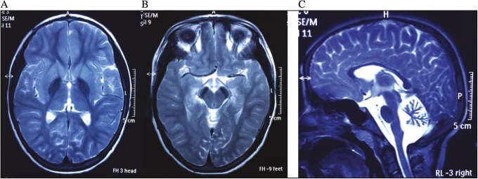
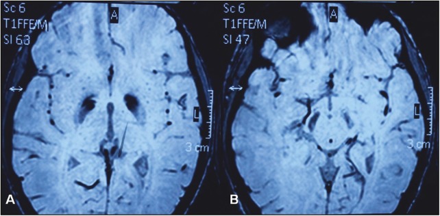
Investigations revealed a normal hemogram, liver and kidney function tests, and thyroid profile. Magnetic resonance imaging (MRI) of the brain showed cerebellar atrophy with hypointense globi pallidi and substantia nigra [Figure 1A and B]. Blooming was observed in these regions in the gradient images, suggesting iron deposition [Figure 1C, Figure 2]. On the basis of clinical and MRI findings, a diagnosis of PLAN was considered. Conventional bidirectional sequencing of the whole coding region of PLA2G6 (NM_003560; NP_003551.2) identified the compound heterozygous mutations, c.1798C>T; p.R546W (rs368008077) + c.2357C>T; p.R741W (rs530348521), each inherited by one healthy heterozygous parent. Both mutations were present in dbSNP146 and in ExAC database but with very low allele frequency and never in the homozygous state. PolyPhen-2 and SIFT tools predicted both variants as damaging. Genetic counseling was offered to the family, and the patient was addressed to physiotherapy and supportive care.
Figure 1.
T2-weighted axial images at the level of the basal ganglia (A) and midbrain (B) showing hypointense globi pallidi (A) and substantia nigra (B), respectively. (C) T2-weighted axial midline sagittal image showing cerebellar atrophy
Figure 2.
Axial gradient images at the level of the basal ganglia (A) and midbrain (B) showing blooming in globi pallidi (A) and substantia nigra (B) suggesting iron deposition in these regions
Discussion
Infantile NAD was first described by Seitelberger[7] in 1952, and the condition was termed INAD by Cowen and Olmstead[8] in 1963 (INAD1/NBIA2A). This starts typically between ages 6 months and 3 years with progressive truncal hypotonia, psychomotor delay, cerebellar ataxia, symmetric pyramidal tract signs, and tetraparesis (usually spastic but sometimes areflexic). Children commonly manifest strabismus, nystagmus, and optic atrophy, and lose the ability to walk shortly after attaining it or never learn to walk.[9,10] Electroencephalography (EEG) shows characteristic high-voltage fast rhythms, with electromyography (EMG) results consistent with chronic denervation.[11] T2-weighted MRI typically shows cerebellar atrophy with signal hyperintensity in the cerebellar cortex and occasionally, hypointensity in the globi pallidi and substantia nigra.[12] Hypertrophy of the clava also has been reported.[13]
Atypical NAD is much rarer than the infantile form, with only few patients reported to date.[1,4,9,14] Age at onset ranges from 1.6 to 6 years. This manifests clinically as gait abnormalities, speech delay or regression, and diminished social interaction. Optic atrophy, nystagmus, and seizures present with comparable frequency to infantile-onset PLAN, but other common features of infantile form such as truncal hypotonia, strabismus, EEG abnormalities, and EMG signs of denervation are much rarer. The disease course is usually characterized by progressive dystonia, dysarthria, speech regression, and neurobehavioral disturbances (impulsivity, hyperactivity, and emotional lability).[10,15] The course is fairly stable during early childhood and resembles static encephalopathy but is followed by neurologic deterioration between ages 7 and 12 years.[16] Besides cerebellar atrophy, iron deposition in the basal ganglia seems to universally develop in the cases so far described, although this is infrequently observed in the infantile-onset form, at least in the early stages of the disease.[3,4,17]
Atypical NAD is rare. In a series of 17 North African patients with NAD, only one had atypical NAD. Despite the early onset (18 months), clinical progression was slower, with behavioral disturbances and dystonia. This patient carried a missense variant predicted to be less deleterious.[3]
Very few reports of atypical INAD have been reported in Indian literature. Of these, only few are genetically proven.[5,6] Our case was also unique because of atypical features, which were the delayed age at presentation, slow progression, and absence of features of typical INAD, namely optic atrophy, nystagmus, seizures, truncal hypotonia, and strabismus. The possibility of atypical PLAN should be kept in mind when a child presents with neuroregression (mainly of the language), spasticity, slow progressive course, and behavioral disturbances, associated to the classical imaging findings. The diagnosis can be confirmed by testing for PLA2G6 mutation studies. Family screening and genetic counseling should be offered to the patients.
Ethics
Ethical clearance was obtained from the institutional ethics committee. Written informed consent was obtained from the caregivers.
Financial support and sponsorship
Nil.
Conflicts of interest
There are no conflicts of interest.
Acknowledgement
This work was supported by the European Research Council starting grant 260888 to Enza Maria Valente.
References
- 1.Salih MA, Mundwiller E, Khan AO, AlDrees A, Elmalik SA, Hassan HH, et al. New findings in a global approach to dissect the whole phenotype of PLA2G6 gene mutations. PLoS One. 2013;8:e76831. doi: 10.1371/journal.pone.0076831. [DOI] [PMC free article] [PubMed] [Google Scholar]
- 2.Paisán-Ruiz C, Guevara R, Federoff M, Hanagasi H, Sina F, Elahi E, et al. Early-onset L-dopa-responsive parkinsonism with pyramidal signs due to ATP13A2, PLA2G6, FBXO7 and spatacsin mutations. Mov Disord. 2010;25:1791–800. doi: 10.1002/mds.23221. [DOI] [PMC free article] [PubMed] [Google Scholar]
- 3.Romani M, Kraoua I, Micalizzi A, Klaa H, Benrhouma H, Drissi C, et al. Infantile and childhood onset PLA2G6-associated neurodegeneration in a large North African cohort. Eur J Neurol. 2015;22:178–86. doi: 10.1111/ene.12552. [DOI] [PubMed] [Google Scholar]
- 4.Illingworth MA, Meyer E, Chong WK, Manzur AY, Carr LJ, Younis R, et al. PLA2G6-associated neurodegeneration (PLAN): further expansion of the clinical, radiological and mutation spectrum associated with infantile and atypical childhood-onset disease. Mol Genet Metab. 2014;112:183–9. doi: 10.1016/j.ymgme.2014.03.008. [DOI] [PMC free article] [PubMed] [Google Scholar]
- 5.Ananthanarayanan K, Ahuja CK, Pratibha S. Spastic paraparesis with basal ganglia changes: infantile neuroaxonal dystrophy. Neurol India. 2018;66:264. doi: 10.4103/0028-3886.222887. [DOI] [PubMed] [Google Scholar]
- 6.Saketh K, Mohd Hussain S, Nivedita S. Genetic analysis of PLA2G6 in 22 Indian families with infantile neuroaxonal dystrophy, atypical late-onset neuroaxonal dystrophy and dystonia parkinsonism complex. PLoS One. 2016;11:e0155605. doi: 10.1371/journal.pone.0155605. [DOI] [PMC free article] [PubMed] [Google Scholar]
- 7.Seitelberger F. Infantile Neuroaxonal Dystrophy. In Proc Ist Int Congr Neuropath Rome. 1952;3:323. [Google Scholar]
- 8.Cowen D, Olmstead EV. Infantile neuroaxonal dystrophy. J Neuropathol Exp Neurol. 1963;22:175–236. doi: 10.1097/00005072-196304000-00001. [DOI] [PubMed] [Google Scholar]
- 9.Gregory A, Westaway SK, Holm IE, Kotzbauer PT, Hogarth P, Sonek S, et al. Neurodegeneration associated with genetic defects in phospholipase A(2) Neurology. 2008;71:1402–9. doi: 10.1212/01.wnl.0000327094.67726.28. [DOI] [PMC free article] [PubMed] [Google Scholar]
- 10.Gregory A, Polster BJ, Hayflick SJ. Clinical and genetic delineation of neurodegeneration with brain iron accumulation. J Med Genet. 2009;46:73–80. doi: 10.1136/jmg.2008.061929. [DOI] [PMC free article] [PubMed] [Google Scholar]
- 11.Aicardi J, Castelein P. Infantile neuroaxonal dystrophy. Brain. 1979;102:727–48. doi: 10.1093/brain/102.4.727. [DOI] [PubMed] [Google Scholar]
- 12.Farina L, Nardocci N, Bruzzone MG, D’Incerti L, Zorzi G, Verga L, et al. Infantile neuroaxonal dystrophy: neuroradiological studies in 11 patients. Neuroradiology. 1999;41:376–80. doi: 10.1007/s002340050768. [DOI] [PubMed] [Google Scholar]
- 13.Solomons J, Ridgway O, Hardy C, Kurian MA, Kurian M, Jayawant S, et al. Infantile neuroaxonal dystrophy caused by uniparental disomy. Dev Med Child Neurol. 2014;56:386–9. doi: 10.1111/dmcn.12327. [DOI] [PubMed] [Google Scholar]
- 14.Morgan NV, Westaway SK, Morton JE, Gregory A, Gissen P, Sonek S, et al. PLA2G6, encoding a phospholipase A2, is mutated in neurodegenerative disorders with high brain iron. Nat Genet. 2006;38:752–4. doi: 10.1038/ng1826. [DOI] [PMC free article] [PubMed] [Google Scholar]
- 15.Kurian MA, McNeill A, Lin JP, Maher ER. Childhood disorders of neurodegeneration with brain iron accumulation (NBIA) Dev Med Child Neurol. 2011;53:394–404. doi: 10.1111/j.1469-8749.2011.03955.x. [DOI] [PubMed] [Google Scholar]
- 16.Kurian MA, Morgan NV, MacPherson L, Foster K, Peake D, Gupta R, et al. Phenotypic spectrum of neurodegeneration associated with mutations in the PLA2G6 gene (PLAN) Neurology. 2008;70:1623–9. doi: 10.1212/01.wnl.0000310986.48286.8e. [DOI] [PubMed] [Google Scholar]
- 17.Wu Y, Jiang Y, Gao Z, Wang J, Yuan Y, Xiong H, et al. Clinical study and PLA2G6 mutation screening analysis in Chinese patients with infantile neuroaxonal dystrophy. Eur J Neurol. 2009;16:240–5. doi: 10.1111/j.1468-1331.2008.02397.x. [DOI] [PubMed] [Google Scholar]


