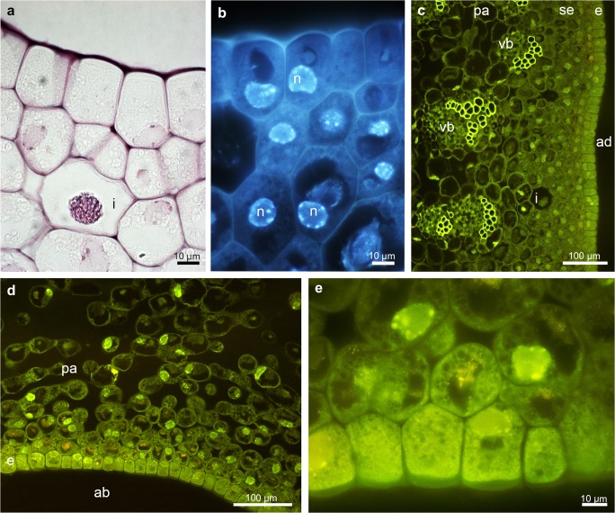
Fig. 4.
Histochemical features of the middle part of the epichile of B. echinolabium.a Transverse section: the epidermis and few layers of subepidermal cells with idioblasts following staining with ruthenium red. b The epidermis and subepidermal tissue with enlarged, nuclei (DAPI). c, d Transverse sections of adaxial (inner) and abaxial (outer) surfaces stained with Auramine O. e Detail of d. ab abaxial surface, ad adaxial surface, e epidermis, i idioblast, n nuclei, pa parenchyma, vb vascular bundle

