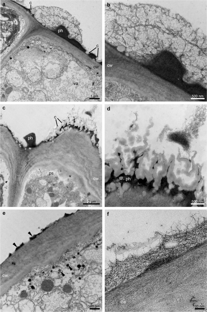
Fig. 6.
Ultrastructural analysis (TEM) of the appendix of the prickle in the middle part of the hypochile. a Sections through the epidermal cell wall with periplasmic spaces beneath and large amounts of heterogeneous residues of secreted material with fragmented pieces of the cuticle on the surface. b Detail of a. c Phenolic secretory material on the cuticle surface and periplasmic spaces beneath the cell wall. d Detail of c. e Phenolic material on the cuticle surface (black arrowheads) and inside the numerous vesicles fusing with the plasmalemma. f Detail of the epidermis cell wall with secretory material on the surface. c cuticle, cw cell wall, ph phenolic secretion, ps periplasmic space, va vacuole

