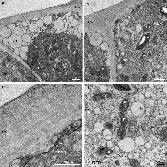
Fig. 7.
Ultrastructural details (TEM) of the prickle (hypochile) showing secretory epidermal cells with dense cytoplasm, numerous small vacuoles in close vicinity of the cell wall (a, b), an abundance of mitochondria, plastids with starch grains and plastoglobuli (a, b, d), fully developed dictyosomes (a, b), profiles of smooth (SER) and rough endoplasmic reticulum (RER) (b–d), lipid droplets (d) and periplasmic space with flocculated secretory material and numerous vesicles building into plasmalemma (white arrowheads) (b, c). cw cell wall, d dictyosome, l lipid droplet, m mitochondrion, p plastid, ps periplasmic space, va vacuole

