Today, most children with acute lymphoblastic leukemia (ALL) can be cured by treatment regimens consisting of combination chemotherapy supplemented in certain patient populations with preventive or therapeutic cranial irradiation (CI) and/or an allogeneic hematopoietic stem cell transplantation (HSCT).1 Unfortunately, after undergoing ALL therapy up to 10% of patients develop secondary malignant neoplasms (SMN)2,3 with cure rates often being dismal.4 Therefore, an improved understanding of the underlying pathobiology of SMN, the development of strategies to identify patients at risk and, ideally, the development of preventive actions for SMN are of great clinical interest.
The role of germline genetic variation in the pathophysiology of SMN is poorly understood. Here, we analyzed three candidate genes, TP53, ETV6 and RUNX1, to characterize their potential contribution to SMN development after treatment for ALL. Besides their frequent genetic aberration in pediatric ALL, the three genes were chosen because of their described roles as predisposition genes for hematologic and/or solid malignancies.5–10
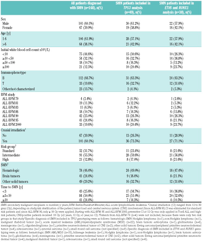
Employing targeted sequencing to characterize the frequency of single nucleotide variants (SNV) in these genes, we analyzed a cohort of patients with SMN after treatment on one of seven consecutive ALL-BFM protocols for treatment of pediatric ALL (ALL-BFM 79 to AIEOP-BFMALL 2000; for trial details see the Online Supplementary Methods). The study was approved by the Ethics Committee of the University Hospital Schleswig-Holstein at Kiel, Germany. In the observation period from 1984 to 2008, 168 patients were identified who developed an SMN after treatment for pediatric ALL; 49 had sufficient material available for analysis. Clinical characteristics of the patients are given in Table 1 and Online Supplementary Table S1. The median follow up for the entire patient cohort was 10.6 years as of April, 2013. DNA was isolated from remission bone marrow and analyzed employing two different multiplex PCR-based Ion AmpliSeq™ Panels (Life Technologies, Darmstadt, Germany) according to the manufacturer’s instructions. The first panel, the Ion AmpliSeq™ TP53 Panel, completely covered all coding exons of TP53 and was applied in 49 patient samples. The second panel interrogated the complete coding regions of ETV6 and RUNX1 and was used for 38 patients with sufficient material available after TP53 analysis. Only SNV with a minimum coverage of 500x and a PHRED quality of 20 were included. Physical positions of the identified variants refer to GRCh37.p13 and the following transcripts: TP53: ENST00000269305 (NM_000546.5); ETV6: ENST00000396373 (NM_001987.4) and RUNX1: ENST00000437180 (NM_001754.4). SPSS (IBM Deutschland GmbH, Ehningen, Germany) was used for computerized calculations and the statistical language R was used to plot the identified variants.
Table 1.
Characteristics of patients diagnosed with secondary malignant neoplasms after treatment on ALL-BFM trials 79 to 2000 included in targeted sequencing analyses in comparison to secondary malignant neoplasm patients not included.
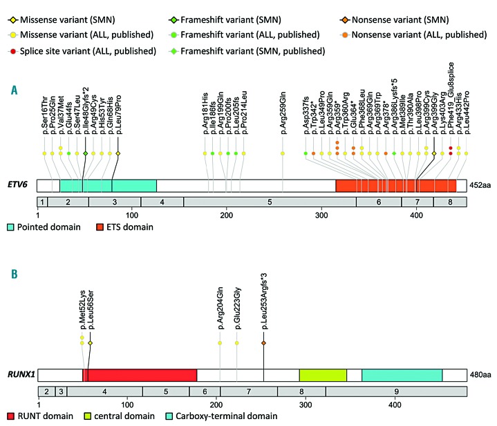
In TP53 analyses, only one of 49 (2%) genotyped SMN patients carried a heterozygous non-synonymous variant within the coding region. This rare missense SNV, p.Asn235Ser (rs144340710), was detected in a patient developing a small round cell sarcoma after T-cell ALL (Online Supplementary Tables S2 and S3 and Online Supplementary Figure S1). The in silico scores from Polyphen and Sift algorithms indicated a benign effect for this change. Regarding ETV6, three of 38 (8%) patients carried a heterozygous non-synonymous SNV; two of them developed an acute myeloid leukemia (AML) and another patient an osteosarcoma (Figure 1A and Online Supplementary Tables S2 and S3). Initially, all three patients were diagnosed with pre-B-cell ALL and only the patient with the solid SMN received CI. One frameshift insertion (p.Ile48Glyfs*2) and a missense variant (p.Leu79Pro) were observed in exon 2, which is part of the pointed domain; an additional missense substitution (p.Arg399Gly) was detected in exon 7 within the C-terminal DNA-binding ETS domain. None of these variants was recorded in the databases dbSNP (build 150), the 1000Genomes Project, the NHLBI GO Exome Sequencing Project, Exome Aggregation Consortium or the Catalogue Of Somatic Mutations In Cancer (COSMIC). For both missense variants the prediction scores implied a probably damaging effect. Molecular modeling11 of p.Arg399Gly suggested that the change might impair the DNA-binding qualities of the ETS domain (Online Supplementary Figure S2). Finally, we identified two of 38 (5%) patients with heterozygous coding variants in RUNX1 (Figure 1B and Online Supplementary Table S2). One patient, initially diagnosed with T-cell ALL, carried a nonsense insertion (p.Leu253Argfs*3) in exon 7. A second patient, initially diagnosed with pre-B-cell ALL, demonstrated a probably damaging (Polyphen) missense SNV (p.Leu56Ser, rs111527738) located in the N-terminal RUNT domain. Both patients with RUNX1 variants developed hematologic SMN and received CI during ALL therapy. The described ETV6 and RUNX1 SNV were mutually exclusive. Further clinical characteristics of the six SMN patients with SNV in either TP53, ETV6 or RUNX1 are shown in Online Supplementary Table S3.
Figure 1.
Location, type and distribution of ETV6 and RUNX1 germline alterations detected in childhood acute lymphoblastic leukemia (ALL) patients. (A) ETV6 and (B) RUNX1 germline variants of the current study, observed in childhood ALL patients developing secondary malignant neoplasms (SMN) are indicated by rhombs and black lines. ALL-related germline alterations reported in the literature7,9,12,13,16 are represented by circles and gray lines. Germline variants published for childhood ALL patients developing SMN12 are indicated by rhombs and gray lines. Only one individual from each kindred, reported in the literature, was included for plotting.9,12,16
The low frequency of germline TP53 SNV in our case series suggests that alterations in this frequently mutated tumor suppressor gene are important, but may not play a major role in the pathobiology of SMN after treatment for ALL on BFM protocols. However, in an analysis from the ALL-BFM relapse study group, four patients carrying a TP53 missense SNV were identified, and two of these patients developed an SMN.6 The rare missense TP53 variant p.Asn235Ser (MAF 0.0003, in individuals of European ancestry, ExAC database) detected in our study is located in the DNA-binding domain. Interestingly, the same variant, which is of uncertain significance, was previously reported in two studies of childhood ALL patients.8,10 In the first study, one of 588 acute leukemia patients in the Pediatric Cancer Genome Project (PCGP) was identified as a carrier.8 In the second study, comprising 3,801 pre-B-cell ALL patients from Children’s Oncology Group (COG) trials, the variant p.Asn235Ser was observed in three patients, one of whom developed an SMN.10 In the later study, five of 34 (15%) SMN patients were identified with pathogenic TP53 SNV within the common isoform of TP53 (NM_000546); all of these patients received HSCT and two of five received irradiation during ALL therapy. The patient observed in our study developed a small round cell sarcoma after treatment for ALL including CI (Online Supplementary Table S3).
Regarding ETV6, a large US study by Moriyama et al. revealed that 35 of 4,405 childhood ALL patients (<1%) harbored germline SNV in this gene.9 Here, we report three of 38 patients (8%) developing an SMN after ALL therapy. Taking the study by Moriyama et al. as a hypothetical reference population for our findings results in an odds ratio (OR) of 10.7 (P=0.0001). This may suggest that ALL patients carrying an ETV6 SNV might be at an increased risk of SMN. A second US study on ETV6 in childhood ALL identified two of 472 patients (<1%) with germline variants.12 Remarkably, one of the affected patients was diagnosed with a secondary vulvar squamous cell carcinoma. The variant p.Arg399Gly detected in our study resides in the ETS domain, as do most of the previously described ETV6 germline variants.9,12,13 Molecular modeling of p.Arg399Gly implied a similar putative effect on the ETV6 binding capacity as it was shown for a different change (p.Arg399Cys13) at the same residue. For the latter, functional assays revealed a diminished binding capacity and a partial cytoplasmic sequestration of the variant ETV6 protein.12,13 In line with previous studies, the ETV6 variants reported here are supposed to confer a damaging effect and the potential to alter the biological function, either due to truncation or by structural modulation of the transcription factor.
Inherited RUNX1 mutations have been related to familial platelet disorder and demonstrated to predispose to myelodysplastic syndrome (MDS), ALL and AML, including therapy-related AML.14 Here, we report two SMN patients carrying monoallelic germline RUNX1 SNV: one patient developed secondary MDS/AML after ALL treatment including CI with 12 Gy, the second a bilineage acute leukemia/AML after similar treatment exposure. Prior to the primary ALL, none of these patients was diagnosed with a previous malignancy. Unfortunately, information on family histories is not available to us. A previous study on acute childhood leukemia patients, including at least 426 ALL, revealed that four ALL patients (<1%) carried a probably damaging RUNX1 germline mutation.8 Compared to this study, we found an approximately 4-fold higher frequency of RUNX1 SNV in patients developing an SMN after ALL treatment on ALL-BFM protocols. Using the study by Zhang et al. as a hypothetical reference population for our findings results in an OR of 4.7 (P=0.071).13
Clearly, there are several limitations to our pilot study. One severe drawback is the lack of an appropriate control group to allow for methodologically sound risk assessments in our population. In addition, due to the sequencing approach used herein, we cannot exclude the potential contribution of larger deletions in our investigated candidate genes to the risk of SMN. There was also no access to neoplastic tissue at ALL or SMN diagnosis to analyze for somatic mutations in the second alleles in any of the patients. Furthermore, employing remission bone marrow samples for analysis, we cannot completely exclude clonal hematopoiesis as an underlying mechanism for our observations; however, the latter phenomenon is rare in younger individuals15 and all reported heterozygous alterations in our study had at least 50% variant sequencing reads.
To conclude, in comparison to published data, we found higher frequencies of ETV6 and RUNX1 alterations in patients developing an SMN after treatment for ALL on ALL-BFM protocols. Further studies employing appropriate control groups need to clarify the potential contribution of germline variants in TP53, ETV6 and RUNX1 to the development of SMN after treatment for ALL on BFM protocols.
Acknowledgments
We thank all patients who took part in the study and colleagues involved in the ALL-BFM studies.
Footnotes
Funding: this work was supported by the Madeleine Schickedanz-Kinderkrebs-Stiftung. We thank Dr. Anna Stengel for her helpful support regarding the development of the R script for mutation plotting.
Information on authorship, contributions, and financial & other disclosures was provided by the authors and is available with the online version of this article at www.haematologica.org.
References
- 1.Möricke A, Zimmermann M, Reiter A, et al. Prognostic impact of age in children and adolescents with acute lymphoblastic leukemia: data from the trials ALL-BFM 86, 90, and 95. Klin Padiatr.2005; 217(6):310–320. [DOI] [PubMed] [Google Scholar]
- 2.Möricke A, Zimmermann M, Reiter A, et al. Long-term results of five consecutive trials in childhood acute lymphoblastic leukemia performed by the ALL-BFM study group from 1981 to 2000. Leukemia. 2010;24(2):265–284. [DOI] [PubMed] [Google Scholar]
- 3.Stanulla M, Schaeffeler E, Möricke A, et al. Thiopurine methyltransferase genetics is not a major risk factor for secondary malignant neoplasms after treatment of childhood acute lymphoblastic leukemia on Berlin-Frankfurt-Munster protocols. Blood. 2009;114(7):1314–1318. [DOI] [PubMed] [Google Scholar]
- 4.Schmiegelow K, Levinsen MF, Attarbaschi A, et al. Second malignant neoplasms after treatment of childhood acute lymphoblastic leukemia. J Clin Oncol. 2013;31(19):2469–2476. [DOI] [PMC free article] [PubMed] [Google Scholar]
- 5.Chiaretti S, Brugnoletti F, Tavolaro S, et al. TP53 mutations are frequent in adult acute lymphoblastic leukemia cases negative for recurrent fusion genes and correlate with poor response to induction therapy. Haematologica. 2013;98(5):e59–61. [DOI] [PMC free article] [PubMed] [Google Scholar]
- 6.Hof J, Krentz S, van Schewick C, et al. Mutations and deletions of the TP53 gene predict nonresponse to treatment and poor outcome in first relapse of childhood acute lymphoblastic leukemia. J Clin Oncol. 2011;29(23):3185–3193. [DOI] [PubMed] [Google Scholar]
- 7.Holmfeldt L, Wei L, Diaz-Flores E, et al. The genomic landscape of hypodiploid acute lymphoblastic leukemia. Nat Genet. 2013; 45(3):242–252. [DOI] [PMC free article] [PubMed] [Google Scholar]
- 8.Zhang J, Walsh MF, Wu G, et al. Germline Mutations in Predisposition Genes in Pediatric Cancer. N Engl J Med. 2015; 373(24):2336–2346. [DOI] [PMC free article] [PubMed] [Google Scholar]
- 9.Moriyama T, Metzger ML, Wu G, et al. Germline genetic variation in ETV6 and risk of childhood acute lymphoblastic leukaemia: a systematic genetic study. Lancet Oncol. 2015;16(16):1659–1666. [DOI] [PMC free article] [PubMed] [Google Scholar]
- 10.Qian M, Cao X, Devidas M, et al. TP53 Germline Variations Influence the Predisposition and Prognosis of B-Cell Acute Lymphoblastic Leukemia in Children. J Clin Oncol. 2018;36(6):591–599. [DOI] [PMC free article] [PubMed] [Google Scholar]
- 11.Biasini M, Bienert S, Waterhouse A, et al. SWISS-MODEL: modelling protein tertiary and quaternary structure using evolutionary information. Nucleic Acids Res. 2014;42(Web Server issue):W252–258. [DOI] [PMC free article] [PubMed] [Google Scholar]
- 12.Topka S, Vijai J, Walsh MF, et al. Germline ETV6 Mutations Confer Susceptibility to Acute Lymphoblastic Leukemia and Thrombocytopenia. PLoS Genet. 2015;11(6):e1005262. [DOI] [PMC free article] [PubMed] [Google Scholar]
- 13.Zhang MY, Churpek JE, Keel SB, et al. Germline ETV6 mutations in familial thrombocytopenia and hematologic malignancy. Nat Genet. 2015;47(2):180–185. [DOI] [PMC free article] [PubMed] [Google Scholar]
- 14.Sood R, Kamikubo Y, Liu P. Role of RUNX1 in hematological malignancies. Blood. 2017;129(15):2070–2082. [DOI] [PMC free article] [PubMed] [Google Scholar]
- 15.Collord G, Park N, Podesta M, et al. Clonal haematopoiesis is not prevalent in survivors of childhood cancer. Br J Haematol. 2018; 181(4):537–539. [DOI] [PMC free article] [PubMed] [Google Scholar]
- 16.Noetzli L, Lo RW, Lee-Sherick AB, et al. Germline mutations in ETV6 are associated with thrombocytopenia, red cell macrocytosis and predisposition to lymphoblastic leukemia. Nat Genet. 2015; 47(5):535–538. [DOI] [PMC free article] [PubMed] [Google Scholar]


