Abstract
Background
Recently, LINC00665 has been reported to be a pivotal regulator in kinds of malignancy, such as lung cancer and liver cancer. However, the functions and underlying mechanisms of LINC00665 in gastric cancer (GC) remain unclear.
Materials and methods
We recruited 49 paired GC tissue to explore LINC00665 expression by qRT-PCR. In vitro function assays were used to explore the roles of LINC00665 in GC progression. Moreover, the interaction among LINC00665, miR-149-3p and RNF2 was explored by bioinformatics analysis and luciferase reporter assay.
Results
In the present study, we found that LINC00665 expression was significantly elevated in GC tissues and cell lines. High LINC00665 expression was associated with TNM stage, histological grade, and poor prognosis of GC patients. Function assays showed that LINC00665 suppression significantly reduced GC cells viability and invasion ability in vitro. Mechanistic analysis showed that LINC00665 might serve as a ceRNA for miR-149-3p to regulate the expression of RNF2.
Conclusion
Our current study revealed the LINC00665/miR-149-3p/RNF2 axis was involved in GC progression, providing novel insights into the treatment for GC.
Keywords: gastric cancer, LINC00665, miR-149-3p, RNF2
Introduction
Gastric cancer (GC), one of the most common malignancies, accounts for almost 10% of all cancer death worldwide.1,2 As a consequence of the negligence regarding self-inspection and regular clinical examination, GC is frequently diagnosed at an advanced stage.3,4 Although a great number of improvements in diagnostic and treatment have been achieved in the past years, the 5-year survival rate of GC remains approximately 25–30%.5 Therefore, it is an urgent need to explore the underlying mechanisms involved in GC development.
Long non-coding RNA (lncRNA), a subset of ncRNAs, is longer than 200 nucleotides and without translation of proteins.6 LncRNAs have gradually got the attention of investigators for their implications in all sorts of biological processes of diseases via molecular mechanisms causing transcriptional, post-transcriptional, and even epigenetic alterations, especially in carcinomas.7,8 For instance, Xiao et al found that lncRNA HMlincRNA717 expression was significantly reduced and associated with poor prognosis of patients with lung cancer.9 Hua et al revealed that lncRNA NNT-AS1 could promote cell proliferation and invasion through Wnt/β-catenin signaling pathway in cervical cancer.10 Gu et al suggested that lncRNA MALAT1 might act as an oncogene in Multiple Myeloma by sponging miR-509-5p to modulate FOXP1 expression.11
LINC00665, located in chromosome 19, has recently been reported to be upregulated and functioned as a putative oncogenic lncRNA in several types of human cancer. For example, Wen et al showed that Overexpression of LINC00665 was involved in the regulation of cell cycle pathways in hepatocellular carcinoma by ten identified hub genes.12 Cong et al showed that linc00665 promoted lung adenocarcinoma progression and functioned as ceRNA to regulate AKR1B10-ERK signaling by sponging miR-98.13 However, the functions and underlying mechanisms of LINC00665 in GC remain unestablished.
In the present study, the functions of LINC00665 in GC were determined and extra mechanisms establishing the basis of the carcinogenic role of LINC00665 were also revealed. Totally, we found that LINC00665 promoted the proliferation and invasion by sponging miR-149-3p to upregulate RNF2 expression in GC.
Materials and methods
Clinical specimens
A total of 49 paired GC tissue specimens were obtained from patients who underwent radical gastrectomy in the Department of Gastroenterology, The Central Hospital of Xinxiang. All patients who underwent surgical treatment without preoperative radiotherapy and/or chemotherapy. All fresh specimens were immediately placed in liquid nitrogen and stored at −80 °C until use. The study was approved by the Medical Ethics Committee of the Central Hospital of Xinxiang. Written informed consent was collected from every participant. Experiments involving human tissues were conducted in accordance with the Declaration of Helsinki.
Cell culture and transfection
Human GC cells (AGS, SGC-7901, HGC27, MGC-803, MKN-45, and BGC-823) and normal gastric epithelial cells (GES-1) were bought from American Type Culture Collection (ATCC, USA) and maintained in RPMI1640 medium (Gibco, NY, USA) supplemented with 10% fetal bovine serum (FBS; Gibco), 100 U/mL penicillin (Gibco), and 100 mg/mL streptomycin (Gibco) at 37 °C in a humidified atmosphere with 5% CO2.
For the knockdown of LINC00665, sh-LINC00665 #1/2/3 were obtained from Ribo Biotech (Guangzhou, China). miR-149-3p mimics, miR-149-3p inhibitors, and overexpression RNF2 vector (pCDNA3.1-RNF2) were obtained from GenePharma (Shanghai, China). GC cells transfected with mimics or plasmid vector by Lipofectamine 2000 (Invitrogen) according to the protocol.
RNA isolation and qRT-PCR
Total RNA was extracted from tissues or cells using TRIzol reagent (Invitrogen) according to the protocol. To detect mRNA expression of LINC00665 and RNF2, extracted RNA was reversely transcribed into cDNA by PrimeScript One-step RT-PCR kit (Takara, China). For miR-149-3p expression analysis, cDNA was synthesized from 50 ng extracted RNA using a miScript reverse transcription kit (Qiagen, Germany). SYBR Green Master Mix (Life Technologies) was used for gene expression level measurement. The qRT-PCR procedures were performed as follows: 50 °C for 2 min, 95 °C for 10 min, 35 cycles at 95 °C for 15 s, and 60 °C for 1 min. The expression levels were calculated using the 2−ΔΔCt method with GAPDH used for the normalization of the mRNA, and U6 for the miRNA.
Cell proliferation assay
For 5-ethynyl-2-deoxyuridine (EdU) assay, 24-well culture plates were applied to culture transfected cells. The newly synthesized DNA was visualized with an EdU imaging kit (Life Technologies) to assess cell proliferation. The number of EdU positive cells was calculated under a fluorescence microscope.
For colony formation assay, transfected cells were resuspended at 1×10³ cells/well and cultured in culture for about 4 days until macroscopic colonies appeared. Finally, the colonies were stained with crystal violet (Beyotime) for 30 mins.
Transwell invasion assay
The invasiveness abilities of GC cells were assessed using an 8-μm pore polycarbonate membrane Boyden chamber insert (Corning, NY, USA). Briefly, the upper chamber precoated with Matrigel was seeded with 5×10⁴ transfected cells in 200 μl FBS-free medium, while the lower chamber was added RPMI-1640 with 20% FBS, which acted as chemoattractant. After 48 h of incubation, the invaded cells were fixed with methanol for 30 min, stained with 0.1% crystal violet for 5 min.
Western blot
Proteins were lysed by RIPA lysis buffer (Beyotime, China) containing 1% PMSF and the concentrations were measured using BCA Kit. E Equal amounts of proteins were resolved on 12% SDS-PAGE gel and transferred into PVDF membranes (Millipore, CA, USA). Following blocked with 5% low fat milk in TBST at 37 °C for 1 h, the membranes were incubated with the primary antibodies overnight at 4 °C. After incubated with corresponding secondary antibodies at room temperature for 1 h, the enhanced chemiluminescence Kit (ECL, Pierce, USA) was used for detection.
Luciferase reporter assay
The fragment of LINC00665 or RNF2 containing the target sequence of miR-149-3p was inserted into a pmirGlO Dual-luciferase miRNA Target Expression Vector (Promega, WI, USA) to form the reporter vector LINC00665-wild-type (LINC00665-Wt) or RNF2-wild-type (RNF2-Wt) while LINC00665-mutated-type (LINC00665-MutT) or RNF2-mutated-type (RNF2-MUT) contained mutated binding site. The cells were co-transfected with WT or MUT and miR-149-3p mimics using Lipofectamine 2000 reagent according to manufacturer’s protocol. Luciferase activity was measured using the Dual Luciferase Reporter Assay System Kit (Promega), normalized to that of Renilla.
Pull-down assay
The experimental methods are based on the previous study.14
Statistical analyses
Statistical software SPSS 19.0 was used for quantitative data analysis. All data in our present study were presented as mean ± standard deviation (SD). Differences between two or more groups were analyzed using Student’s t-test or one-way ANOVA. P<0.05 were considered statistically significant.
Results
LINC00665 was upregulated in GC
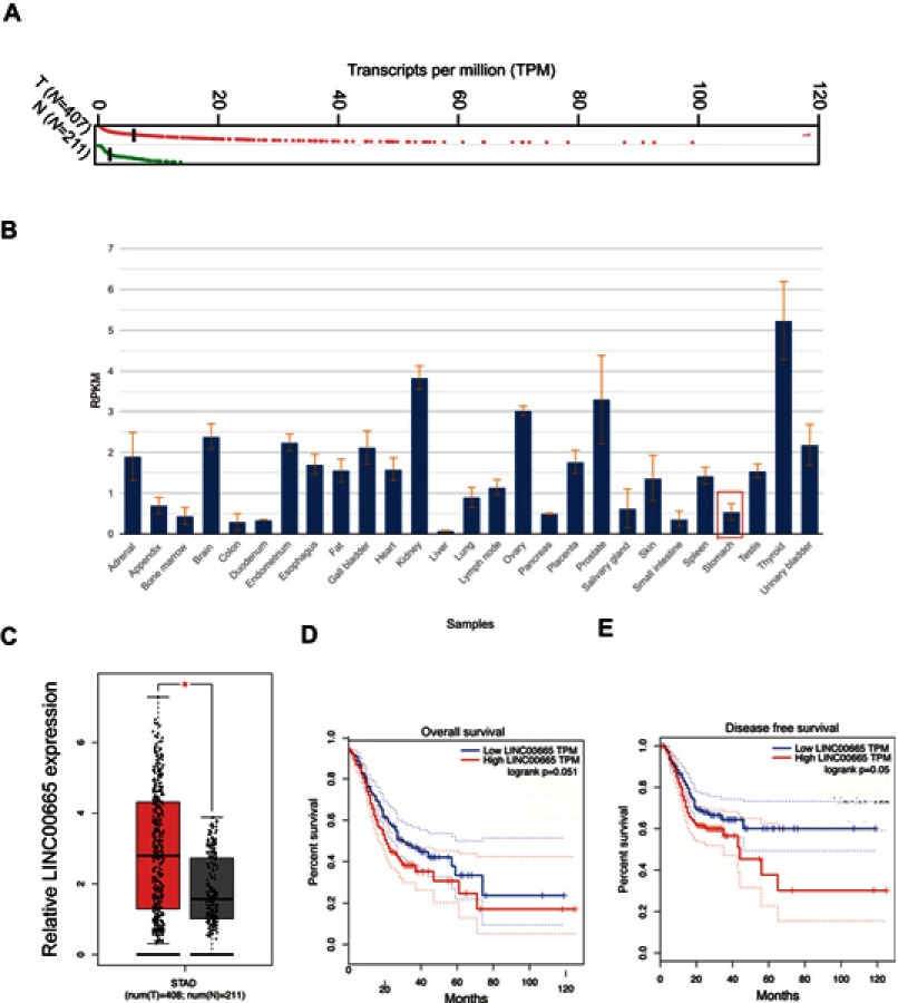
To probe the lncRNA-mediated GC progression, we analyzed TCGA database, results showed that LINC00665 was significantly upregulated in GC (Figure 1A and C). Subsequently, National Center for Biotechnology Information results showed that LINC00665 expression was decreased in normal gastric tissues (Figure 1B). Moreover, Kaplan-Meier analysis showed that high LINC00665 expression was associated with poor overall survival and disease free survival rate of patients with GC (Figure 1D and E). These data indicated that LINC00665 might serve act as a tumor oncogenic lncRNA in GC tumorigenesis.
Figure 1.
LINC00665 was upregulated in gastric cancer (GC). (A) The expression of LINC00665 in GC was determined by The Cancer Genome Atlas (TCGA) database. (B) The expression of LINC00665 in normal tissues explored by National Center for Biotechnology Information. (C) LINC00665 expression was significantly increased in GC tissues compared to normal tissues. (D, E) High LINC00665 expression was significantly associated with poor overall survival (D) and disease free survival rate (E) of GC patients. *P<0.05.
LINC00665 promoted GC cells proliferation and invasion
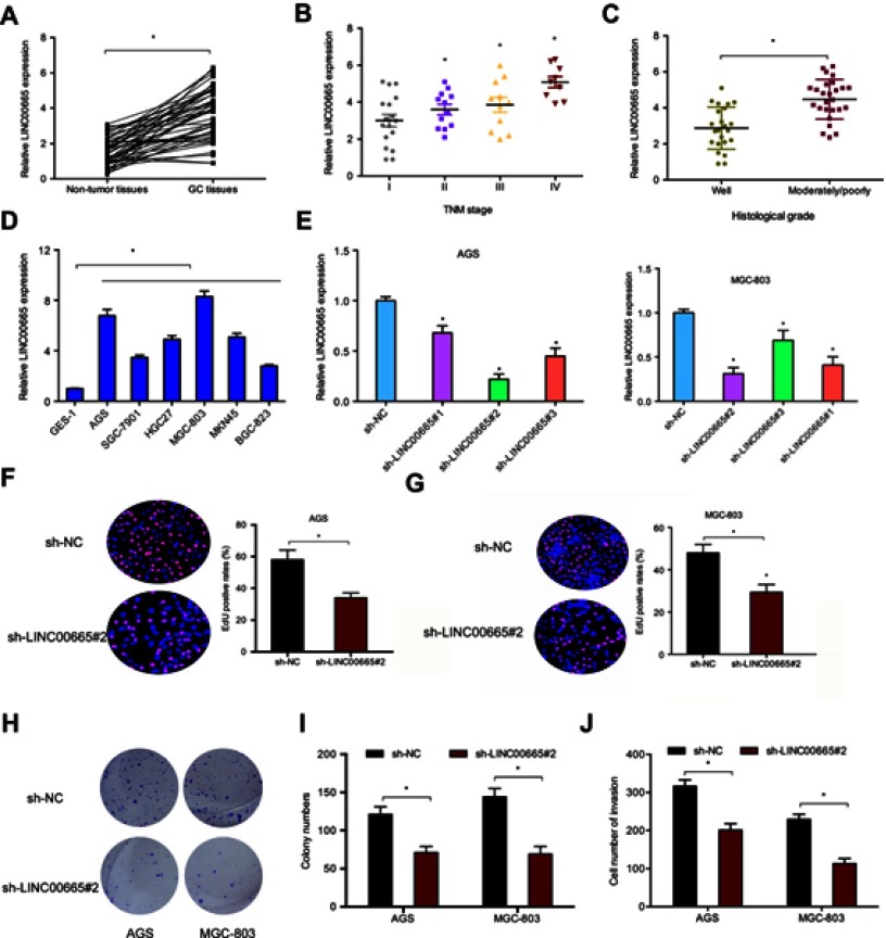
We further detected the expression of LINC00665 expression in GC. qRT-PCR revealed that LINC00665 expression was significantly increased in GC tissues compared to adjacent non-tumor tissues (Figure 2A). High LINC00665 expression was obviously associated with advanced TNM stage and histological grade of GC patients (Figure 2B and C). Moreover, we found that LINC00665 expression in GC cells (AGS, SGC-7901, HGC27, MGC-803, MKN-45, and BGC-823) was significantly increased compared to GES-1 cells (Figure 2D).
Figure 2.
LINC00665 promoted gastric cancer (GC) cells proliferation and invasion. (A) Expression of LINC00665 in GC tissues was measured by qRT-PCR. (B) High LINC00665 expression was associated with advanced TNM stage. (C) High LINC00665 expression was associated with advanced histological grade. (D) Expression of LINC00665 in GC cell lines was measured by qRT-PCR. (E) sh-LINC00665 significantly reduced LINC00665 expression in AGS and MGC-803 cells. (F–I) Cell viabilities were measured in AGS and MGC-803 cells transfected with sh-LINC00665 or sh-NC by EdU assay (F, G) and colony formation assay (H, I). (J) Cell invasion was measured in AGS and MGC-803 cells transfected with sh-LINC00665 or sh-NC by Transwell assay. *P<0.05.
To further determine the roles of LINC00665 in GC tumorigenesis, AGS and MGC-803 cells were transfected with sh-LINC00665 (Figure 2E). Subsequently, EdU and colony formation assays were used to determine the LINC00665 effects on GC cells viability. Results showed that LINC00665 inhibition significantly reduced the proliferation ability (Figure 2F and G) and colony formation ability in AGS and MGC-803 cells (Figure 2H and I). Moreover, transwell assay showed that the invasion ability of AGS and MGC-803 cells transfected with sh-LINC00665 was obviously suppressed (Figure 2J). These results demonstrated that LINC00665 might promote GC cells proliferation and invasion in vitro.
MiR-149-3p was a target of LINC00665
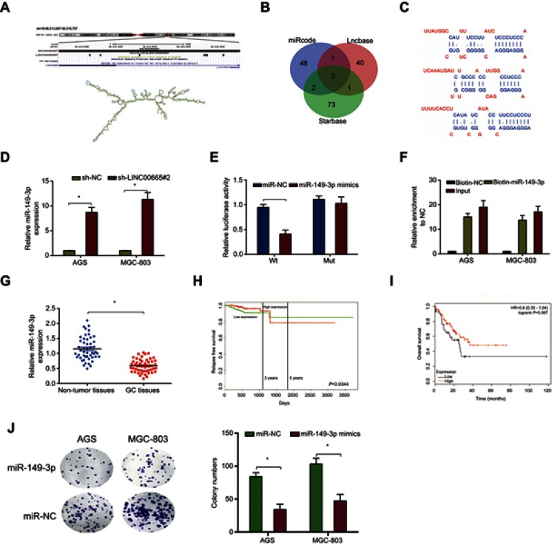
To determine the underlying mechanism of LINC00665 in GC progression, we predicted the potential target of LINC00665 by online bioinformatics analysis (LncBase, miRcode, and Starbase). MiR-149-3p was found to be a target of LINC00665, the LINC00665 information, secondary structure, and possible binding sites were shown in Figure 3A–C. To test this prediction, we further confirmed the correlations between LINC00665 and miR-149-3p. QRT-PCR showed that LINC00665 inhibition significantly increased miR-149-3p expression in GC cells (Figure 3D). Moreover, luciferase reporter and pull-down assays suggested that LINC00665 could regulate miR-149-3p expression via a ceRNA manner (Figure 3E and F).
Figure 3.
MiR-149-3p was a target of LINC00665. (A) Architecture of LINC00665 genomic region according to the University of California, Santa Cruz database. (B, C) Target sequences of miR-149-3p in LINC00665. (D) LINC00665 inhibition increased miR-149-3p expression in gastric cancer (GC) cells. (E, F) The interaction between LINC00665 and miR-149-3p was confirmed by luciferase reporter assay (E) and pull-down assay (F). (G) Expression of miR-149-3p in GC tissues was measured by qRT-PCR. (H, I) Low miR-149-3p expression was associated with poor relapse free survival (H) and overall survival (I) of GC patients. (J) miR-149-3p mimics reduced GC cells colony formation ability. *P<0.05.
Next, we explored the expression of miR-149-3p in GC. QRT-PCR showed that miR-149-3p expression was significantly reduced in GC tissues compared to non-tumor tissues (Figure 3G). Kaplan-Meier analysis showed that low miR-149-3p expression was associated with relapse free survival and overall survival of GC patients (Figure 3H and I). Moreover, we explored the effects of miR-149-3p mimics on GC cells viability, colony formation assays showed that miR-149-3p overexpression significantly reduced the colony numbers of AGS and MGC-803 cells (Figure 3J). These data suggested that miR-149-3p might act as a target of LINC00665 in GC progression.
LINC00665 regulated RNF2 by absorbing miR-149-3p
By using bioinformatics predication tools, we found a potential site of miR-149-3p binding in 3ʹ UTR of RNF2 (Figure 4A). To further confirm this, luciferase reporter assay was used. Results showed that miR-149-3p mimics significantly reduced the luciferase activity in RNF2-Wt group, but not of RNF2-Mut group (Figure 4B). Western blot showed that miR-149-3p mimics significantly reduced RNF2 expression in GC cells (Figure 4C).
Figure 4.

LINC00665 regulated RNF2 by absorbing miR-149-3p. (A) miR-149-3p and its putative binding sequence sites in the RNF2 3′UTR. (B) miR-149-3p mimics reduced the luciferase activity of RNF2-Wt group, but not RNF2-Mut group. (C) miR-149-3p mimics reduced RNF2 protein expression in gastric cancer (GC) cells. (D) LINC00665 expression was positively associated with RNF2 expression in GC tissues. (E) RNF2 protein expression was suppressed in sh-LINC00665 group, while miR-149-3p inhibitors rescued the effects. (F) IHC determined RNF2 expression in GC tissues and normal tissues. (G) RNF2 expression in GC tissues was determined by GEPIA database. (H, I) High RNF2 expression was associated with poor relapse free survival (H) and overall survival (I) of GC patients. *P<0.05.
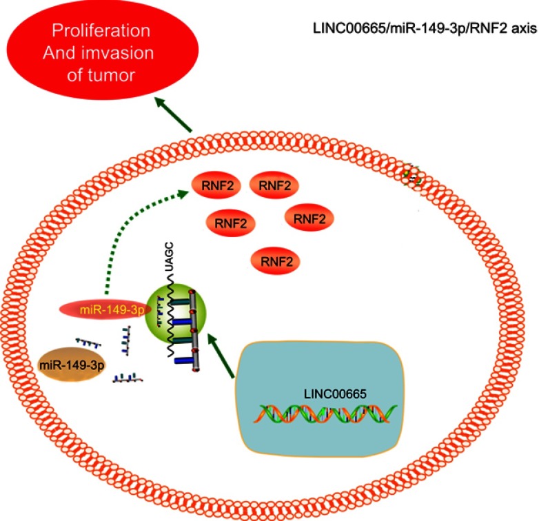
To further confirm the LINC00665/miR-149-3p/RNF2 axis in GC. Correlation analysis revealed that LINC00665 expression was positively associated with RNF2 expression in GC tissues (Figure 4D). Western blot assay showed that relative expression of RNF2 was down-regulated in LINC00665 shRNA group, while miR-149-3p inhibitors rescued the effects (Figure 4E). In addition, we found that RNF2 expression was significantly increased in GC tissues and positively correlated with TNM stage (Figure 4F and G). High RNF2 expression was associated with poor relapse free survival and overall survival of GC patients by TCGA database (Figure 4H and I). These results determined that LINC00665 might promote the proliferation and invasion of GC cells by regulating miR-149-3p/RNF2 axis (Figure 5).
Figure 5.
Schematic representation of LINC00665 in gastric cancer.
Discussion
Recently, increasing evidence revealed that dysregulation lncRNAs could play important roles in GC tumorigenesis and progression by regulating cell proliferation, apoptosis, and metastasis.15 For example, Li et al showed that lncRNA CASC2 reduced GC cells proliferation via regulating the MAPK pathway.16 Liu et al found that lncRNA FLVCR1-AS1 could sponge miR-155 to promote the tumorigenesis of GC via regulating c-Myc.17 Li et al found that lncRNA SLC25A5-AS1 reduced GC cells growth by regulating miR-19a-3p/PTEN/PI3K/AKT axis.18 However, the roles and underlying mechanisms of lncRNAs in GC remain unclear.
In the current study, we found that LINC00665 was increased in GC tissues and cell lines. High LINC00665 expression was significantly correlated with advanced TNM stage, histological grade, and poor prognosis of patients with GC. Function assays revealed that LINC00665 inhibition restricted GC cells proliferation and invasion abilities in vitro. Hence, we suggested that LINC00665 might serve as a tumor oncogenic lncRNA in GC progression.
Previous studies showed that miR-149-3p play important roles in tumor progression. For example, Yang et al showed that miR-149-3p inhibited bladder cancer cells proliferation and invasion by targeting S100A4.19 Ma et al found that lncRNA ZEB1-AS1 promotes GC progression via targeting miR-149-3p in GC.20 However, the roles of miR-149-3 in GC remain unclear. In the present study, we observed that miR-149-3p was significantly reduced in GC tissues and associated with poor prognosis. MiR-149-3p overexpression reduced GC cells viability. Furthermore, miR-149-3p was predicted as the target of LINC00665. LINC00665 inhibition significantly increased miR-149-3p expression in GC cells. Luciferase reporter assay and pull-down assay were further confirmed the correlation between LINC00665 and miR-149-3p in GC. Thus, we suggested that LINC00665 could function as a ceRNA to sponge miR-149-3p in GC.
RING finger protein 2 (RNF2) was first identified as an important interactor of Bmi1, which is a member of polycomb group.21 Recent studies showed that RNF2 was upregulated in various tumors and play critical roles in tumorigenesis.22 For example, Yang et al knockdown of RNF2 enhanced radio-sensitivity of lung squamous cell carcinoma cells.23 Li et al found that overexpression of RNF2 was an independent predictor of outcome in patients with urothelial carcinoma of the bladder undergoing radical cystectomy.24 Chen et al suggested that miR-139-5p overexpression combined with inactivation of the MAPK signaling pathway can reverse the cisplatin resistance of ovarian cancer by suppressing RNF2.25 In the present study, we found that RNF2 expression was significantly increased in GC tissues and positively correlated with TNM stage and poor prognosis. Bioinformatic prediction and experimental verification showed that RNF2 served as a target of miR-149-3p in GC. Moreover, LINC00665 expression was positively associated with RNF2 expression in GC tissues. RNF2 was suppressed in sh-LINC00665 group, while miR-149-3p inhibitors rescued the effects. Therefore, we demonstrated that LINC00665 might promote GC progression via regulating miR-149-3p/RNF2 axis.
However, this study has several limitations. For example, the number of clinical samples is not large enough. In addition, the roles of LINC00665/miR-149-3p/RNF2 axis in vivo remain unclear. Furthermore, due to the difficulties in conducting clinical experiments, we did not clinically verify our conclusions.
In conclusion, the present work elucidated that LINC00665 was significantly up-regulated in GC. Knockdown of LINC00665 reduced GC cells proliferation and invasion by sponging miR-149-3p/RNF2 axis, which providing a novel therapeutic target for GC treatment.
Acknowledgment
We are appreciating to every author in this study for their assistance with the experiments and constructive comments on the manuscript.
Ethics approval
Ethical approval was given by the Ethics Committee of The Central Hospital of Xinxiang.
Availability of data and materials
The dataset supporting the conclusions of this article is included within the article.
Disclosure
The authors report no conflicts of interest in this work.
References
- 1.Torre LA, Bray F, Siegel RL, Ferlay J, Lortet-Tieulent J, Jemal A. Global cancer statistics, 2012. CA Cancer J Clin. 2015;65(2):87–108. doi: 10.3322/caac.21262 [DOI] [PubMed] [Google Scholar]
- 2.Guggenheim DE, Shah MA. Gastric cancer epidemiology and risk factors. J Surg Oncol. 2013;107(3):230–236. doi: 10.1002/jso.23262 [DOI] [PubMed] [Google Scholar]
- 3.Brenner H, Rothenbacher D, Arndt V. Epidemiology of Stomach cancer[M]//Cancer Epidemiology. Totowa, NJ: Humana Press; 2009:467–477. [DOI] [PubMed] [Google Scholar]
- 4.Karimi P, Islami F, Anandasabapathy S, Freedman ND, Kamangar F. Gastric cancer: descriptive epidemiology, risk factors, screening, and prevention. Cancer Epidemiol Biomarkers Prev. 2014;23(5):700–713. doi: 10.1158/1055-9965.EPI-13-1057 [DOI] [PMC free article] [PubMed] [Google Scholar]
- 5.Van Cutsem E, Sagaert X, Topal B, et al. Gastric cancer. Lancet. 2016;388(10060):2654–2664. doi: 10.1016/S0140-6736(16)30354-3 [DOI] [PubMed] [Google Scholar]
- 6.Mercer TR, Dinger ME, Mattick JS. Long non-coding RNAs: insights into functions. Nat Rev Genet. 2009;10(3):155. doi: 10.1038/nrg2521 [DOI] [PubMed] [Google Scholar]
- 7.Quinn JJ, Chang HY. Unique features of long non-coding RNA biogenesis and function. Nat Rev Genet. 2016;17(1):47. doi: 10.1038/nrg.2015.10 [DOI] [PubMed] [Google Scholar]
- 8.Shi X, Sun M, Liu H, Yao Y, Song Y. Long non-coding RNAs: a new frontier in the study of human diseases. Cancer Lett. 2013;339(2):159–166. doi: 10.1016/j.canlet.2013.06.013 [DOI] [PubMed] [Google Scholar]
- 9.Xie X, H T L, Mei J, et al. LncRNA HMlincRNA717 is down-regulated in non-small cell lung cancer and associated with poor prognosis. Int J Clin Exp Pathol. 2014;7(12):8881. [PMC free article] [PubMed] [Google Scholar]
- 10.Hua F, Liu S, Zhu L, Ma N, Jiang S, Yang J. Highly expressed long non-coding RNA NNT-AS1 promotes cell proliferation and invasion through Wnt/β-catenin signaling pathway in cervical cancer. Biomed Pharmacother. 2017;92:1128–1134. doi: 10.1016/j.biopha.2017.03.057 [DOI] [PubMed] [Google Scholar]
- 11.Gu Y, Xiao X, Yang S. LncRNA MALAT1 acts as an oncogene in multiple myeloma through sponging miR-509-5p to modulate FOXP1 expression. Oncotarget. 2017;8(60):101984. doi: 10.18632/oncotarget.v8i60 [DOI] [PMC free article] [PubMed] [Google Scholar]
- 12.Wen DY, Lin P, Pang YY, et al. Expression of the long intergenic non-protein coding RNA 665 (LINC00665) gene and the cell cycle in hepatocellular carcinoma using the Cancer Genome Atlas, the gene expression omnibus, and quantitative real-time polymerase chain reaction. Med Sci Monit. 2018;24:2786. doi: 10.12659/MSM.907389 [DOI] [PMC free article] [PubMed] [Google Scholar]
- 13.Cong Z, Diao Y, Xu Y, et al. Long non-coding RNA linc00665 promotes lung adenocarcinoma progression and functions as ceRNA to regulate AKR1B10-ERK signaling by sponging miR-98. Cell Death Dis. 2019;10(2):84. doi: 10.1038/s41419-019-1361-3 [DOI] [PMC free article] [PubMed] [Google Scholar]
- 14.S D L, Yang JM, Xia Y, Fan Q, Yang K-P. Long noncoding RNA NEAT1 promotes proliferation and invasion via targeting miR-181a-5p in non-small cell lung cancer. Oncol Res Featuring Preclin Clin Cancer Ther. 2018;26(2):289–296. doi: 10.3727/096504017X15009404458675 [DOI] [PMC free article] [PubMed] [Google Scholar]
- 15.Xia T, Liao Q, Jiang X, et al. Long noncoding RNA associated-competing endogenous RNAs in gastric cancer. Sci Rep. 2014;4:6088. doi: 10.1038/srep06088 [DOI] [PMC free article] [PubMed] [Google Scholar]
- 16.Li P, Xue WJ, Feng Y, et al. Long non-coding RNA CASC2 suppresses the proliferation of gastric cancer cells by regulating the MAPK signaling pathway. Am J Transl Res. 2016;8(8):3522. [PMC free article] [PubMed] [Google Scholar]
- 17.Liu Y, Guo G, Zhong Z, et al. Long non-coding RNA FLVCR1-AS1 sponges miR-155 to promote the tumorigenesis of gastric cancer by targeting c-Myc. Am J Transl Res. 2019;11(2):793. [PMC free article] [PubMed] [Google Scholar]
- 18.Li X, Yan X, Wang F, et al. Down‐regulated lncRNA SLC25A5‐AS1 facilitates cell growth and inhibits apoptosis via miR‐19a‐3p/PTEN/PI3K/AKT signalling pathway in gastric cancer. J Cell Mol Med. 2019;23(4):2920–2932. [DOI] [PMC free article] [PubMed] [Google Scholar]
- 19.Yang D, Du G, Xu A, et al. Expression of miR-149-3p inhibits proliferation, migration, and invasion of bladder cancer by targeting S100A4. Am J Cancer Res. 2017;7(11):2209. [PMC free article] [PubMed] [Google Scholar]
- 20.Ma MH, An JX, Zhang C, et al. ZEB1-AS1 initiates a miRNA-mediated ceRNA network to facilitate gastric cancer progression. Cancer Cell Int. 2019;19(1):27. doi: 10.1186/s12935-019-0742-0 [DOI] [PMC free article] [PubMed] [Google Scholar]
- 21.Lorick KL, Jensen JP, Fang S, et al. RING fingers mediate ubiquitin-conjugating enzyme (E2)-dependent ubiquitination. Proc Natl Acad Sci. 1999;96(20):11364–11369. doi: 10.1073/pnas.96.20.11364 [DOI] [PMC free article] [PubMed] [Google Scholar]
- 22.Rai K, Akdemir KC, Kwong LN, et al. Dual roles of RNF2 in melanoma progression. Cancer Discov. 2015;5(12):1314–1327. doi: 10.1158/2159-8290.CD-15-0493 [DOI] [PMC free article] [PubMed] [Google Scholar]
- 23.Yang J, Yu F, Guan J, et al. Knockdown of RNF2 enhances radiosensitivity of lung squamous cell carcinoma cells. Biochem Cell Biol. Epub 2019. Jan 23. doi:10.1139/bcb-2018-0252 [DOI] [PubMed] [Google Scholar]
- 24.LI XD, Chen SL, Dong P, et al. Overexpression of RNF2 is an independent predictor of outcome in patients with urothelial carcinoma of the bladder undergoing radical cystectomy. Sci Rep. 2016;6:20894. doi: 10.1038/srep20894 [DOI] [PMC free article] [PubMed] [Google Scholar]
- 25.Chen Y, Cao XY, Li YN, et al. Reversal of cisplatin resistance by microRNA-139-5p-independent RNF2 downregulation and MAPK inhibition in ovarian cancer. Am J Physiol Cell Physiol. 2018;315(2):C225–C235. doi: 10.1152/ajpcell.00283.2017 [DOI] [PubMed] [Google Scholar]
Associated Data
This section collects any data citations, data availability statements, or supplementary materials included in this article.
Data Availability Statement
The dataset supporting the conclusions of this article is included within the article.


