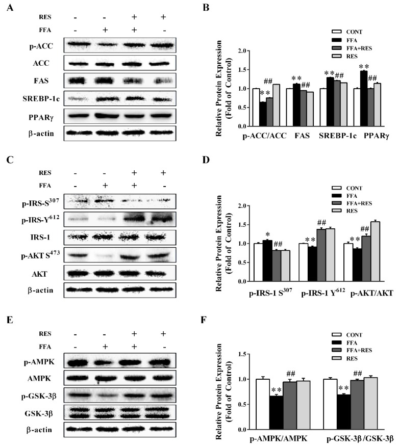
Figure 2.
RES ameliorates FFA-triggered hepatic glucolipid metabolism disorders in HepG2 cells. HepG2 cells were pretreated with RES (100 µM) for 6 h and with FFA (100 µM) for 24 h. (A) The effect of RES on FFA-induced abnormal lipid metabolism was determined by western blot analysis. phospho-acetyl-CoA carboxylase (p-ACC), total acetyl-CoA carboxylase (ACC), fatty acid synthase (FAS), sterol regulatory element-binding protein 1c (SREBP-1c), and peroxisome proliferator activated receptor gamma (PPARγ) were detected in HepG2 cells, and β-actin was used as a loading control. (C) The effect of RES on FFA-induced insulin signaling changes was determined by western blot analysis. p-IRS-1 Tyr612, p-IRS-1 Ser307, total insulin receptor substrate 1 (IRS-1), p-AKT Ser473, and total protein kinase B (AKT) were detected in cells, and β-actin was used as a loading control. (E) Representative western blot of p-AMPK, p-GSK3β, total AMP-activated protein kinase (AMPK), and total GSK3β after treatment with RES and FFA in HepG2 cells. (B), (D), and (F) are the densitometric analysis of the blots shown in (A), (C), and (E), respectively. Data were presented as the mean ± SD, n ≥ 3. (∗) p < 0.05 and (∗∗) p < 0.01, versus control group; (#) p < 0.05 and (##) p < 0.01, versus FFA group.

