Abstract
The unique stereoelectronic properties of sp2-iminosugars enable their participation in glycosylation reactions, thereby behaving as true carbohydrate chemical mimics. Among sp2-iminosugar conjugates, the sp2-iminosugar glycolipids (sp2-IGLs) have shown a variety of interesting pharmacological properties ranging from glycosidase inhibition to antiproliferative, antiparasitic, and anti-inflammatory activities. Developing strategies compatible with molecular diversity-oriented strategies for structure–activity relationship studies was therefore highly wanted. Here we show that a reaction sequence consisting in stereoselective C-allylation followed by thiol-ene “click” coupling provides a very convenient access to α-C-glycoside sp2-IGLs. Both the glycone moiety and the aglycone tail can be modified by using sp2-iminosugar precursors with different configurational profiles (d-gluco or d-galacto in this work) and varied thiols, as well as by oxidation of the sulfide adducts (to the corresponding sulfones in this work). A series of derivatives was prepared in this manner and their glycosidase inhibitory, antiproliferative and antileishmanial activities were evaluated in different settings. The results confirm that the inhibition of glycosidases, particularly α-glucosidase, and the antitumor/leishmanicidal activities are unrelated. The data are also consistent with the two later activities arising from the ability of the sp2-IGLs to interfere in the immune system response in a cell line and cell context dependent manner.
Keywords: sp2-Iminosugars, C-glycosides, glycolipids, glycomimetics, glycosidase inhibitors, Leishmaniasis, cancer
1. Introduction
Since their conception in the mid-1990s, sp2-iminosugars have consolidated as a unique class of glycomimetics in terms of chemical and structural versatility. Examples on record include piperidine [1,2,3,4], pyrrolidine [5,6], pyrrolizidine [7,8], indolizidine [9,10,11,12,13,14], and nor-tropane cores [15,16,17] with varied hydroxylation profiles. The presence of a pseudoamide-type nitrogen, with substantial sp2-hybridized character, at the position of the ring oxygen in monosaccharides greatly facilitates the incorporation of substituents at the endocyclic heteroatom, providing a very convenient manner to modulate their properties as regulators of carbohydrate processing enzymes. This strategy has been exploited for the development of glycosidase activity enhancers as pharmacological chaperone candidates against several lysosomal storage disorders [18,19,20], including Gaucher [21,22,23,24,25,26], Fabry [27,28], GM1-gangliosidosis [29,30], Tay–Sachs [31], and α-mannosidosis [32] diseases. The pseudoamide functional group additionally influences the stereoelectronic properties at the pseudoanomeric region, which translates into an exacerbated anomeric effect that imparts a high chemical stability to axially-oriented heteroatom substituents. Thus, sp2-iminosugars exist in reducing form (anomeric OH) in aqueous solution and can engage in glycosylation reactions, thereby behaving as true chemical sugar mimics wherein the pseudoamide function exerts strict control of the stereochemical outcome to provide exclusively the α-anomer [33,34]. A variety of O-, S-, N-, and even C-glycoside derivatives [35,36], including sp2-iminosugar disaccharide mimetics [37], multivalent systems [38,39,40,41], glycopeptides (sp2-IGPs) [42], and glycolipids (sp2-IGLs) [43], have been prepared in this manner in single diastereomeric form. For comparison, C-glycosides are the only representatives that are stable in the case of classical iminosugars, but their stereoselective synthesis generally requires rather elaborated reaction sequences [44,45,46,47,48].
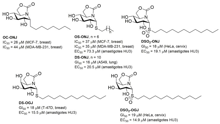
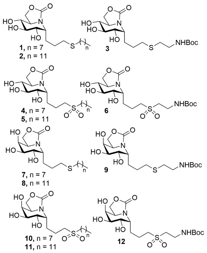
The compatibility of the synthetic methodologies for the preparation of sp2-iminosugar conjugates with structural diversity-oriented strategies has contributed decisively to expand their range of biological activities. Notably, sp2-IGLs have shown to be potent antitumor, antileishmanial, and anti-inflammatory agents [49,50] depending on the nature of the aglycone moiety. For instance, the C-octyl pseudo-α-glycoside 5N,6O-oxomethylidenenojirimycin (ONJ) derivative OC-ONJ (Figure 1) exhibited selective antimitotic, proapoptotic, and antimetastatic activities against breast carcinoma in vitro (noninvasive MCF-7 and invasive MDA-MB-231 cell lines) and in vivo (mice) [51,52]. Replacement of the pseudoanomeric carbon atom by sulfur (OS-ONJ; Figure 1) additionally led to modest antiparasitic activity against intracellular amastigotes of Leishmania donovani (EC50 = 73.3 μM) [53]. Increasing the length of the S-alkyl chain up to twelve carbon atoms (DS-ONJ) and oxidation to the corresponding sulfone derivative (DSO2-ONJ) considerably improved the potency as leishmanicidal (up to 5-fold) without reducing the antiproliferative efficiency against different human tumor cell lines (GI50 < 20 μM). Although initially these behaviors were ascribed to the ability of inhibiting α-glucosidase, further evidence suggested that this might not be the case, as illustrated by the fact that the 5N,6O-oxomethylidenenojirimycin (OGJ) epimers (DS-OGJ and DSO2-OGJ, respectively, displayed similar antiproliferative and antiparasitic trends (Figure 1). Here we report the synthesis of new sp2-IGLs that combine the key structural features previously encountered responsible for the mentioned biological activities, namely a C-glycosidic linkage and the presence of sulfide or sulfone functionalities in the aglycone moiety by making use of the thiol-ene “click” reaction [54,55,56,57,58,59]. Precisely, the ONJ (1–6) and OGJ (7–12) derivatives (Figure 2) have been prepared and evaluated in parallel for their glycosidase inhibitory, antiproliferative and antileishmanial activities in an attempt to ascertain whether or not the mechanisms at play are concurrent.
Figure 1.
Chemical structures and biological activities of featured sp2-IGLs.
Figure 2.
Chemical structures of the pseudo-α-C-glycoside sp2-IGLs prepared in this work.
2. Results and Discussion
2.1. Synthesis
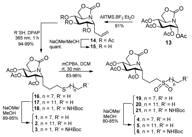
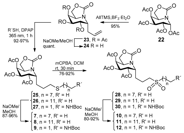
The C-allylation reaction of the ONJ tetraacetate 13 [33] by treatment with an excess of allyltrimethylsilane [60] and boron trifluoride etherate (BF3·OEt2) at 80 °C proceeded with total α-stereoselectivity to give the pivotal precursor 14 in high yield (Scheme 1). An aliquot of 14 was deacetylated to afford the unprotected C-allyl ONJ 15, used as control in subsequent pharmacological tests. The corresponding NMR data were in full agreement with the proposed structure. Particularly, the vicinal coupling constant value between the anomeric H-1 proton and H-2 (6.0 and 6.6 Hz for 14 and 15, respectively) support their gauche relative disposition characteristic of α-gluco arrangements in the 4C1 chair conformation. In order to access the target sp2-IGLs, photoinduced thiol-ene coupling reactions [61] of 14 with octanethiol, dodecanethiol, and 2-[N-(tert-butoxycarbonyl)amino]ethanethiol were conducted. The reaction conditions implied irradiation at 365 nm in the presence of a catalytic amount of 2,2-dimethoxy-2-phenyl-acetophenone (DPAP) in dry DMF, leading to the expected anti-Markovnikov sulfide adducts 16, 17, and 18, respectively, in over 90% yield. Conventional deacetylation reactions provided the fully unprotected α-C-glycoside derivatives 1, 2, and 3 (Scheme 1). Compounds 16–18 were further oxidized with m-chloroperoxybenzoic acid (mCPBA) [53] to the corresponding sulfones 19–21, which after removal of the acetate groups gave the amphiphilic sp2-IGLs 4, 5, and 6 (Scheme 1). A parallel reaction sequence starting from the OGJ tetraacetate 22, let access the corresponding α-C-allyl glycoside (23, 24) and α-C-glycoside sp2-IGL sulfide (25–27 and 7–9) and sulfone derivatives (28–30 and 10–12) with a substitution pattern of stereochemical complementarity to α-C-galactopyranosides (Scheme 2).
Scheme 1.
Synthesis of new ONJ sp2-IGLs 1–6.
Scheme 2.
Synthesis of new OGJ sp2-IGLs (7–12).
2.2. Inhibitory Properties against Commercial Enzymes
The inhibitory properties of the new sp2-IGLs 1–12 and the α-C-allyl ONJ and OGJ glycosides 15, and 24 were examined against a panel of commercial glycosidases that included the α-glucosidases maltase (Saccharomyces cerevisiae) and amyloglucosidase (Aspergillus niger), the β-glucosidases from almonds and bovine liver, α-galactosidase from green coffee beans, β-galactosidase from Escherichia coli, α-mannosidase from Jack beans, and β-mannosidase from Helix pomatia. The corresponding inhibition constant (Ki) values are collected in Table 1.
Table 1.
Glycosidase inhibitory activities (Ki, μM) of the new ONJ (1–6, 15) and OGJ (7–12, 24) C-glycoside derivatives. Values represent the mean ± SD (three independent determinations). Inhibition was competitive in all cases.
| Compound a | α-Glcase (Yeast Maltase) |
β-Glcase (Bovine Liver) |
|---|---|---|
| 15 | 79 ± 8 | n.i. |
| 1 | 0.34 ± 0.02 | 400 ± 20 |
| 2 | 0.74 ± 0.03 | 54 ± 5 |
| 3 | 0.28 ± 0.02 | 151 ± 10 |
| 4 | 2.6 ± 0.3 | 172 ± 14 |
| 5 | 2.5 ± 0.2 | 85 ± 18 |
| 6 | 0.75 ± 0.02 | 342 ± 20 |
| 7 | n.i. | n.i. |
| 8 | n.i. | 53 ± 12 |
| 9 | n.i. | n.i. |
| 10 | n.i. | 422 ± 22 |
| 11 | n.i. | 134 ± 10 |
| 12 | n.i. | 770 ± 45 |
| 24 | n.i. | n.i. |
a No inhibition was observed for any compound at 1 mM concentration on Aspergillus niger amyloglucosidase, almond β-glucosidase, green coffee bean α-galactosidase, E. coli β-galactosidase, Jack bean α-mannosidase, and Helix pomatia β-mannosidase.
None of ONJ sp2-IGLs 1–6 showed inhibition towards α- or β-mannosidase or α- or β-galactosidase, in agreement with their d-gluco configurational pattern. They behaved instead as low-micromolar to nanomolar inhibitors of yeast maltase (Ki 2.6–0.28 μM). The presence of the lipidic aglycone was determinant for the inhibitory potency, since the parent α-C-allyl derivative 15 was a 30- to 100-fold weaker inhibitor of this enzyme (Ki 79 μM). The sulfone derivatives 4–6 behaved as less potent maltase inhibitors than the corresponding sulfides 1–3, probably because of their moderated lipophilicity. Compounds 1–6 were much weaker inhibitors of β-glucosidase (bovine liver), with Ki values in the 54 to 400 μM range, meaning α:β anomeric selectivity ratios about 100:1. Neither epimeric OGJ sp2-IGLs 7–12 nor α-allyl OGJ glycoside 24 behaved as inhibitors of the α-galactosidase used in this assay, in spite of the configurational complementarity with the natural substrates. This is consistent with previous reports on the inhibitory properties of galacto-configured sp2-iminosugars showing that α-galactosidase inhibition is totally cancelled after engaging the primary hydroxyl in closing de five-membered ring [62]. As expected, the OGJ α-C-glycosides did not inhibit α-glucosidase, β-galactosidase or α- or β-mannosidase, although some representatives exhibited modest inhibition of the mammalian β-glucosidase (Ki 53 to 422 μM), an enzyme known to accept both β-d-gluco and β-d-galacto substrates [63].
2.3. Antiproliferative Activity
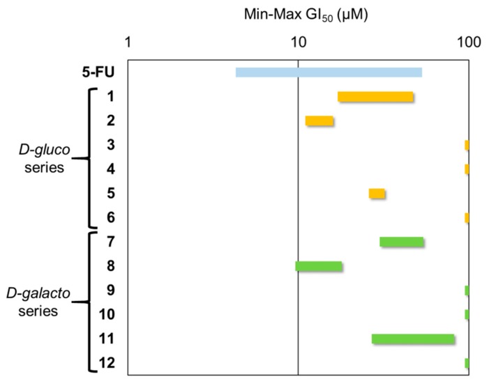
The antiproliferative activity of all the sp2-iminosugar C-glycosides (1–12) was evaluated against a panel of six representative human solid tumor cell lines including lung (A549 and SW1573), breast (HBL-100, T-47D), cervix (HeLa), and colon (WiDr). The results, expressed as the concentration to achieve 50% growth inhibition (GI50), are shown in Figure 3; only GI50 values below 100 μM are considered significant.
Figure 3.
GI50 range plot of tested compounds. 5-Fluorouracil (5-FU) was used as reference drug.
The new α-C-glycoside sp2-IGLs included in this study were found not to affect the viability and mortality of the breast normal cell line (MCF-10A) at concentrations up to 100 μM. The ensemble of data revealed a strong dependence of the antiproliferative activity against tumor cells on the nature of the aglycone substituent. Comparatively, the effect of the configurational hydroxylation profile of the glycone moiety (d-gluco for the ONJ derivatives 1–6; d-galacto for the OGJ derivatives 7–12) is much less marked. The carbamate-containing sp2-IGLs 3, 6, 9, and 12 did not show any significant antiproliferative activity in this assay. Conversely, the octyl (1,7) and dodecyl sulfides (2,8) were active against all the assayed tumor cell lines, with GI50 values ranging from 14 to 54 μM for the first pair and from 9.6 to 19 μM for the second pair, highlighting an increase in the antiproliferative activity with lipophilicity. This trend becomes much more pronounced in the sulfone series: whereas the octylsufonyl sp2-IGLs 4 and 10 did not achieve 50% tumor cell growth inhibition below the 100 μM concentration threshold, dodecylsulfonyl homologs 5 and 11 exhibited GI50 values in the range of 26 to 82 μM.
Inhibition of the α-glycosidases involved in N-glycoprotein biosynthesis, including α-glucosidases, has been invoked to explain the antitumor effects of some classical iminosugar glycomimetics [64,65]. Although the α-glucosidase inhibitory potency and antiproliferative activities of the ONJ derivatives both increased with aglycone lipophilicity, at least for the sulfide and sulfone representatives, close inspection of the data discards any direct relationship between them in the case of the sp2-IGLs. For instance, compounds 2 and 8 have very similar broad range antiproliferative profiles (Figure 3), but whereas 2 is a nanomolar competitive inhibitor of maltase, 8 did not inhibit this enzyme (Table 1). Even more revealing, compound 3, the most potent α-glucosidase inhibitor of the library (Ki 0.28 μM), was out of range in the antiproliferative assay. These evidences strongly suggest that the mechanism responsible for the antiproliferative outcome of sp2-IGLs in cancer cell lines is glycosidase-independent.
2.4. Antileishmanial Activity and Cellular Toxicity
The leishmanicidal activity of all the synthesized sp2-iminosugar C-glycosides (1–12) was evaluated against intracellular amastigotes of Leishmania donovani MHOM/ET/67/HU3 line with luciferase gene integrated into the parasite genome [66]. To this end, the concentration of compound required to inhibit the growth of parasites by 50% (50% effective concentration; EC50) was determined, with a threshold established at 20 μM (Table 2). A strong influence of the nature of the lipid chain α-linked to the pseudoanomeric position of the bicyclic core in the sp2-IGLs was inferred from the corresponding data. Thus, the 3-thiadodecyl (1 and 7) or the 3-thia-6-[N-(tert-butoxycarbonyl)amino]hexyl α-C-glycosides (3 and 9) promoted potent inhibition of the parasite growth, with EC50 values in the low micromolar range (7–15 μM), regardless of the configurational pattern of the sp2-iminosugar aglycone moiety (ONJ for 1 and 3; DGJ for 7 and 9). The longer-chain sp2-IGLs 2 and 8 were over the 20 μM concentration threshold in this assay. In the sulfone series, the results evidenced some disparity between the ONJ and OGJ epimers. Thus, the octylsulfonyl and dodecylsulfonyl segment-bearing derivatives 5 (EC50 11.44 ± 5.08 μM) and 10 (EC50 13.81 ± 1.72 μM) were the only representatives with EC50 values below the 20 μM threshold in the first and second case, respectively. Although the antileishmanial activity is modest as compared to the reference drug amphotericin B (AmB, Table 2), all sp2-IGLs exhibited lower toxicities in the monocytic leukemia (THP-1) cell line, broadly used as a model for human monocytes to assess potential toxic side-effects of antileishmanial drug candidates, and in human lung fibroblast MRC-5 cells (Table 2).
Table 2.
Drug susceptibility profile of the C-glycosides sp2-IGLs 1–12 for intracellular amastigotes of L. donovani (EC50 values, μM) and cellular toxicity (MTT assay) in THP-1 and MRC-5 cells (EC50 values, μM). Data for the reference drug AmB are also shown a.
| Compound | Intracellular Amastigotes | THP-1 (SI) | MRC-5 (SI) |
|---|---|---|---|
| 1 | 7.68 ± 0.29 | 130.96 ± 4.29 (17.06) | 32.20 ± 1.03 (4.19) |
| 2 | >20 | 72.24 ± 4.18 (<3.61) | 19.92 ± 2.76 (<1) |
| 3 | 14.96 ± 1.28 | >200 | 133.89 ± 6.02 (8.95) |
| 4 | >20 | >200 | 188.98 ± 15.59 (<9.45) |
| 5 | 11.44 ± 5.08 | 100.43 ± 10.87 (8.78) | 71.49 ± 3.75 (6.25) |
| 6 | >20 | >200 | 155.18 ± 3.10 (<7.76) |
| 7 | 10.58 ± 1.51 | 127.55 ± 5.71 (12.06) | 60.44 ± 2.80 (5.72) |
| 8 | >20 | 68.30 ± 3.04 (<3.42) | 32.08 ± 1.94 (<1.60) |
| 9 | 13.39 ± 1.13 | >200 | 139.92 ± 17.31 (10.44) |
| 10 | 13.81 ± 1.72 | >200 | 184.97 ± 21.25 (13.40) |
| 11 | >20 | >200 | 161.89 ± 20.18 (<8.10) |
| 12 | >20 | >200 | 181.51 ± 14.93 (<9.08) |
| AmB | 0.15 ± 0.01 | 20.07 ± 4.43 (133.80) | 16.93 ± 2.44 (112.87) |
a Parasites were grown as described in the Experimental section for 72 h at 37 °C in the presence of increasing concentrations of compounds. THP-1 and MRC-5 cells were grown as described in the Experimental section for 72 h at 37 °C, in the presence of increasing concentrations of compounds. Cell viability was determined using the luciferase assay (intracellular amastigotes). AmB was used as the reference antileishmanial agent. Data are means of EC50 ± SD from three independent experiments. SI: Selectivity Index (EC50 THP-1 or MRC-5/EC50 parasite).
While it is apparent that the antiparasitic activity data in Table 2 do not correlate with the α-glucosidase inhibitory activity data in Table 1, it is less obvious to discern from the ensemble of results whether or not the antiproliferative and antileishmanial activities of the sp2-IGLs share common molecular mechanisms. The fact that both cancer and parasite infection lead to subversion of the innate immune system suggests that the compounds act as immunoregulators, as reported for other drugs displaying anticancer and antileishmanial behaviors, e.g., the alkyl lipid ethers miltefosine and perifosine [67]. Indeed, the sp2-IGL representative DSO2-ONJ (Figure 1), which also displayed antiproliferative and antileishmanial activities [43,53], was found to behave as a toll-like receptor-4 (TLR4) antagonist in LPS-induced inflammation models in microglia and dendritic cells, as well as in a mouse model of acute inflammation, displaying potent anti-inflammatory activity [49]. Biochemical and computational data pointed at the p38 mitogen activated protein kinase (MAPK), a master regulator of the immune response, as the putative target. Considering that p38 is also involved in cancer and parasite infection, it seems likely that sp2-IGL binding to p38 is responsible for the ensemble of the observed pharmacological properties. Notwithstanding, a comparative analysis of the results collected in Figure 3 and Table 2 reveals significant differences in the antiproliferative and antileishmanial activity trends of the new sp2-IGL 1–12 as a function of their structure. In both cases, the efficacy depends essentially of the aglycone nature, whereas the configuration of the glycone configuration—ONJ or OGJ—is much less influential. Yet, whereas the antitumor potency follows the tendency dictated by aglycone lipophilicity, the antiparasitic efficiency is less predictable and more sensitive to the aglycone structure. For instance, the derivatives combining a sulfide and a carbamate group in the lipid chain (3 and 9), which did not display significant antiproliferative activity against any of the solid tumor cells assayed (see previous section), are low micromolar antileishmanial agents (EC50 14.96 ± 1.28 and 13.39 ± 1.13 μM, respectively). In contrast, the oxidized sulfone counterparts 6 and 12 were over the 20 μM threshold in the antileishmanial assayed. It should be considered that other factors, such as membrane-crossing or membrane-insertion capabilities, can strongly influence the biological activities of glycolipids in a cell line and cell-context dependent manner and can be also at the origin of the differences observed between both assays.
In summary, the results here presented demonstrate the suitability of the C-allylation of sp2-iminosugars in combination with the thiol-ene click reaction to access α-C-glycoside sp2-IGLs in high yield and with total stereocontrol, providing a versatile strategy to assess their pharmacological properties. The glycosidase inhibitory, antiproliferative and antileishmanial activities can be then optimized either individually or concertedly. The biological data support that the capabilities to inhibit glycosidases and the abilities to act as immunoregulators in the context of cancer or parasite infection are independent, opening the possibility of purposely designing multitarget compounds. It is conceivable, for instance, that sp2-IGLs behaving concomitantly as immunostimulants and inhibitors of a glycosidase involved in cancer or parasite cell metabolism will lead to more efficient therapeutics. Work in this direction is currently sought in our laboratories.
3. Materials and Methods
3.1. General Methods
Reagents and solvents were purchased from commercial sources and used without further purification. Optical rotations were measured with a JASCO P-2000 polarimeter, using a sodium lamp (λ = 589 nm) at 22 °C in 1 cm or 1 dm tubes. NMR experiments were performed at 300 (75.5), 400 (100.6) and 500 (125.7) MHz. 1-D TOCSY, as well as 2-D COSY and HMQC, experiments were carried out to assist signal assignment. For ESI mass spectra, 0.1 pm sample concentrations were used, the mobile phase consisting of 50% aq MeCN at 0.1 mL/min. Thin-layer chromatography was performed on precoated TLC plates, silica gel 30F-245, with visualization by UV light and also with 10% H2SO4 or 0.2% w/v cerium (IV) sulfate–5% ammonium molybdate in 2 m H2SO4, or 0.1% ninhydrin in EtOH. Column chromatography was performed on Chromagel (silice 60 AC.C 70–200 μm). Deacetylation reactions were carried out by using Zemplén procedure, e.i., addition of NaOMe (0.1 equiv. per mol of acetate) in MeOH at room temperature and neutralization with solid CO2, evaporation of the solvent and purification by column chromatography. All compounds were purified to ≥95% purity as determined by elemental microanalysis results obtained on a CHNS-TruSpect® Micro elemental analyzer (Instituto de Investigaciones Químicas de Sevilla, Spain) from vacuum-dried samples. The analytical results for C, H, N and S were within ± 0.4 of the theoretical values. (1R)-1,2,3,4-Tetra-O-acetyl-5N,6O-oxomethylidenegluco(galacto)nojirimycin (13 and 22, respectively) were prepared according reported procedures [33,68].
Inhibition constant (Ki) values were determined by spectrophotometrically measuring the residual hydrolytic activities of the glycosidases against the respective o- (for β-galactosidase from E. coli) or p-nitrophenyl α- or β-d-glycopyranoside (for other glycosidases) in the presence of the iminosugars. Each assay was performed in phosphate buffer or phosphate-citrate buffer (for α- or β-mannosidase and amyloglucosidase) at the optimal pH for the enzymes. The reactions were initiated by addition of the enzyme to a solution of the substrate in the absence or presence of various inhibitor concentrations. The mixture was incubated for 10–30 min at 37 °C or 55 °C (for amyloglucosidase) and the reaction was quenched by addition of 1 m Na2CO3. Reaction times were appropriate to obtain 10–20% conversion of the substrate in order to achieve linear rates. The absorbance of the resulting mixture was determined at 405 nm. Approximate value of Ki was determined from the slope of Dixon plots using a fixed concentration of substrate (around the Km value for the different glycosidases) and various concentrations of inhibitor. Full Ki determinations and enzyme inhibition mode were determined from the slope of Lineweaver–Burk plots and double reciprocal analysis. Representative examples of Dixon and Lineweaver–Burk plots are shown in the Supplementary materials.
3.2. Statistical Analysis
All results are expressed as mean ± SD of three independent experiments, each conducted in triplicate. The measurements were statistically analyzed using the Student’s t-test for comparing 2 groups. The level of significance was set at p < 0.05.
3.3. General Procedure for the Synthesis of the Allylated ONJ and OGJ Precursors 14 and 23
To a solution of 13/22 (2.20 mmol) in anhydrous CH3CN (36 mL) under Ar atmosphere, allyltrimethylsilane (3.5 mL, 22 mmol) and BF3.Et2O (2.8 mL) were added at rt and the mixture was then heated at 80 °C for 20 min. The reaction mixture was diluted with EtOAc (50 mL) and quenched with saturated aqueous NaHCO3 (2 × 30 mL). The organic phase was separated and the aqueous phase extracted with EtOAc (3 × 25 mL). The combined organic layer was washed with water (25 mL) and brine (25 mL), dried (MgSO4) and concentrated under reduced pressure. The resulting residue was purified by column chromatography using the solvent indicated in each case to give the corresponding C-allylated pseudo-α-glycosides.
(1R)-2,3,4-Tri-O-acetyl-1-allyl-5N,6O-oxomethylidene-1-deoxynojirimycin (14). Purification by column chromatography (1:1 EtOAc-cyclohexane) afforded 14. Yield: 712 mg (91%); Rf = 0.60 (2:1 EtOAc-cyclohexane). [α]D + 34.0 (c 1.0 in DCM); 1H NMR (500 MHz, CDCl3) δ 5.72 (dddd, 1 H, Jc,d = 17.0 Hz, Jc,e = 10.0 Hz, Jc,b = 8.0 Hz, Jc,a = 5.5 Hz, CH(c)=CH2), 5.34 (t, 1 H, J2,3 = J3,4 = 10.0 Hz, H-3), 5.14 (d, 1 H, CH=CH2(d)), 5.11 (d, 1 H, CH=CH2(e)), 5.06 (dd, 1 H, J1,2 = 6.0 Hz, H-2), 4.93 (t, 1 H, J4,5 = 10.0 Hz, H-4), 4.41–4.33 (m, 2 H, H-1, H-6a), 4.22 (dd, 1 H, J6a,6b = 9.5 Hz, J5,6b = 5.0 Hz, H-6b), 3.81 (ddd, 1 H, J5,6a = 8.5 Hz, H-5), 2.52 (dt, 1 H, Ja,b = 15.0 Hz, Ja,1 = 5.0 Hz, CH2(a)CH=CH2), 2.34 (ddd, 1 H, Jb,1 = 11.5 Hz, CH2(b)CH=CH2), 2.05–2.02 (3 s, 9 H, MeCO); 13C NMR (125.7 MHz, CDCl3) δ 170.0-169.2 (CO ester), 156.2 (CO carbamate), 132.8 (CH=CH2), 118.5 (CH=CH2), 72.5 (C-4), 69.9–69.5 (C-2, C-3), 65.7 (C-6), 52.1 (C-5), 50.0 (C-1), 30.7 (CH2CH=CH2), 20.6 (MeCO); ESIMS: m/z 378.28 [M + Na]+; Anal. Calcd for C16H21NO8: C 54.08, H 5.96, N 3.94; found: C 54.22, H 6.08, N 3.71.
(1R)-2,3,4-Tri-O-acetyl-1-allyl-5N,6O-oxomethylidene-1-deoxygalactonojirimycin (23). Purification by column chromatography (1:2 → 2:1 EtOAc-cyclohexane) afforded 23. Yield: 344 mg (95%); Rf = 0.63 (2:1 EtOAc-cyclohexane); [α]D + 64.7 (c 1.0 in DCM); 1H NMR (300 MHz, CDCl3) δ 5.71 (dddd, 1 H, Jc,d = 17.1 Hz, Jc,e = 10.2 Hz, Jc,b = 8.1 Hz, Jc,a = 5.4 Hz, CH(c)=CH2), 5.38 (t, 1 H, J3,4 = J4,5 = 2.1 Hz, H-4), 5.30 (dd, 1 H, J2,3 = 10.8 Hz, J1,2 = 6.6 Hz, H-2), 5.12 (m, 3 H, H-3, CH=CH2), 4.49 (ddd, 1 H, J1,b = 11.1 Hz, J1,a = 4.5 Hz, H-1), 4.31 (t, 1 H, J6a,6b = J5,6a = 8.5 Hz, H-6a), 4.02 (m, 2 H, H-5, H-6b), 2.48 (m, 1 H, CH2(a)CH=CH2), 2.27 (ddd, 1 H, Ja,b = 15.0 Hz, Jb,H1 = 11.5 Hz, CH2(b)CH=CH2), 2.15–1.98 (3 s, 9 H, MeCO); 13C NMR (75.5 MHz, CDCl3) δ 170.5-169.3 (CO ester), 156.6 (CO carbamate), 133.2 (CH=CH2), 118.3 (CH=CH2), 68.9 (C-4), 68.5 (C-3), 66.5 (C-2), 62.6 (C-6), 51.1 (C-5), 50.3 (C-1), 30.4 (CH2CH=CH2), 20.7-20.6 (MeCO); ESIMS: m/z 378.26 [M + Na]+; Anal. Calcd for C16H21NO8: C 54.08, H 5.96, N 3.94. Found: C 54.11, H 6.03, N 3.90.
(1R)-1-Allyl-5N,6O-oxomethylidene-1-deoxynojirimycin (15). Compound 15 was obtained by conventional O-deacetylation of 14 (51 mg, 0.14 mmol), as described in general methods, followed by purification by column chromatography (25:1 EtOAc-MeOH). Yield: 33 mg (quant); Rf = 0.26 (25:1 EtOAc-MeOH); [α]D + 22.0 (c 1.1 in MeOH); 1H NMR (300 MHz, CD3OD) δ 5.76 (dddd, 1 H, Jc,d = 17.1 Hz, Jc,e = 10.2 Hz, Jc,a = 8.1 Hz, Jc,b = 5.7 Hz, CH(c)=CH2), 5.14 (dd, 1 H, Jd,b = 1.2 Hz, CH=CH2(d)), 5.05 (d, 1 H, CH=CH2(e)), 4.43 (t, 1 H, J6a,6b = J5,6a = 8.5 Hz, H-6a), 4.27 (dd, 1 H, J5,6b = 4.2 Hz, H-6b), 4.00 (ddd, 1 H, J1,a = 12.3 Hz, J1,2 = 5.4 Hz, J1,b = 3.6 Hz, H-1), 3.65 (ddd, 1 H, J4,5 = 9.5 Hz, H-5), 3.57–3.43 (m, 2 H, H-2, H-3), 3.24 (t, 1 H, J3,4 = 9.0 Hz, H-4), 2.66 (dddd, 1 H, Ja,b = 15.0 Hz, CH2(b)CH=CH2), 2.21 (ddd, 1 H, CH2(a)CH=CH2); 13C NMR (75.5 MHz, CD3OD) δ 159.5 (CO), 136.0 (CH=CH2), 117.7 (CH=CH2), 75.5 (C-4), 74.7–72.0 (C-2, C-3), 67.5 (C-6), 55.2–55.0 (C-1, C-5), 30.5 (CH2CH=CH2); ESIMS: m/z 252.2 [M + Na]+; Anal. Calcd for C10H15NO5: C 52.40, H 6.60, N 6.11. Found: C 52.28, H 6.73, N 5.87.
(1R)-1-Allyl-5N,6O-oxomethylidene-1-deoxygalactonojirimycin (24). Compound 24 was obtained by conventional O-deacetylation of 23 (33 mg, 0.09 mmol) followed by purification by column chromatography (30:1 EtOAc-MeOH). Yield: 21 mg (quant); Rf = 0.22 (30:1 EtOAc-MeOH); [α]D − 3.0 (c 1.0 in MeOH); 1H NMR (300 MHz, CDCl3) δ 5.78 (dddd, 1 H, Jc,d = 18.3 Hz, Jc,e = 10.2 Hz, Jc,b = 8.3 Hz, Jc,a = 5.6 Hz, CH(c)=CH2), 5.15 (m, 1 H, CH=CH2(d)), 5.06 (m, 1 H, CH=CH2(e)), 4.41 (dd, 1 H, J6a,6b = 8.6 Hz, J5,6a = 4.6 Hz, H-6a), 4.34 (t, 1 H, J5,6b = 8.6 Hz, H-6b), 4.08 (m, 1 H, H-1), 4.02 (ddd, 1 H, J4,5 = 2.3 Hz, H-5), 3.96 (dd, 1 H, J2,3 = 9.9 Hz, J1,2 = 6.4 Hz, H-2), 3.82 (t, 1 H, J3,4 = 2.3 Hz, H-4), 3.65 (dd, 1 H, H-3), 2.65 (m, 1 H, CH2(b)CH=CH2), 2.24 (ddd, 1 H, Ja,b = 14.6 Hz, Jb,H1 = 12.0 Hz, CH2(a)CH=CH2); 13C NMR (75.5 MHz, CDCl3) δ 160.3 (CO), 136.4 (CH=CH2), 117.6 (CH=CH2), 71.5 (C-3), 71.1 (C-4), 68.1 (C-2), 64.8 (C-6), 54.9 (C-5), 54.5 (C-1), 30.1 (CH2CH=CH2); ESIMS: m/z 252.20 [M + Na]+; Anal. Calcd for C10H15NO5: C 52.40, H 6.60, N 6.11. Found: C 52.09, H 6.38, N 5.85.
3.4. General Procedure for the Synthesis of ONJ (1–3) and OGJ Derivatives (7–9) by Photoinduced Thiol-Ene Reaction
To a stirred solution of the corresponding pseudo-α-C-allyl glycosides 14/23 (142 mg, 0.40 mmol) and 2,2-dimethoxy-2-phenyl-acetophenone (DPAP) (31 mg, 0.12 mmol) in anhydrous DMF (2.5 mL), the corresponding thiol (1.20 mmol) was added. The mixture was deoxygenated for 30 min and then irradiated at rt under UVA lamp (λ 365 nm) for 1 h. The solvent was removed and the residue was purified by column chromatography using (1:5 → 1:3 →1:1 EtOAc:cyclohexane) as eluent.
(1R)-2,3,4-Tri-O-acetyl-1-[3-(octylthio)propyl]-5N,6O-oxomethylidene-1-deoxynojirimycin (16). Yield: 70 mg (99%); Rf = 0.74 (2:1 EtOAc-cyclohexane); [α]D + 42.2 (c 1.0 in DCM); 1H NMR (300 MHz, CDCl3) δ 5.31 (t, 1 H, J2,3 = J3,4 = 10.0 Hz, H-3), 5.01 (dd, 1 H, J1,2 = 6.3 Hz, H-2), 4.94 (t, 1 H, J4,5 = 10.0 Hz, H-4), 4.38 (dd, 1 H, J6a,6b = 9.0 Hz, J5,6a = 8.1 Hz, H-6a), 4.31–4.20 (m, 1 H, H-1), 4.25 (dd, 1 H, J5,6b = 4.5 Hz, H-6b), 3.85 (1 H, ddd, H-5), 2.66–2.43 (m, 4 H, CH2SCH2), 2.00–1.96 (3 s, 9 H, MeCO), 1.90–1.20 (m, 16 H, CH2), 0.87 (t, 3 H, 3JH,H = 7.0 Hz, CH3); 13C NMR (75.5 MHz, CDCl3) δ 169.9–169.2 (CO ester), 156.3 (CO carbamate), 72.3 (C-4), 69.8 (C-3), 69.6 (C-2), 65.6 (C-6), 52.1 (C-5), 50.7 (C-1), 32.2–22.6 (CH2), 20.6 (MeCO), 14.1 (CH3); ESIMS: m/z 524.39 [M + Na]+; Anal. Calcd for C24H39NO8S: C 57.46, H 7.84, N 2.79, S, 6.39. Found: C 57.61, H 8.00, N 2.48, S, 6.13.
(1R)-2,3,4-Tri-O-acetyl-1-[3-(dodecylthio)propyl]-5N,6O-oxomethylidene-1-deoxynojirimycin (17). Yield: 118 mg (96%); Rf = 0.58 (1:1 EtOAc-cyclohexane). [α]D + 37.5 (c 1.1 in DCM); 1H NMR (300 MHz, CDCl3) δ 5.30 (t, 1 H, J2,3 = J3,4 = 9.6 Hz, H-3), 5.00 (dd, 1 H, J1,2 = 6.3 Hz, H-2), 4.94 (t, 1 H, J4,5 = 9.6 Hz, H-4), 4.37 (dd, 1 H, J6a,6b = 9.0 Hz, J5,6a = 8.1 Hz, H-6a), 4.30–4.20 (m, 1 H, H-1), 4.23 (dd, 1 H, J5,6b = 4.5 Hz, H-6b), 3.84 (1 H, ddd, H-5), 2.65–2.42 (m, 4 H, CH2SCH2), 2.05–2.01 (3 s, 9 H, MeCO), 1.90–1.20 (m, 24 H, CH2), 0.86 (t, 3 H, 3JH,H = 7.0 Hz, CH3); 13C NMR (75.5 MHz, CDCl3) δ 169.9–169.2 (CO ester), 156.3 (CO carbamate), 72.3 (C-4), 69.8 (C-3), 69.5 (C-2), 65.6 (C-6), 52.1 (C-5), 50.7 (C-1), 32.2–22.7 (CH2), 20.6–20.5 (MeCO), 14.1 (CH3); ESIMS: m/z 580.43 [M + Na]+; Anal. Calcd for C28H47NO8S: C 60.30, H 8.49, N 2.51, S 5.75. Found: C 60.54, H 8.63, N 2.36, S 5.49.
(1R)-2,3,4-Tri-O-acetyl-1-[3-(N-tert-butoxycarbonylaminoethylthio)propyl]-5N,6O-oxomethylidene-1-deoxynojirimycin (18). Yield: 218 mg (94%); Rf = 0.39 (1:1 EtOAc-cyclohexane); [α]D + 36.5 (c 1.1 in DCM); 1H NMR (300 MHz, CDCl3) δ 5.30 (t, 1 H, J2,3 = J3,4 = 9.6 Hz, H-3), 5.00 (dd, 1 H, J1,2 = 6.0 Hz, H-2), 4.95 (t, 1 H, J4,5 = 9.6 Hz, H-4), 4.40 (dd, 1 H, J6a,6b = 9.3 Hz, J5,6a = 8.1 Hz, H-6a), 4.30–4.20 (m, 2 H, H-1, H-6b), 3.84 (ddd, 1 H, J5,6b = 4.2 Hz, H-5), 3.28 (q, 2 H, J = 6.5 Hz, CH2NHBoc), 2.70–2.46 (m, 4 H, CH2SCH2), 2.06–2.01 (3 s, 9 H, MeCO), 1.90–1.57 (m, 4 H, CH2), 1.43 (s, 9 H, CMe3); 13C NMR (75.5 MHz, CDCl3) δ 169.9–169.2 (CO ester), 156.3, 155.8 (CO carbamate), 79.4 (CMe3), 72.3 (C-4), 69.8 (C-3), 69.6 (C-2), 65.6 (C-6), 52.0 (C-5), 50.5 (C-1), 39.8 (CH2NHBoc), 32.1, 30.9 (CH2), 28.4 (CMe3), 25.3, 24.0 (CH2), 20.7–20.6 (MeCO); ESIMS: m/z 555.28 [M + Na]+; Anal. Calcd for C23H36N2O10S: C 51.87, H 6.81, N 5.26, S 6.02. Found: C 51.71, H 6.99, N 4.97, S 5.78.
(1R)-2,3,4-Tri-O-acetyl-1-[3-(octylthio)propyl]-5N,6O-oxomethylidene-1-deoxygalactonojirimycin (25). Yield: 188 mg (94%); Rf = 0.59 (1:1 EtOAc-cyclohexane); [α]D + 77.7 (c 1.0 in DCM); 1H NMR (300 MHz, CDCl3) δ 5.37 (t, 1 H, J3,4 = J4,5 = 2.7 Hz, H-4), 5.25 (dd, 1 H, J2,3 = 10.9 Hz, J1,2 = 6.3 Hz, H-2), 5.10 (dd, 1 H, H-3), 4.34 (m, 1 H, H-1), 4.33 (t, 1 H, J6a,6b = J5,6a = 9.0 Hz, H-6a), 4.10 (m, 1 H, H-5), 4.01 (dd, 1 H, J5,6b = 3.5 Hz, H-6b), 2.52 (m, 4 H, CH2SCH2), 2.14–1.95 (3 s, 9 H, MeCO), 1.83–1.47 (m, 6 H, CH2), 1.32 (m, 10 H, CH2), 0.84 (t, 3 H, 3JH,H = 7.0 Hz, CH3); 13C NMR (75.5 MHz, CDCl3) δ 170.4–169.4 (CO ester), 156.8 (CO carbamate), 69.0 (C-4), 68.4 (C-3), 66.5 (C-2), 62.3 (C-6), 51.0 (C-5), 50.8 (C-1), 32.1–22.6 (CH2), 20.7–20.5 (MeCO), 14.1 (CH3); ESIMS: m/z 524.33 [M + Na]+; Anal. Calcd for C24H39NO8S: C 57.46, H 7.84, N 2.79. S, 6.39. Found: C 57.57, H 8.03, N 2.56, S 6.04.
(1R)-2,3,4-Tri-O-acetyl-1-[3-(dodecylthio)propyl]-5N,6O-oxomethylidene-1-deoxygalactonojirimycin (26). Yield: 205 mg (92%); Rf = 0.69 (1:1 EtOAc-cyclohexane); [α]D + 63.8 (c 1.0 in DCM); 1H NMR (300 MHz, CDCl3) δ 5.34 (t, 1 H, J3,4 = J4,5 = 2.7 Hz, H-4), 5.23 (dd, 1 H, J2,3 = 10.9 Hz, J1,2 = 6.4 Hz, H-2), 5.07 (dd, 1 H, H-3), 4.30 (m, 1 H, H-1), 4.29 (t, 1 H, J6a,6b = J5,6a = 8.9 Hz, H-6a), 4.04 (m, 1 H, H-5), 3.97 (dd, 1 H, J5,6b = 3.5 Hz, H-6b), 2.49 (m, 4 H, CH2S), 2.11–1.93 (3 s, 9 H, MeCO), 1.85–1.45 (m, 6 H, CH2), 1.25 (m, 18 H, CH2), 0.81 (t, 3 H, 3JH,H = 7.0 Hz, CH3); 13C NMR (75.5 MHz, CDCl3) δ 170.5–169.4 (CO ester), 156.8 (CO carbamate), 69.0 (C-4), 68.4 (C-3), 66.5 (C-2), 62.8 (C-6), 51.1 (C-5), 50.9 (C-1), 32.2–22.7 (CH2), 20.7–20.5 (MeCO), 14.1 (CH3); ESIMS: m/z 580.38 [M + Na]+; Anal. Calcd for C28H47NO8S: C 60.30, H 8.49, N 2.51, S 5.75. Found: C, 60.17, H 8.42, N 2.29, S 5.60.
(1R)-2,3,4-Tri-O-acetyl-1-[3-(N-tert-butoxycarbonylaminoethylthio)propyl]-5N,6O-oxomethylidene-1-deoxygalactonojirimycin (27). Yield: 207 mg (97%); Rf = 0.44 (2:1 EtOAc-cyclohexane); [α]D + 64.1 (c 1.0 in DCM); 1H NMR (300 MHz, CDCl3) δ 5.34 (t, 1 H, J3,4 = J4,5 = 2.7 Hz, H-4), 5.23 (dd, 1 H, J2,3 = 10.9 Hz, J1,2 = 6.4 Hz, H-2), 5.10 (dd, 1 H, H-3), 4.88 (bs, 1 H, NH), 4.33 (m, 1 H, H-1), 4.33 (t, 1 H, J6a,6b = J5,6a = 8.9 Hz, H-6a), 4.04 (m, 1 H, H-5), 3.97 (dd, 1 H, J5,6b = 3.5 Hz, H-6b), 3.23 (q, 2 H, 3JH,H = 6.5 Hz, CH2NHBoc), 2.50 (m, 4 H, CH2SCH2), 2.11–1.93 (3 s, 9 H, MeCO), 1.80–1.50 (m, 4 H, CH2), 1.38 (s, 9 H, CMe3); 13C NMR (75.5 MHz, CDCl3) δ 170.4–169.4 (CO ester), 156.8, 155.8 (CO carbamate), 79.4 (CMe3), 69.0 (C-4), 68.3 (C-3), 66.5 (C-2), 62.8 (C-6), 51.1 (C-5), 50.7 (C-1), 39.8 (CH2NHBoc), 32.1, 30.9 (CH2), 28.4 (CMe3), 25.4, 23.6 (CH2), 20.8-20.6 (MeCO); ESIMS: m/z 555.30 [M + Na]+. Anal. Calcd for C23H36N2O10S: C 51.87, H 6.81, N 5.26, S 6.02. Found: C 51.60, H 6.95, N 5.09, S 5.71.
(1R)-1-[3-(Octylthio)propyl)-5N,6O-oxomethylidene-1-deoxynojirimycin (1). Compound 1 was obtained by conventional O-deacetylation of 16 (70 mg, 0.14 mmol), as described in general methods, followed by purification by column chromatography (40:1 EtOAc-MeOH). Yield: 50 mg (95%); Rf = 0.47 (30:1 EtOAc-MeOH); [α]D + 43.9 (c 1.1 in MeOH); 1H NMR (300 MHz, CD3OD) δ 4.45 (t, 1 H, J6a,6b = J5,6a = 9.0 Hz, H-6a), 4.29 (dd, 1 H, J5,6b = 4.0 Hz, H-6b), 3.96–3.86 (m, 1 H, H-1), 3.66 (ddd, 1 H, J4,5 = 9.6 Hz, H-5), 3.53–3.40 (m, 2 H, H-2, H-3), 3.28–3.18 (m, 1 H, H-4), 2.66–2.46 (m, 4 H, CH2SCH2), 2.07–1.93 (m, 1 H, CH2), 1.72–1.23 (m, 15 H, CH2), 0.90 (t, 3 H, 3JH,H = 7.0 Hz, CH3); 13C NMR (75.5 MHz, CD3OD) δ 159.6 (CO), 75.4 (C-4), 74.6–72.1 (C-2, C-3), 67.5 (C-6), 55.3 (C-1, C-5), 33.0–23.7 (CH2), 14.4 (CH3); ESIMS: m/z 398.38 [M + Na]+; Anal. Calcd for C18H33NO5S: C 57.57, H 8.86, N 3.73, S 8.54. Found: C 57.22, H 8.76, N 3.52, S 8.16.
(1R)-1-[3-(Dodecylthio)propyl]-5N,6O-oxomethylidene-1-deoxynojirimycin (2). Compound 2 was obtained by conventional O-deacetylation of 17 (59 mg, 0.10 mmol), as described in general methods, followed by purification by column chromatography (30:1 EtOAc-MeOH). Yield: 41mg (89%); Rf = 0.47 (30:1 EtOAc-MeOH); [α]D + 42.6 (c 1.0 in MeOH); 1H NMR (400 MHz, CD3OD) δ 4.45 (dd, 1 H, J6a,6b = 9.0 Hz, J5,6a = 8.4 Hz, H-6a), 4.29 (dd, 1 H, J5,6b = 4.0 Hz, H-6b), 3.95–3.88 (m, 1 H, H-1), 3.66 (ddd, 1 H, J4,5 = 9.5 Hz, H-5), 3.51–3.41 (m, 2 H, H-2, H-3), 3.23 (bt, 1 H, H-4), 2.65–2.48 (m, 4 H, CH2SCH2), 2.06–1.95 (m, 1 H, CH2), 1.70–1.25 (m, 23 H, CH2), 0.90 (t, 3 H, 3JH,H = 7.0 Hz, CH3); 13C NMR (100.6 MHz, CD3OD) δ 159.7 (CO), 75.4 (C-4), 74.6–72.2 (C-2, C-3), 67.5 (C-6), 55.3 (C-1, C-5), 33.1–23.7 (CH2), 14.4 (CH3); ESIMS: m/z 454.40 [M + Na]+; Anal. Calcd for C22H41NO5S: C 61.22, H 9.57, N 3.25, S 7.43. Found: C 60.88, H 9.33, N 2.99, S 7.15.
(1R)-1-[3-(N-tert-Butoxycarbonylaminoethylthio)propyl]-5N,6O-oxomethylidene-1-deoxynojirimycin (3). Compound 3 was obtained by conventional O-deacetylation of 18 (66 mg, 0.12 mmol), as described in general methods, followed by purification by column chromatography (30:1 EtOAc-MeOH). Yield: 47 mg (92%); Rf = 0.45 (30:1 EtOAc-MeOH); [α]D + 36.2 (c 1.0 in MeOH); 1H NMR (300 MHz, CD3OD) δ 4.47 (t, 1 H, J6a,6b = J5,6a = 8.5 Hz, H-6a), 4.29 (dd, 1 H, J5,6b = 4.0 Hz, H-6b), 3.97–3.82 (m, 1 H, H-1), 3.66 (ddd, 1 H, J4,5 = 9.6 Hz, H-5), 3.53–3.40 (m, 2 H, H-2, H-3), 3.28–3.11 (m, 3 H, H-4, CH2NHBoc), 2.70–2.50 (m, 4 H, CH2SCH2), 2.05–1.91 (m, 1 H, CH2), 1.72–1.49 (m, 3 H, CH2), 1.44 (s, 9 H, CMe3); 13C NMR (75.5 MHz, CD3OD) δ 159.7, 158.3 (CO), 80.1 (CMe3), 75.4 (C-4), 74.6–72.1 (C-2, C-3), 67.5 (C-6), 55.3 (C-1, C-5), 41.4 (CH2NHBoc), 32.5–24.5 (CH2), 28.8 (CMe3); ESIMS: m/z 429.31 [M + Na]+; Anal. Calcd for C17H30N2O7S: C 50.23, H 7.44, N 6.89, S 7.89. Found: C 50.37, H 7.60, N 6.58, S 7.61.
(1R)-1-[3-(Octylthio)propyl]-5N,6O-oxomethylidene-1-deoxygalactonojirimycin (7). Compound 7 was obtained by conventional O-deacetylation of 25 (103 mg, 0.21 mmol) followed by purification by column chromatography (30:1 EtOAc-MeOH). Yield: 74 mg (96%); Rf = 0.19 (30:1 EtOAc-MeOH); [α]D + 31.0 (c 1.1 in MeOH); 1H NMR (300 MHz, CD3OD) δ 4.31 (dd, 1 H, J6a,6b = 8.6 Hz, J5,6a = 4.6 Hz, H-6a), 4.26 (t, 1 H, J5,6b = 8.6 Hz, H-6b), 3.91 (m, 2 H, H-1, H-5), 3.81 (dd, 1 H, J2,3 = 9.7 Hz, J1,2 = 6.4 Hz, H-2), 3.71 (t, 1 H, J3,4 = J4,5 = 2.3 Hz, H-4), 3.51 (dd, 1 H, H-3), 2.46 (m, 4 H, CH2SCH2), 1.95–1.36 (m, 6 H, CH2), 1.26 (m, 10 H, CH2), 0.80 (t, 3 H, 3JH,H = 6.9 Hz, CH3); 13C NMR (75.5 MHz, CD3OD) δ 160.4 (CO), 71.4 (C-3), 71.1 (C-4), 68.1 (C-2), 64.9 (C-6), 55.1 (C-5), 54.6 (C-1), 33.0–23.7 (CH2), 14.5 (CH3); ESIMS: m/z 398.36 [M + Na]+; Anal. Calcd for C18H33NO5S: C 57.57, H 8.86, N 3.73, S 8.54. Found: C 57.26, H 8.64, N 3.46, S 8.23.
(1R)-1-[3-(Dodecylthio)propyl]-5N,6O-oxomethylidene-1-deoxygalactonojirimycin (8). Compound 8 was obtained by conventional O-deacetylation of 26 (86 mg, 0.15 mmol) followed by purification by column chromatography (30:1 EtOAc-MeOH). Yield: 58 mg (87%); Rf = 0.31 (30:1 EtOAc-MeOH); [α]D + 27.2 (c 1.1 in MeOH); 1H NMR (300 MHz, CD3OD) δ 4.43 (dd, 1 H, J6a,6b = 8.6 Hz, J5,6a = 4.4 Hz, H-6a), 4.37 (t, 1 H, J5,6b = 8.6 Hz, H-6b), 4.02 (m, 2 H, H-1, H-5), 3.93 (dd, 1 H, J2,3 = 9.7 Hz, J1,2 = 6.4 Hz, H-2), 3.82 (t, 1 H, J3,4 = J4,5 = 2.3 Hz, H-4), 3.62 (dd, 1 H, H-3), 2.65–2.49 (m, 4 H, CH2SCH2), 2.04–1.38 (m, 6 H, CH2), 1.37 (m, 18 H, CH2), 0.98 (t, 3 H, 3JH,H = 6.9 Hz, CH3); 13C NMR (75.5 MHz, CD3OD) δ 160.4 (CO), 71.4 (C-3), 71.1 (C-4), 68.1 (C-2), 64.9 (C-6), 55.1 (C-5), 54.6 (C-1), 33.1–23.7 (CH2), 14.4 (CH3); ESIMS: m/z 454.39 [M + Na]+; Anal. Calcd for C22H41NO5S: C 61.22, H 9.57, N 3.25, S 7.43. Found: C 60.95, H 9.28, N 2.98, S 7.14.
(1R)-1-[3-(N-tert-Butoxycarbonylaminoethylthio)propyl]-5N,6O-oxomethylidene-1-deoxygalactonojirimycin (9). Compound 9 was obtained by conventional O-deacetylation of 27 (72 mg, 0.14 mmol) followed by purification by column chromatography (30:1 EtOAc-MeOH). Yield: 52 mg (95%); Rf = 0.15 (30:1 EtOAc-MeOH); [α]D + 26.2 (c 1.0 in MeOH); 1H NMR (300 MHz, CD3OD) δ 4.31 (dd, 1 H, J6a,6b = 8.5 Hz, J5,6a = 4.9 Hz, H-6a), 4.27 (t, 1 H, J5,6b = 8.5 Hz, H-6b), 3.95 (m, 2 H, H-1, H-5), 3.82 (dd, 1 H, J2,3 = 9.7 Hz, J1,2 = 6.4 Hz, H-2), 3.71 (t, 1 H, J3,4 = J4,5 = 2.3 Hz, H-4), 3.51 (dd, 1 H, H-3), 3.11 (t, 2 H, 3JH,H = 6.9 Hz, CH2NHBoc), 2.59–2.41 (m, 4 H, CH2SCH2), 1.93–1.40 (m, 6 H, CH2), 1.34 (s, 9 H, CMe3); 13C NMR (75.5 MHz, CD3OD) δ 160.4, 158.3 (CO), 80.1 (CMe3), 71.4 (C-3), 71.1 (C-4), 68.1 (C-2), 64.9 (C-6), 55.1 (C-5), 54.6 (C-1), 41.4 (CH2NHBoc), 32.5, 32.2 (CH2), 28.8 (CMe3), 27.5, 24.0 (CH2); ESIMS: m/z 429.26 [M + Na]+; Anal. Calcd for C17H30N2O7S: C 50.23, H 7.44, N 6.89, S 7.89. Found: C 50.01, H 7.33, N 6.58, S 7.62.
3.5. General Procedure for the Synthesis of Sulfone Derivatives of ONJ 4–6 and OGJ 10–12
To a solution of the corresponding sulfide precursor (16–18, 25–27) (0.13 mmol) in DCM (3 mL), 70% mCPBA (0.26 mmol) was added at 0 °C. The reaction mixture was stirred for 30–60 min, diluted with DCM (50 mL), washed with aqueous NaHCO3 (2 × 10 mL), brine (10 mL), dried (MgSO4), and concentrated. The resulting residue was purified by column chromatography using the solvent indicated in each case.
(1R)-2,3,4-Tri-O-acetyl-1-[3-(octylsulfonyl)propyl]-5N,6O-oxomethylidene-1-deoxynojirimycin (19). Column chromatography (1:1 EtOAc-cyclohexane). Yield: 179 mg (96%); Rf = 0.50 (2:1 EtOAc-cyclohexane); [α]D + 41.3 (c 0.9 in DCM); 1H NMR (300 MHz, CDCl3) δ 5.27 (t, 1 H, J2,3 = J3,4 = 9.6 Hz, H-3), 5.03 (dd, 1 H, J1,2 = 6.0 Hz, H-2), 4.96 (t, 1 H, J4,5 = 9.6 Hz, H-4), 4.40 (t, 1 H, J6a,6b = J5,6a = 8.5 Hz, H-6a), 4.32–4.20 (m, 1 H, H-1), 4.24 (dd, 1 H, J5,6b = 4.5 Hz, H-6b), 3.86 (ddd, 1 H, H-5), 3.14–2.86 (m, 4 H, CH2SO2CH2), 2.04–1.99 (3 s, 9 H, MeCO), 1.97–1.18 (m, 16 H, CH2), 0.85 (t, 3 H, 3JH,H = 7.0 Hz, CH3); 13C NMR (75.5 MHz, CDCl3) δ 169.9–169.3 (CO ester), 156.5 (CO carbamate), 72.2 (C-4), 69.6 (C-3), 69.3 (C-2), 65.8 (C-6), 53.4 (CH2SO2), 52.0 (C-5), 50.9 (CH2SO2), 50.2 (C-1), 31.7–18.2 (CH2), 20.6–20.5 (MeCO), 14.1 (CH3); ESIMS: m/z 556.29 [M + Na]+; Anal. Calcd for C24H39NO10S: C 54.02, H 7.37, N 2.62, S 6.01. Found: C 54.24, H 7.50, N 2.36, S 5.77.
(1R)-2,3,4-Tri-O-acetyl-1-[3-(dodecylsulfonyl)propyl]-5N,6O-oxomethylidene-1-deoxynojirimycin (20). Column chromatography (1:1 EtOAc-cyclohexane). Yield: 46.5 mg (87%); Rf = 0.52 (2:1 EtOAc-cyclohexane); [α]D + 30.7 (c 1.1 in DCM); 1H NMR (300 MHz, CDCl3) δ 5.27 (t, 1 H, J2,3 = J3,4 = 10.0 Hz, H-3), 5.03 (dd, 1 H, J1,2 = 6.0 Hz, H-2), 4.95 (t, 1 H, J4,5 = 9.6 Hz, H-4), 4.40 (t, 1 H, J6a,6b = J5,6a = 8.5 Hz, H-6a), 4.33–4.20 (m, 1 H, H-1), 4.24 (dd, 1 H, J5,6b = 4.5 Hz, H-6b), 3.86 (ddd, 1 H, H-5), 3.15–2.87 (m, 4 H, CH2SO2CH2), 2.04–2.00 (3 s, 9 H, MeCO), 1.96–1.18 (m, 24 H, CH2), 0.85 (t, 3 H, 3JH,H = 7.0 Hz, CH3); 13C NMR (75.5 MHz, CDCl3) δ 169.9–169.3 (CO ester), 156.5 (CO carbamate), 72.2 (C-4), 69.6 (C-3), 69.3 (C-2), 65.8 (C-6), 53.4 (CH2SO2), 52.0 (C-5), 50.9 (CH2SO2), 50.2 (C-1), 31.9–18.2 (CH2), 20.6–20.5 (MeCO), 14.1 (CH3); ESIMS: m/z 612.47 [M + Na]+; Anal. Calcd for C28H47NO10S: C 57.03, H 8.03, N 2.38, S 5.44. Found: C 57.28, H 8.17, N 2.06, S 5.24.
(1R)-2,3,4-Tri-O-acetyl-1-[3-(N-tert-butoxycarbonylaminoethylsulfonyl)propyl]-5N,6O-oxomethylidene-1-deoxynojirimycin (21). Column chromatography (1:1 EtOAc-cyclohexane). Yield: 60 mg (83%); Rf = 0.23 (2:1 EtOAc-cyclohexane); [α]D + 30.7 (c 1.1 in DCM); 1H NMR (300 MHz, CDCl3) δ 5.30 (t, 1 H, J2,3 = J3,4 = 10.2 Hz, H-3), 5.04 (dd, 1 H, J1,2 = 6.3 Hz, H-2), 4.97 (t, 1 H, J4,5 = 9.5 Hz, H-4), 4.43 (t, 1 H, J6a,6b = J5,6a = 8.4 Hz, H-6a), 4.34–4.22 (m, 2 H, H-1, H-6b), 3.87 (ddd, 1 H, J5,6b = 4.5 Hz, H-5), 3.60 (q, 2 H, J = 6.0 Hz, CH2NHBoc), 3.23–2.93 (m, 4 H, CH2SO2CH2), 2.06–2.01 (3 s, 9 H, MeCO), 1.98–1.76 (m, 4 H, CH2), 1.43 (s, 9 H, CMe3); 13C NMR (75.5 MHz, CDCl3) δ 169.9–169.3 (CO ester), 156.6, 155.7 (CO carbamate), 80.0 (CMe3), 72.2 (C-4), 69.6 (C-3), 69.3 (C-2), 65.9 (C-6), 53.0 (CH2SO2), 52.0, 51.9 (C-5, CH2SO2), 50.0 (C-1), 34.4 (CH2NHBoc), 28.3 (CMe3), 23.9 (CH2), 20.6–20.5 (MeCO), 18.1 (CH2); ESIMS: m/z 587.22 [M + Na]+; Anal. Calcd for C23H36N2O12S: C 48.93, H 6.43, N 4.96, S 5.68. Found: C 49.14, H 6.50, N 4.79, S 5.46.
(1R)-2,3,4-Tri-O-acetyl-1-[3-(octylsulfonyl)propyl]-5N,6O-oxomethylidene-1-deoxygalactonojirimycin (28). Column chromatography (1:1 → 2:1 EtOAc-cyclohexane). Yield: 79 mg (92%); Rf = 0.34 (2:1 EtOAc-cyclohexane); [α]D + 5.6 (c 1.0 in DCM); 1H NMR (300 MHz, CDCl3) δ 5.42 (t, 1 H, J3,4 = J4,5 = 2.7 Hz, H-4), 5.32 (dd, 1 H, J2,3 = 10.9 Hz, J1,2 = 6.5 Hz, H-2), 5.13 (dd, 1 H, H-3), 4.43 (m, 1 H, H-1), 4.42 (t, 1 H, J6a,6b = J5,6a = 8.9 Hz, H-6a), 4.14 (m, 1 H, H-5), 4.07 (dd, 1 H, J5,6b = 3.5 Hz, H-6b), 3.16–2.93 (m, 4 H, CH2SO2CH2), 2.20–2.02 (3 s, 9 H, MeCO), 2.12–1.78 (m, 6 H, CH2), 1.37 (m, 10 H, CH2), 0.90 (t, 3 H, 3JH,H = 7.0 Hz, CH3); 13C NMR (75.5 MHz, CDCl3) δ 170.4–169.5 (CO ester), 157.0 (CO carbamate), 68.9 (C-4), 68.3 (C-3), 66.4 (C-2), 63.0 (C-6), CH2SO2 (53.4), 51.0 (C-5), 50.9 (C-1), 50.4 (CH2SO2), 31.9–21.9 (CH2), 20.7–20.6 (MeCO), 18.4 (CH2), 14.0 (CH3); ESIMS: m/z 556.27 [M + Na]+; Anal. Calcd for C24H39NO10S: C 54.02, H 7.37, N 2.62, S 6.01. Found: C 53.88, H 7.42, N 2.41, S 5.69.
(1R)-2,3,4-Tri-O-acetyl-1-[3-(dodecylsulfonyl)propyl]-5N,6O-oxomethylidene-1-deoxygalactonojirimycin (29). Column chromatography (1:1 → 2:1 EtOAc-cyclohexane). Yield: 72 mg (76%); Rf = 0.45 (2:1 EtOAc-cyclohexane); [α]D + 5.8 (c 1.0 in DCM); 1H NMR (300 MHz, CDCl3) δ 5.34 (m, 1 H, H-4), 5.23 (dd, 1 H, J2,3 = 10.9 Hz, J1,2 = 6.4 Hz, H-2), 5.10 (dd, 1 H, J3,4 = 2.6 Hz, H-3), 4.34 (m, 1 H, H-1), 4.33 (t, 1 H, J6a,6b = J5,6a = 9.0 Hz, H-6a), 4.08 (m, 1 H, H-5), 3.98 (dd, 1 H, J5,6b = 3.4 Hz, H-6b), 3.13–2.85 (m, 4 H, CH2SO2CH2), 2.11–1.93 (3 s, 9 H, MeCO), 1.84–1.30 (m, 6 H, CH2), 1.19 (m, 18 H, CH2), 0.81 (t, 3 H, 3JH,H = 6.9 Hz, CH3); 13C NMR (75.5 MHz, CDCl3) δ 170.4, 170.0, 169.5 (CO ester), 157.0 (CO carbamate), 68.9 (C-4), 68.2 (C-3), 66.4 (C-2), 62.9 (C-6), 53.5 (CH2SO2), 51.0 (C-5), 50.8 (C-1), 50.4 (CH2SO2), 31.9–21.9 (CH2), 20.7–20.5 (MeCO), 18.4 (CH2), 14.1 (CH3); ESIMS: m/z 612.26 [M + Na]+; Anal. Calcd for C24H47NO10S: C 57.03, H 8.03, N 2.38, S 5.44. Found: C 57.14, H 8.23, N 2.17, S 5.16.
(1R)-2,3,4-Tri-O-acetyl-1-[3-(N-tert-butoxycarbonylaminoethylsulfonyl)propyl]-5N,6O-oxomethylidene-1-deoxygalactonojirimycin (30). Column chromatography (1:1 → 2:1 → 3:1 EtOAc-cyclohexane). Yield: 77 mg (85%); Rf = 0.24 (2:1 EtOAc-cyclohexane); [α]D + 44.8 (c 1.1 in DCM); 1H NMR (300 MHz, CDCl3) δ 5.34 (m, 1 H, H-4), 5.23 (dd, 1 H, J2,3 = 10.9 Hz, J1,2 = 6.4 Hz, H-2), 5.05 (dd, 1 H, J3,4 = 2.6 Hz, H-3), 4.37 (m, 1 H, H-1), 4.36 (t, 1 H, J6a,6b = J5,6a = 9.0 Hz, H-6a), 4.08 (m, 1 H, H-5), 3.98 (dd, 1 H, J5,6b = 3.4 Hz, H-6b), 3.54 (q, 2 H, 3JH,H = 6.0 Hz, CH2NHBoc), 3.15–2.90 (m, 4 H, CH2SO2CH2), 2.12–1.93 (3 s, 9 H, MeCO), 1.99–1.66 (m, 4 H, CH2), 1.37 (s, 9 H, CMe3); 13C NMR (75.5 MHz, CDCl3) δ 170.4–169.5 (CO ester), 157.0, 155.8 (CO carbamate), 80.0 (CMe3), 68.9 (C-4), 68.2 (C-3), 66.3 (C-2), 63.0 (C-6), 53.0, 51.9 (CH2SO2), 51.0 (C-5), 50.3 (C-1), 34.5 (CH2NHBoc), 29.7 (CH2), 28.3 (CMe3), 23.5 (CH2), 20.7–20.5 (MeCO); ESIMS: m/z 587.24 [M + Na]+; Anal. Calcd for C23H36N2O12S: C 48.93, H 6.43, N 4.96, S 5.68. Found: C 49.11, H 6.51, N 4.72, S 5.46.
(1R)-1-[3-(Octylsulfonyl)propyl]-5N,6O-oxomethylidene-1-deoxynojirimycin (4). Compound 4 was obtained by conventional O-deacetylation of 19 (96 mg, 0.18 mmol), as described in general methods, followed by purification by column chromatography (30:1 EtOAc-MeOH). Yield: 62 mg (85%); Rf = 0.27 (30:1 EtOAc-MeOH); [α]D + 51.8 (c 0.8 in MeOH); 1H NMR (300 MHz, CD3OD) δ 4.48 (t, 1 H, J6a,6b = J5,6a = 8.5 Hz, H-6a), 4.30 (dd, 1 H, J5,6b = 3.9 Hz, H-6b), 3.93 (ddd, 1 H, J1,CH = 11.7 Hz, J1,2 = 5.7 Hz, J1,CH´ = 3.3 Hz, H-1), 3.69 (ddd, 1 H, J4,5 = 9.6 Hz, H-5), 3.55–3.40 (m, 2 H, H-2, H-3), 3.29–3.00 (m, 5 H, H-4, CH2SO2CH2), 2.10–1.58 (m, 6 H, CH2), 1.52–1.22 (m, 10 H, CH2), 0.91 (t, 3 H, 3JH,H = 7.0 Hz, CH3); 13C NMR (75.5 MHz, CD3OD) δ 159.7 (CO), 75.3 (C-4), 74.6, 72.0 (C-2, C-3), 67.6 (C-6), 55.2 (C-5), 55.0 (C-1), 53.5, 52.6 (CH2SO2CH2), 32.9–19.8 (CH2), 14.4 (CH3); ESIMS: m/z 430.32 [M + Na]+; Anal. Calcd for C18H33NO7S: C 53.05, H 8.16, N 3.44, S 7.87. Found: C 52.81, H 7.98, N 3.11, S 7.49.
(1R)-1-[3-(Dodecylsulfonyl)propyl]-5N,6O-oxomethylidene-1-deoxynojirimycin (5). Compound 5 was obtained by conventional O-deacetylation of 20 (34 mg, 0.058 mmol), as described in general methods, followed by purification by column chromatography (50:1 → 30:1 → 15:1 EtOAc-MeOH). Yield: 22 mg (83%); Rf = 0.33 (30:1 EtOAc-MeOH); [α]D + 34.4 (c 0.9 in MeOH); 1H NMR (300 MHz, CD3OD) δ 4.48 (t, 1 H, J6a,6b = J5,6a = 8.5 Hz, H-6a), 4.30 (dd, 1 H, J5,6b = 3.9 Hz, H-6b), 3.93 (ddd, 1 H, 3J1,CH = 11.7 Hz, J1,2 = 5.7 Hz, 3J1,CH´ = 3.0 Hz, H-1), 3.69 (ddd, 1 H, J4,5 = 10.0 Hz, H-5), 3.55–3.39 (m, 2 H, H-2, H-3), 3.29–3.14 (m, 2 H, H-4, CH2SO2), 3.12–3.00 (m, 3 H, CH2SO2CH2), 2.10–1.25 (m, 24 H, CH2), 0.90 (t, 3 H, 3JH,H = 7.0 Hz, CH3); 13C NMR (75.5 MHz, CD3OD) δ 159.8 (CO), 75.4 (C-4), 74.6, 72.0 (C-2, C-3), 67.6 (C-6), 55.3 (C-5), 55.1 (C-1), 53.5, 52.7 (CH2SO2CH2), 33.1–19.8 (CH2), 14.4 (CH3); ESIMS: m/z 486.41 [M + Na]+. Anal. Calcd for C22H41NO7S: C 56.99, H 8.91, N 3.02, S 6.91. Found: C 56.67, H 8.73, N 2.78, S 6.53.
(1R)-1-[3-(N-tert-Butoxycarbonylaminoethylsulfonyl)propyl]-5N,6O-oxomethylidene-1-deoxynojirimycin (6). Compound 6 was obtained by conventional O-deacetylation of 21 (35.6 mg, 0.06 mmol), as described in general methods, followed by purification by column chromatography (30:1 → 10:1 EtOAc:MeOH). Yield: 22 mg (80%); Rf = 0.33 (10:1 EtOAc-MeOH); [α]D + 26.5 (c 1.0 in MeOH); 1H NMR (300 MHz, CD3OD) δ 4.49 (dd, 1 H, J6a,6b = 9.0 Hz, J5,6a = 8.1 Hz, H-6a), 4.30 (dd, 1 H, J5,6b = 3.9 Hz, H-6b), 3.93 (ddd, 1 H, 3J1,CH = 11.7 Hz, J1,2 = 5.7 Hz, 3J1,CH´ = 3.0 Hz, H-1), 3.69 (ddd, 1 H, J4,5 = 9.6 Hz, H-5), 3.54–3.39 (m, 4 H, H-2, H-3, CH2NHBoc), 3.29–3.05 (m, 5 H, H-4, CH2SO2CH2), 2.10–1.58 (m, 4 H, CH2), 1.44 (s, 9 H, CMe3); 13C NMR (75.5 MHz, CD3OD) δ 159.8 (CO), 80.6 (CMe3), 75.3 (C-4), 74.6, 72.0 (C-2, C-3), 67.6 (C-6), 55.3 (C-5), 55.1 (C-1), 53.5, 53.1 (CH2SO2CH2), 35.3 (CH2NHBoc), 28.7 (CMe3), 24.6–19.8 (CH2); ESIMS: m/z 461.22 [M + Na]+; Anal. Calcd for C17H30N2O9S: C 46.57, H 6.90, N 6.39, S 7.31. Found: C 46.23, H 6.69, N 6.08, S 6.95.
(1R)-1-[3-(Octylsulfonyl)propyl]-5N,6O-oxomethylidene-1-deoxygalactonojirimycin (10). Compound 10 was obtained by conventional O-deacetylation of 28 (73 mg, 0.14 mmol) followed by purification by column chromatography (50:1 → 30:1 → 15:1 EtOAc-MeOH). Yield: 50 mg (90%); Rf = 0.30 (9:1 EtOAc-MeOH); [α]D + 29.6 (c 1.0 in MeOH); 1H NMR (500 MHz, 3:7 CDCl3-CD3OD) δ 4.43 (dd, 1 H, J6a,6b = 8.6 Hz, J5,6a = 4.3 Hz, H-6a), 4.39 (t, 1 H, J5,6b = 8.6 Hz, H-6b), 4.00 (m, 2 H, H-1, H-5), 3.95 (dd, 1 H, J2,3 = 10.0 Hz, J1,2 = 6.5 Hz, H-2), 3.82 (t, 1 H, J3,4 = 2.8 Hz, H-4), 3.60 (dd, 1 H, H-3), 3.21 (ddd, 1 H, 2JH,H = 15.1 Hz, 3JH,H = 9.7 Hz, 3JH,H = 5.6 Hz,CHSO2), 3.03 (m, 3 H, CHSO2, CH2SO2), 2.06–1.61 (m, 6 H, CH2), 1.50–1.28 (m, 10 H, CH2), 0.90 (t, 3 H, 3JH,H = 7.1 Hz, CH3); 13C NMR (125.7 MHz, 3:7 CDCl3-CD3OD) δ 160.0 (CO), 71.0 (C-3), 70.5 (C-4), 67.5 (C-2), 64.5 (C-6), 55.3 (C-5), 54.1 (C-1), 53.4, 52.4 (CH2SO2CH2), 32.5–19.6 (CH2), 14.3 (CH3); ESIMS: m/z 430.32 [M + Na]+; Anal. Calcd for C18H33NO7S: C 53.05, H 8.16, N 3.44, S 7.87. Found: C 52.74, H 7.89, N 3.19, S 7.51.
(1R)-1-[3-(Dodecylsulfonyl)propyl]-5N,6O-oxomethylidene-1-deoxygalactonojirimycin (11). Compound 11 was obtained by conventional O-deacetylation of 29 (56 mg, 0.09 mmol) followed by purification by column chromatography (50:1 → 30:1 → 15:1 EtOAc-MeOH). Yield: 35 mg (80%); Rf = 0.30 (30:1 EtOAc-MeOH); [α]D + 26.0 (c 1.1 in MeOH); 1H NMR (500 MHz, 3:7 CDCl3-CD3OD) δ 4.44 (dd, 1 H, J6a,6b = 8.6 Hz, J5,6a = 4.2 Hz, H-6a), 4.39 (t, 1 H, J5,6b = 8.6 Hz, H-6b), 4.00 (m, 2 H, H-1, H-5), 3.95 (dd, 1 H, J2,3 = 10.0 Hz, J1,2 = 6.5 Hz, H-2), 3.82 (t, 1 H, J3,4 = 2.8 Hz, H-4), 3.60 (dd, 1 H, H-3), 3.21 (ddd, 1 H, 2JH,H = 15.1 Hz, 3JH,H = 9.6 Hz, 3JH,H = 5.6 Hz,CHSO2), 3.03 (m, 3 H, CHSO2, CH2SO2), 2.03–1.63 (m, 6 H, CH2), 1.50–1.29 (m, 18 H, CH2), 0.89 (t, 3 H, 3JH,H = 6.8 Hz, CH3); 13C NMR (125.7 MHz, 3:7 CDCl3-CD3OD) δ 160.0 (CO), 71.1 (C-3), 70.6 (C-4), 67.5 (C-2), 64.6 (C-6), 54.4 (C-5), 54.2 (C-1), 53.4, 52.4 (CH2SO2CH2), 32.7–19.6 (CH2), 14.4 (CH3); ESIMS: m/z 486.38 [M + Na]+; Anal. Calcd for C22H41NO7S: C 56.99, H 8.91, N 3.02, S 6.91. Found: C 56.87, H 8.79, N 2.85, S 6.70.
(1R)-1-[3-(N-tert-Butoxycarbonylaminoethylsulfonyl)propyl]-5N,6O-oxomethylidene-1-deoxygalactonojirimycin (12). Compound 12 was obtained by conventional O-deacetylation of 30 (66 mg, 0.12 mmol) followed by purification by column chromatography (30:1 → 10:1 → 5:1 EtOAc-MeOH). Yield: 47 mg (92%); Rf = 0.29 (9:1 EtOAc-MeOH); [α]D + 20.7 (c 1.0 in MeOH); 1H NMR (500 MHz, 3:7 CDCl3-CD3OD) δ 4.35 (dd, 1 H, J6a,6b = 8.5 Hz, J5,6a = 4.7 Hz, H-6a), 4.31 (t, 1 H, J5,6b = 8.5 Hz, H-6b), 3.92 (m, 2 H, H-1, H-5), 3.85 (dd, 1 H, J2,3 = 9.7 Hz, J1,2 = 6.5 Hz, H-2), 3.72 (t, 1 H, J3,4 = 2.7 Hz, H-4), 3.50 (dd, 1 H, H-3), 3.44 (t, 2 H, 3JH,H = 6.7 Hz, CH2NHBoc), 3.15 (m, 3 H, CHSO2, CH2SO2), 2.98 (ddd, 1 H, 2JH,H = 14.2 Hz, 3JH,H = 9.3 Hz, 3JH,H = 5.9 Hz,CHSO2), 1.99–1.47 (m, 4 H, CH2), 1.35 (s, 9 H, CMe3); 13C NMR (75.5 MHz, 3:7 CDCl3-CD3OD) δ 160.0, 157.6 (CO), 80.5 (CMe3), 71.0 (C-3), 70.5 (C-4), 67.5 (C-2), 64.5 (C-6), 54.3 (C-5), 54.1 (C-1), 53.2, 52.9 (CH2SO2CH2), 35.0 (CH2NHBoc), 28.6 (CMe3), 23.7, 19.5 (CH2); ESIMS: m/z 461.25 [M + Na]+; Anal. Calcd for C17H30N2O9S: C 46.57, H 6.90, N 6.39, S 7.31. Found: C 46.28, H 6.69, N 6.11, S 7.03.
3.6. General Procedure for Antiproliferative Assays
All reagents were used as purchased from commercial suppliers without further purification. The human solid tumor cell lines A549 (lung), HBL-100 (breast), HeLa (cervix), SW1573 (lung), T-47D (breast), and WiDr (colon) were used in this study. These cell lines were a kind gift from Prof. G. J. Peters (VU Medical Center, Amsterdam, The Netherlands).
Chemosensitive Testing
Cells were inoculated onto 96-well microtiter plates in a volume of 100 μL per well at densities of 2500 (A549, HBL-100, HeLa, and SW1573) and 5000 (T-47D and WiDr) cells per well, based on their doubling times. Compounds were initially dissolved in DMSO at 400 times the desired final maximum test concentration. Control cells were exposed to an equivalent concentration of DMSO (0.25% v/v, negative control). Each agent was tested in triplicate at different dilutions in the range of 1 to 100 μM. The drug treatment started on day 1 after plating. Drug incubation times were 48 h, after which cells were precipitated with 25 μL ice-cold TCA (50% w/v) and fixed for 60 min at 4 °C. Then the SRB assay was performed. The optical density (OD) of each well was measured at 530 nm, using BioTek’s PowerWave XS Absorbance Microplate Reader. Values were corrected for background OD from wells only containing medium. The antiproliferative activity for each compound, expressed as GI50 values, was calculated according to NCI formulas [69].
3.7. General Procedure for Antileishmanial Assays
3.7.1. Leishmania Culture Conditions
For the biological assays, stock solutions of the synthesized compounds in DMSO at 10 mM were prepared. Triton X-100, paraformaldehyde, 3-(4,5-dimethyltriazol-2-yl)-2,5-diphenyltetrazolium bromide (MTT), resazurin and phorbol 12-myristate 13-acetate (PMA), were purchased from Sigma-Aldrich (St. Louis, MO, USA). Amphotericine was purchased from Zentaris GmbH (Frankfurt am Main, Germany). DMNPE-luciferin {D-luciferin-1[-(4,5-dimethoxy-2-nitrophenyl) ethyl ester]}, hygromycin B, and 4,6-diamidino-2-phenylindole dihydrochloride (DAPI) were purchased from Invitrogen (Carlsbad, CA, USA). Kit Luciferase Assay System was purchased from Promega. L-glutamine and penicillin/streptomycin were obtained from Gibco.
3.7.2. Cell lines Culture and Determination of Cellular Toxicity
Human myelomonocytic cell line THP-1 was grown at 37 °C and 5% CO2 in RPMI-1640 supplemented with 10% iFBS, 2 mM glutamate, 100 U/mL penicillin, and 100 μg/mL streptomycin; 3 × 10⁴ cells/well in 96-well plates were differentiated to macrophages with 20 ng/mL of PMA treatment for 48 h followed by 24 h of culture in fresh medium [70]. Cellular toxicity of all compounds was determined using the colorimetric MTT-based assay after incubation at 37 °C for 72 h in the presence of increasing concentrations of compounds [71]. The results are expressed as EC50 values, as the concentration of compound that reduce cell growth by 50% versus untreated control cells.
3.7.3. Susceptibility Analysis in Intracellular Leishmania Amastigotes.
Macrophage-differentiated THP-1 cells, which are considered a suitable model for human macrophages, were plated at a density of 3 × 10⁴ or 3 × 10⁵ macrophages/well in 96-well white polystyrene microplates or 24-well tissue culture chamber slides, respectively, and were infected at a macrophage/parasite ratio of 1:10 with L. donovani promastigotes. Twenty-four hours after infection at 35 °C and 5% CO2, extracellular parasites were removed by washing with serum-free medium. Infected cell cultures were then incubated at different compound concentrations in RPMI 1640 medium plus 10% iFBS at 37 °C with 5% CO2 for 72 h. To determine the susceptibility of L. donovani LUC amastigotes, infected macrophages maintained in 96-well plates were lysed and then luminescence intensity was measured as indicative of the intracellular parasite growth, using the Luciferase Assay System Kit (Promega, Madison, Wis.) according to the instructions of the supplier. To determine the susceptibility of L. donovani HU3 amastigotes, infected macrophages maintained in 24-well plates were fixed for 30 min at 4 °C with 2.5% paraformaldehyde phosphate-buffered saline (PBS; 1.2 mM KH2PO4, 8.1 mM Na2HPO4, 130 mM NaCl, and 2.6 mM KCl adjusted to pH 7) and permeabilized with 0.1% Triton X-100 in PBS for 30 min. Intracellular parasites and macrophages were detected by nuclear staining with ProLong® Gold antifade reagent plus DAPI (Invitrogen). The percentage infection and mean number of amastigotes from infected macrophages were determined with 200 macrophages/well.
Supplementary Materials
The following are available online at https://www.mdpi.com/1420-3049/24/16/2882/s1. Figures S1–S28: 1H and 13C NMR spectra of the new compounds 1–12, 15, 24, Figures S29–S45: selected Dixon and Lineweaver–Burk plots for Ki determinations and Figure S46: Antiproliferative activity (GI50) of new compounds.
Author Contributions
E.M.S.-F. and C.O.M. conceived and designed the experiments; M.I.G.-M., J.M.P., R.G.-H., and F.G. performed the experiments; J.M.P., F.G., J.M.G.F., and C.O.M. analyzed the data; E.M.S.-F., J.M.G.F., and C.O.M. wrote the paper.
Funding
This study was supported by the Spanish Ministerio de Economía y Competitividad (contract numbers RTI2018-093940-B-I00 and SAF2016-76083-R), the Spanish Ministerio de Ciencia, Innovación y Universidades (RTI2018-097210-B-I00 to FG), the European Regional Development Funds (FEDER and FSE). J.M.P. thanks the Spanish Government for financial support through project PGC2018-094503-B-C22 (MCIU/AEI/FEDER, UE). Technical assistance from the research support services of the University of Seville (CITIUS) is also acknowledged.
Conflicts of Interest
The authors declare no conflicts of interest. The founding sponsors had no role in the design of the study; in the collection, analyses, or interpretation of data; in the writing of the manuscript, and in the decision to publish the results.
Footnotes
Sample Availability: Samples of the compounds 1–12 are available from the authors.
References
- 1.García-Moreno M.I., Ortiz Mellet C., García Fernández J.M. Polyhydroxylated N-(thio)carbamoylpiperidines: Nojirimycin-type glycomimetics with controlled anomeric configuration. Tetrahedron: Asymmetry. 1999;10:4271–4275. doi: 10.1016/S0957-4166(99)00462-0. [DOI] [Google Scholar]
- 2.Aguilar-Moncayo M., Díaz-Pérez P., García-Moreno M.I., Ortiz Mellet C., García Fernández J.M. Synthesis and biological evaluation of guanidine-type iminosugars. J. Org. Chem. 2008;73:1995–1998. doi: 10.1021/jo702374f. [DOI] [PubMed] [Google Scholar]
- 3.Sevšek A., Sastre Toraño J., van Ufford L.Q., Moret E.E., Pieters R.J., Martin N.I. Orthoester functionalized N-guanidino derivatives of 1,5-dideoxy-1,5-imino-d-xylitol as pH-responsive inhibitors of β-glucocerebrosidase. Med. Chem. Commun. 2017;8:2050–2054. doi: 10.1039/C7MD00480J. [DOI] [PMC free article] [PubMed] [Google Scholar]
- 4.Sevšek A., Šrot L., Rihter J., Celan M., van Ufford L.Q., Moret E.E., Martin N.I., Pieters R.J. N-Guanidino Derivatives of 1,5-Dideoxy-1,5-imino-d-xylitol are Potent, Selective, and Stable Inhibitors of β-Glucocerebrosidase. ChemMedChem. 2017;12:483–486. doi: 10.1002/cmdc.201700050. [DOI] [PubMed] [Google Scholar]
- 5.Mena Barragán T., García Moreno M.I., Nanba E., Higaki K., Lisa Concia A., Clapés P., García Fernández J.M., Ortiz Mellet C. Inhibitor versus chaperone behaviour of fagomine, DAB and LAB sp2-iminosugar conjugates against glycosidases: A structure-activity relationship study in Gaucher fibroblasts. Eur. J. Med. Chem. 2016;121:880–891. doi: 10.1016/j.ejmech.2015.08.038. [DOI] [PubMed] [Google Scholar]
- 6.Miyawaki S., Hirokami Y., Kinami K., Hoshino M., Minehira D., Daiki Miyamoto D., Nash R.J., Fleet G.W.J., Adachi I., Toyooka N., et al. Strategy for designing selective α-L-rhamnosidase inhibitors: Synthesis and biological evaluation of L-DMDP cyclic isothioureas. Bioorg. Med. Chem. 2017;25:107–115. doi: 10.1016/j.bmc.2016.10.015. [DOI] [PubMed] [Google Scholar]
- 7.García-Moreno M.I., Rodríguez-Lucena D., Ortiz Mellet C., García Fernández J.M. Pseudoamide-type pyrrolidine and pyrrolizidine glycomimetics and their inhibitory activities against glycosidases. J. Org. Chem. 2004;69:3578–3581. doi: 10.1021/jo0499221. [DOI] [PubMed] [Google Scholar]
- 8.Li Y.-L., Shimada Y., Adachi I., Kato A., Jia Y.-M., Fleet G.W.J., Xiao M., Yu C.-Y. Fluorinated and Conformationally Fixed Derivatives of L-Homo DMDP: Synthesis and Glycosidase Inhibition. J. Org. Chem. 2015;80:5151–5158. doi: 10.1021/acs.joc.5b00571. [DOI] [PubMed] [Google Scholar]
- 9.Díaz-Pérez P., García-Moreno M.I., Ortiz Mellet C., García Fernández J.M. Synthesis of (1S,2S,3R,8S,8aR)-1,2,3,8-tetrahydroxy-6-oxa-5-thioindolizidine: A stable reducing swainsonine analog with controlled anomeric configuration. Synlett. 2003;3:341–344. doi: 10.1055/s-2003-37109. [DOI] [Google Scholar]
- 10.Benltifa M., García-Moreno M.I., Ortiz Mellet C., García Fernández J.M., Wadouachi A. Synthesis and evaluation of sulfamide-type indolizidines as glycosidase inhibitors. Bioorg. Med. Chem. Lett. 2008;18:2805–2808. doi: 10.1016/j.bmcl.2008.04.004. [DOI] [PubMed] [Google Scholar]
- 11.Aguilar-Moncayo M., García-Moreno M.I., Ortiz Mellet C., García Fernández J.M. Synthesis of thiohydantoine-castanospermine glycomimetics as glycosidase inhibitors. J. Org. Chem. 2009;74:3595–3598. doi: 10.1021/jo900231b. [DOI] [PubMed] [Google Scholar]
- 12.Silva S., Sánchez-Fernández E.M., Ortiz Mellet C., Tatibouët A., Rauter A.P., Rollin P. N-Thiocarbonyl iminosugars: Synthesis and evaluation of castanospermine analogues bearing oxazole-2(3H)-thione moieties. Eur. J. Org. Chem. 2013:7941–7951. doi: 10.1002/ejoc.201300720. [DOI] [Google Scholar]
- 13.Sánchez-Fernández E.M., Álvarez E., Ortiz Mellet C., García Fernández J.M. Synthesis of Multibranched Australine Derivatives from Reducing Castanospermine Analogues through the Amadori Rearrangement of gem-Diamine Intermediates: Selective Inhibitors of β-Glucosidase. J. Org. Chem. 2014;79:11722–11728. doi: 10.1021/jo5025283. [DOI] [PubMed] [Google Scholar]
- 14.Sevšek A., Čelan M., Erjavec B., van Ufford L.Q., Sastre Toraño J., Moret E.E., Pieters R.J., Martin N.I. Bicyclic isoureas derived from 1-deoxynojirimycinare potent inhibitors of β-glucocerebrosidase. Org. Biomol. Chem. 2016;14:8670–8673. doi: 10.1039/C6OB01735E. [DOI] [PubMed] [Google Scholar]
- 15.García-Moreno M.I., Benito-Hernández J.M., Ortiz Mellet C., García Fernández J.M. Synthesis and evaluation of calystegine B2 analogues as glycosidase inhibitors. J. Org. Chem. 2001;66:7604–7614. doi: 10.1021/jo015639f. [DOI] [PubMed] [Google Scholar]
- 16.García Fernández J.M., Ortiz Mellet C., Benito-Hernández J.M., Fuentes-Mota J. Synthesis of calystegine B2 analogs by tandem tautomerization-intramolecular glycosylation of thioureidosugars. Synlett. 1998;3:316–318. doi: 10.1055/s-1998-1631. [DOI] [Google Scholar]
- 17.Aguilar-Moncayo M., Gloster T.M., García-Moreno M.I., Ortiz Mellet C., Davies G.J., Llebaria-Soldevilla A., Casas-Brugulat J., Egido-Gabás M., García Fernández J.M. Molecular basis for β-glucosidase inhibition by ring-modified calystegine analogues. ChemBioChem. 2008;9:2612–2618. doi: 10.1002/cbic.200800451. [DOI] [PubMed] [Google Scholar]
- 18.Sánchez Fernández E.M., García Fernández J.M., Ortiz Mellet C. Glycomimetic-based pharmacological chaperones for lysosomal storage disorders: Lessons from Gaucher, GM1-gangliosidosis and Fabry diseases. Chem. Commun. 2016;52:5497–5515. doi: 10.1039/C6CC01564F. [DOI] [PubMed] [Google Scholar]
- 19.Platt F.M., Boland B., van der Spoel A.C. The cell biology of disease: lysosomal storage disorders: the cellular impact of lysosomal dysfunction. J. Cell. Biol. 2012;199:723–734. doi: 10.1083/jcb.201208152. [DOI] [PMC free article] [PubMed] [Google Scholar]
- 20.García Fernández J.M., Ortiz Mellet C. Novel therapies for orphan diseases. ACS Med. Chem. Lett. 2019;10:1020–1023. doi: 10.1021/acsmedchemlett.9b00242. [DOI] [PMC free article] [PubMed] [Google Scholar]
- 21.Luan Z., Higaki K., Aguilar-Moncayo M., Ninomiya H., Ohno K., García-Moreno M.I., Ortiz Mellet C., García Fernández J.M., Suzuki Y. Chaperone Activity of Bicyclic Nojirimycin Analogues for Gaucher Mutations in Comparison with N-(n-nonyl)-Deoxynojirimycin. ChemBioChem. 2009;10:2780–2792. doi: 10.1002/cbic.200900442. [DOI] [PubMed] [Google Scholar]
- 22.Luan Z., Higaki K., Aguilar-Moncayo M., Li L., Ninomiya H., Nanba E., Ohno K., García-Moreno M.I., Ortiz Mellet C., García Fernández J.M., et al. A Fluorescent sp2-Iminosugar with Pharmacological Chaperone Activity for Gaucher Disease: Synthesis and Intracellular Distribution Studies. ChemBioChem. 2010;11:2453–2464. doi: 10.1002/cbic.201000323. [DOI] [PubMed] [Google Scholar]
- 23.Alfonso P., Andreu V., Pino-Ángeles A., Moya-García A.A., García-Moreno M.I., Rodríguez-Rey J.C., Sánchez-Jiménez F., Pocoví M., Ortiz Mellet C., García Fernández J.M., et al. Bicyclic derivatives of L-idonojirimycin as pharmacological chaperones for neuronopathic forms of Gaucher disease. ChemBioChem. 2013;14:943–949. doi: 10.1002/cbic.201200708. [DOI] [PubMed] [Google Scholar]
- 24.Tiscornia G., Lorenzo Vivas E., Matalonga L., Berniakovich I., Barragán Monasterio M., Eguizábal Argaiz C., Gort L., González F., Ortiz Mellet C., García Fernández J.M., et al. Neuronopathic Gaucher’s disease: Induced pluripotent stem cells for disease modelling and testing chaperone activity of small compounds. Hum. Mol. Genet. 2013;22:633–645. doi: 10.1093/hmg/dds471. [DOI] [PubMed] [Google Scholar]
- 25.De la Mata M., Cotán D., Oropesa-Ávila M., Garrido-Maraver J., Cordero M.D., Villanueva Paz M., Delgado Pavón A., Alcocer-Gómez E., de Lavera I., Ybot-González P., et al. Pharmacological Chaperones and Coenzyme Q10 Treatment Improves Mutant β-Glucocerebrosidase Activity and Mitochondrial Function in Neuronopathic Forms of Gaucher Disease. Sci. Rep. 2015;5:10903. doi: 10.1038/srep10903. [DOI] [PMC free article] [PubMed] [Google Scholar]
- 26.Mena-Barragán T., García-Moreno M.I., Sevšek A., Okazaki T., Nanba E., Higaki K., Martin N.I., Pieters R.J., García Fernández J.M., Ortiz Mellet C. Probing the Inhibitor versus Chaperone Properties of sp2-Iminosugars towards Human β-Glucocerebrosidase: A Picomolar Chaperone for Gaucher Disease. Molecules. 2018;23:927. doi: 10.3390/molecules23040927. [DOI] [PMC free article] [PubMed] [Google Scholar]
- 27.Yu Y., Mena-Barragán T., Higaki K., Johnson J., Drury J., Lieberman R., Nakasone N., Ninomiya H., Tsukimura T., Sakuraba H., et al. Molecular basis of 1-deoxygalactonojirimycin arylthiourea binding to human α-galactosidase: Pharmacological chaperoning efficacy on Fabry disease mutants. ACS. Chem. Biol. 2014;9:1460–1469. doi: 10.1021/cb500143h. [DOI] [PubMed] [Google Scholar]
- 28.Mena-Barragán T., Narita A., Matias D., Tiscornia G., Nanba E., Ohno K., Suzuki Y., Higaki K., García Fernández J.M., Ortiz Mellet C. pH-Responsive Pharmacological Chaperones for Rescuing Mutant Glycosidases. Angew. Chem. Int. Ed. 2015;54:11696–11700. doi: 10.1002/anie.201505147. [DOI] [PubMed] [Google Scholar]
- 29.Takai T., Higaki K., Aguilar-Moncayo M., Mena-Barragán T., Hirano Y., Yura K., Yu L., Ninomiya H., García-Moreno M.I., Sakakibara Y., et al. A Bicyclic 1 Deoxygalactonojirimycin Derivative as Novel Pharmacological Chaperone for GM1 Gangliosidosis. Mol. Ther. 2013;21:526–532. doi: 10.1038/mt.2012.263. [DOI] [PMC free article] [PubMed] [Google Scholar]
- 30.Suzuki H., Ohto U., Higaki K., Mena-Barragán T., Aguilar-Moncayo M., Ortiz Mellet C., Nanba E., García Fernández J.M., Suzuki Y., Shimizu T. Structural basis of pharmacological chaperoning for human β-galactosidase. J. Biol. Chem. 2014;289:14560–14568. doi: 10.1074/jbc.M113.529529. [DOI] [PMC free article] [PubMed] [Google Scholar]
- 31.De la Fuente A., Rísquez Cuadro R., Verdaguer X., García Fernández J.M., Nanba K., Higaki K., Ortiz Mellet C., Riera A. Efficient Stereoselective Synthesis of 2-Acetamido-1,2-dideoxyallonojirimycin (DAJNAc) and sp2-Iminosugar Conjugates: Novel Hexosaminidase Inhibitors with Discrimination Capabilities between the Mature and Precursor Forms of the Enzyme. Eur. J. Med. Chem. 2016;121:926–938. doi: 10.1016/j.ejmech.2015.10.038. [DOI] [PubMed] [Google Scholar]
- 32.Rísquez-Cuadro R., Matsumoto R., Ortega-Caballero F., Nanba E., Katsumi H., García Fernández J.M., Ortiz Mellet C. Pharmacological Chaperones for the Treatment of α-Mannosidosis. J. Med. Chem. 2019;62:5832–5843. doi: 10.1021/acs.jmedchem.9b00153. [DOI] [PubMed] [Google Scholar]
- 33.Díaz-Pérez V., García-Moreno M.I., Ortiz Mellet C., Fuentes-Mota J., Díaz-Arribas J.C., Cañada J., García Fernández J.M. Generalized anomeric effect in action: synthesis and evaluation of stable reducing indolizidine glycomimetics as glycosidase inhibitors. J. Org. Chem. 2000;65:136–143. doi: 10.1021/jo991242o. [DOI] [PubMed] [Google Scholar]
- 34.García-Moreno M.I., Díaz-Pérez P., Ortiz Mellet C., García Fernández J.M. Castanospermine-trehazoline hybrids: a new family of glycomimetics with tuneable glycosidase inhibitory properties. Chem. Commun. 2002;8:848–849. doi: 10.1039/b200162d. [DOI] [PubMed] [Google Scholar]
- 35.Sánchez-Fernández E.M., Rísquez-Cuadro R., Aguilar-Moncayo M., García-Moreno M.I., Ortiz Mellet C., García Fernández J.M. Generalized anomeric effect in gem-diamines: Stereoselective synthesis of α-N-linked disaccharide mimics. Org. Lett. 2009;11:3306–3309. doi: 10.1021/ol901125n. [DOI] [PubMed] [Google Scholar]
- 36.Sánchez-Fernández E.M., Rísquez-Cuadro R., Chasseraud M., Ahidouch A., Ortiz Mellet C., Ouadid-Ahidouch H., García Fernández J.M. Synthesis of N-, S-, and C-Glycoside castanospermine analogues with selective neutral α-glucosidase inhibitory activity as antitumor agents. Chem. Commun. 2010;46:5328–5330. doi: 10.1039/c0cc00446d. [DOI] [PubMed] [Google Scholar]
- 37.Sánchez Fernández E.M., Rísquez-Cuadro R., Ortiz Mellet C., García Fernández J.M., Nieto P.M., Angulo J. sp2-Iminosugar O-, S- and N-glycosides as conformational mimics of α-linked disaccharides: Implications for glycosidase inhibition. Chem. Eur. J. 2012;18:8527–8539. doi: 10.1002/chem.201200279. [DOI] [PubMed] [Google Scholar]
- 38.Rísquez-Cuadro R., García Fernández J.M., Nierengarten J.-F., Ortiz Mellet C. Fullerene-sp2-iminosugarballs as multimodal ligands for lectins and glycosidases: A mechanistic hypothesis for the inhibitory multivalent. Chem. Eur. J. 2013;19:16791–16803. doi: 10.1002/chem.201303158. [DOI] [PubMed] [Google Scholar]
- 39.García Fernández J.M., Nierengarten J.-F., Ortiz Mellet C. Multivalency as an action principle in multimodal lectin recognition and glycosidase inhibition: A paradigm shift driven by carbon-based glyconanomaterials. J. Mater. Chem. B. 2017;5:6428–6436. doi: 10.1039/c7tb00860k. [DOI] [PubMed] [Google Scholar]
- 40.García-Moreno M.I., Ortega-Caballero F., Rísquez-Cuadro R., Ortiz Mellet C., García Fernández J.M. The Impact of Heteromultivalency in Lectin Recognition and Glycosidase Inhibition: An Integrated Mechanistic Study. Chem. Eur. J. 2017;23:6295–6304. doi: 10.1002/chem.201700470. [DOI] [PubMed] [Google Scholar]
- 41.Abellán Flos M., García-Moreno M.I., Ortiz Mellet C., García Fernández J.M., Nierengarten J.-F., Vincent S.P. Potent glycosidase inhibition with heterovalent fullerenes: Unveiling the binding modes triggering multivalent inhibition. Chem. Eur. J. 2016;22:11450–11460. doi: 10.1002/chem.201601673. [DOI] [PubMed] [Google Scholar]
- 42.Sánchez-Fernández E.M., Navo C., Martínez-Saez N., Gonçalves-Pereira R., Somovilla V.J., Avenoza A., Busto J.H., Bernardes G.J.L., Jiménez-Osés G., Corzana F., et al. Tn Antigen Mimics based on sp2-Iminosugars with Affinity for an anti-MUC1 Antibody. Org. Lett. 2016;18:3890–3893. doi: 10.1021/acs.orglett.6b01899. [DOI] [PubMed] [Google Scholar]
- 43.Alcalde-Estévez E., Arroba A.I., Sánchez-Fernández E.M., Ortiz Mellet C., García Fernández J.M., Masgrau L., Valverde A.M. The sp2-iminosugar glycolipid 1-dodecylsulfonyl-5N,6O-oxomethylidenenojirimycin (DSO2-ONJ) as selective anti-inflammatory agent by modulation of hemeoxygenase-1 in Bv.2 microglial cells and retinal explants. Food. Chem. Toxicol. 2018:454–466. doi: 10.1016/j.fct.2017.11.050. [DOI] [PubMed] [Google Scholar]
- 44.Fontelle N., Yamamoto A., Arda A., Jiménez-Barbero J., Kato A., Désiré J., Blériot Y. 2-Acetamido-2-deoxy-L-iminosugar C-Alkyl and C-Aryl Glycosides: Synthesis and Glycosidase Inhibition. Eur. J. Org. Chem. 2018:5477–5488. doi: 10.1002/ejoc.201800678. [DOI] [Google Scholar]
- 45.Compain P., Chagnault V., Martin O.R., Auberger N., Onfroy B., Fontelle N., Foucart Q., Blériot Y. Lewis acid-catalysed nucleophilic opening of a bicyclic hemiaminal followed by ring contraction: Access to functionalized L-idonojirimycin derivatives. Carbohydr. Res. 2019;472:65–71. doi: 10.1016/j.carres.2018.11.008. [DOI] [PubMed] [Google Scholar]
- 46.Bergeron-Brlek M., Meanwell M., Britton R. Direct synthesis of imino-C-nucleoside analogues and other biologically active iminosugars. Nat. Commun. 2015;6:6903. doi: 10.1038/ncomms7903. [DOI] [PMC free article] [PubMed] [Google Scholar]
- 47.Bergeron-Brlek M., Goodwin-Tindall J., Cekic N., Roth C., Zandberg W.F., Shan X., Varghese V., Chan S., Davies G.J., Vocadlo D.J., et al. A Convenient Approach to Stereoisomeric Iminocyclitols: Generation of Potent Brain-Permeable OGA Inhibitors. Angew. Chem. Int. Ed. 2015;54:15429–15433. doi: 10.1002/anie.201507985. [DOI] [PubMed] [Google Scholar]
- 48.Compain P., Compain O.R. In: Iminosugars: From Synthesis to Therapeutic Applications. Martin, editor. Wiley & Sons; Chichester, UK: 2007. pp. 63–86. [Google Scholar]
- 49.Schaeffer E., Sánchez-Fernández E.M., Gonçalves-Pereira R., Flacher V., Lamon D., Duval M., Fauny J.-D., García Fernández J.M., Mueller C.G., Ortiz Mellet C. sp2-Iminosugar glycolipids as inhibitors of lipopolysaccharide mediated human dendritic cell activation in vitro and of acute inflammation in mice in vivo. Eur. J. Med. Chem. 2019;169:111–120. doi: 10.1016/j.ejmech.2019.02.078. [DOI] [PubMed] [Google Scholar]
- 50.Arroba A., Alcalde-Estévez E., García-Ramírez M., Cazzoni D., de la Villa P., Sánchez-Fernández E.M., Ortiz Mellet C., García Fernández J.M., Hernández C., Simo R., et al. Modulation of microglia polarization dynamics during diabetic retinopathy in db/db mice. BBA Mol. Basis. Dis. 2016;1862:1663–1674. doi: 10.1016/j.bbadis.2016.05.024. [DOI] [PubMed] [Google Scholar]
- 51.Allan G., Ouadid-Ahidouch H., Sánchez-Fernández E.M., Rísquez Cuadro R., García Fernández J.M., Ortiz Mellet C., Ahidouch A. New Castanospermine glycoside analogues inhibit breast cancer cell proliferation and induce apoptosis without affecting normal cells. PLoS ONE. 2013;8:e76411. doi: 10.1371/journal.pone.0076411. [DOI] [PMC free article] [PubMed] [Google Scholar]
- 52.Allan G., Gueder N., Telliez M.-S., Hague F., Sánchez-Fernández E.M., García Fernández J.M., Ortiz Mellet C., Ahidouch A., Ouadid-Ahidouch H. sp2-Iminosugar α-glucosidase inhibitor 1-C-octyl-2-oxa-3-oxocastanospermine specifically affected breast cancer cell migration through Stim1, beta1-integrin, and FAK signaling pathways. J. Cell. Physiol. 2017;232:3631–3640. doi: 10.1002/jcp.25832v. [DOI] [PubMed] [Google Scholar]
- 53.Sánchez-Fernández E.M., Gómez-Pérez V., García-Hernández R., García Fernández J.M., Plata G.B., Padrón J.M., Ortiz Mellet C., Castanys S., Gamarro F. Antileishmanial activity of sp2-iminosugar derivatives. RSC Adv. 2015;5:21812–21822. doi: 10.1039/C5RA02627J. [DOI] [Google Scholar]
- 54.Sinha A.K., Equbal D. Thiol-Ene Reaction: Synthetic Aspects and Mechanistic Studies of an Anti-Markovnikov-Selective Hydrothiolation of Olefins. Asian. J. Org. Chem. 2019;8:32–47. doi: 10.1002/ajoc.201800639. [DOI] [Google Scholar]
- 55.McSweeney L., Denes F., Scanlan E.M. Thiyl-Radical Reactions in Carbohydrate Chemistry: From Thiosugars to Glycoconjugate Synthesis. Eur. J. Org. Chem. 2016:2080–2095. doi: 10.1002/ejoc.201501543. [DOI] [Google Scholar]
- 56.Dondoni A., Marra A. Recent applications of thiol–ene coupling as a click process for glycoconjugation. Chem. Soc. Rev. 2012;41:573–586. doi: 10.1039/C1CS15157F. [DOI] [PubMed] [Google Scholar]
- 57.Lázár L., Borbás A., Somsák L. Synthesis of thiomaltooligosaccharides by a thio-click approach. Carbohydr. Res. 2018;470:8–12. doi: 10.1016/j.carres.2018.09.005. [DOI] [PubMed] [Google Scholar]
- 58.József J., Juhász L., Somsák L. Thio-click reaction of 2-deoxy-exo-glycals towards new glycomimetics: stereoselective synthesis of C-2-deoxy-D-glycopyranosyl compounds. New J. Chem. 2019;43:5670. doi: 10.1039/C8NJ06138F. [DOI] [Google Scholar]
- 59.Eszenyi D., Kelemen V., Balogh F., Bege M., Csávás M., Herczegh P., Borbás A. Promotion of a Reaction by Cooling: Stereoselective 1,2-cis-α-Thioglycoconjugation by Thiol-Ene Coupling at -80 °C. Chem. Eur. J. 2018;24:4532–4536. doi: 10.1002/chem.201800668. [DOI] [PubMed] [Google Scholar]
- 60.Horton D., Miyake T. Stereoselective syntheses of C-(D-glucopyranosyl)alkenes and alkadienes. Carbohydr. Res. 1988;184:221–229. doi: 10.1016/0008-6215(88)80020-X. [DOI] [PubMed] [Google Scholar]
- 61.Dondoni A., Massi A., Nanni P., Roda A. A New Ligation Strategy for Peptide and Protein Glycosylation: Photoinduced Thiol–Ene Coupling. Chem. Eur. J. 2009;15:11444–11449. doi: 10.1002/chem.200901746. [DOI] [PubMed] [Google Scholar]
- 62.Aguilar-Moncayo M., Takai T., Higaki K., Mena-Barragán T., Hirano Y., Yura K., Li L., Yu Y., Ninomiya H., García-Moreno M.I., et al. Tuning glycosidase inhibition through aglycone interactions: pharmacological chaperones for Fabry disease and GM1 gangliosidosis. Chem. Commun. 2012;48:6514–6516. doi: 10.1039/c2cc32065g. [DOI] [PubMed] [Google Scholar]
- 63.Rodríguez J.A., Cabezas J.A., Calvo P. β-Fucosidase, β-glucosidase and β-galactosidase activities associated in bovine liver. Int. J. Biochem. 1982;14:695–698. doi: 10.1016/0020-711X(82)90003-9. [DOI] [PubMed] [Google Scholar]
- 64.Hossain F., Andreana P.R. Developments in Carbohydrate-Based Cancer Therapeutics. Pharmaceuticals. 2019;12:84. doi: 10.3390/ph12020084. [DOI] [PMC free article] [PubMed] [Google Scholar]
- 65.Wrodnigg T.M., Steiner A.J., Ueberbache B.J. Natural and Synthetic Iminosugars as Carbohydrate Processing Enzyme Inhibitors for Cancer Therapy. Anti-Cancer Agents Med. Chem. 2008;8:77–85. doi: 10.2174/187152008783330851. [DOI] [PubMed] [Google Scholar]
- 66.García-Hernández R., Gómez-Pérez V., Castanys S., Gamarro F. Fitness of Leishmania donovani parasites resistant to drug combinations. PLoS. Negl. Trop. Dis. 2015;9:e0003704. doi: 10.1371/journal.pntd.0003704. [DOI] [PMC free article] [PubMed] [Google Scholar]
- 67.Ríos-Marco P., Marco C., Gálvez X., Jiménez-López J.M., Carrasco M.P. Alkylphospholipids: An update on molecular mechanisms and clinical relevance. Biochim. Biphys. Acta-Biomembr. 2017;1859:1657–1667. doi: 10.1016/j.bbamem.2017.02.016. [DOI] [PubMed] [Google Scholar]
- 68.Díaz-Pérez P., García-Moreno M.I., Ortiz Mellet C., García Fernández J.M. Synthesis and comparative glycosidase inhibitory properties of reducing castanospermine analogues. Eur. J. Org. Chem. 2005;14:2903–2913. doi: 10.1002/ejoc.200500071. [DOI] [Google Scholar]
- 69.Monks A., Scudiero D., Skehan P., Shoemaker R., Paull K., Vistica D., Hose C., Langley J., Cronise P., Vaigro-Wolff A., et al. Feasibility of a high-flux anticancer drug screen using a diverse panel of cultured human tumor cell lines. J. Natl. Cancer Inst. 1991;83:757–766. doi: 10.1093/jnci/83.11.757. [DOI] [PubMed] [Google Scholar]
- 70.El Fadili K., Imbeault M., Messier N., Roy G., Gourbal B., Bergeron M., Tremblay M.J., Légaré D., Ouellette D.M. Modulation of gene expression in human macrophages treated with the anti-Leishmania pentavalent antimonial drug sodium stibogluconate. Antimicrob. Agents Chemother. 2008;52:526. doi: 10.1128/AAC.01183-07. [DOI] [PMC free article] [PubMed] [Google Scholar]
- 71.Gómez-Pérez V., Manzano J.I., García-Hernández R., Castanys S., Campos J.M., Gamarro F. 4-Amino bis-pyridinium derivatives as novel antileishmanial agents. Antimicrob. Agents Chemother. 2014;58:4103–4112. doi: 10.1128/AAC.02481-13. [DOI] [PMC free article] [PubMed] [Google Scholar]
Associated Data
This section collects any data citations, data availability statements, or supplementary materials included in this article.


