. 2019 Aug 21;8:e46112. doi: 10.7554/eLife.46112

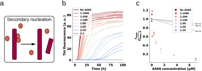
Figure 6. AS69 inhibits α-synuclein fibril amplification.

(a) Schematic representation of fibril amplification through secondary nucleation Buell et al. (2014a). (b) Change in ThT fluorescence intensity when a 70 μM solution of monomeric α-synuclein was incubated with increasing concentrations of AS69 in acetate buffer (pH 5.0) under quiescent conditions and weak seeding. (c) Relative rate of fibril amplification as a function of the concentration of AS69. The solid lines correspond to simulations based on the assumption that AS69 acts only through monomer sequestration, for different values of the monomer dependence (reaction order) of secondary nucleation (see Appendix 2 for details).

Figure 6.

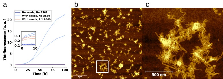
Figure 6—figure supplement 1. Seeds are required for aggregation under quiescent conditions.

Figure 6—figure supplement 1.

(a) Change in ThT fluorescence intensity when a 70 μM solution of monomeric α-synuclein was incubated either with or without pre-formed seeds, and with or without AS69 in acetate buffer (pH 5.0) under quiescent conditions. The inset shows the initial fluorescence intensity values. (b) AFM image of the seeds that were used for the experiment shown in (a), note the inhomogeneity in the dispersion of the fibrils, caused by the solution conditions that favour higher order assembly. The white square shows the approximate area of the zoom-in shown in (c).
Figure 6—figure supplement 2. Weakly seeded aggregation experiments at pH 5.0.
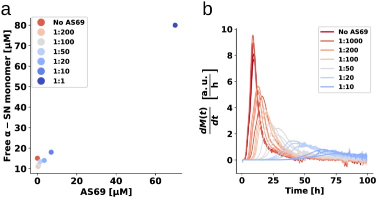
Figure 6—figure supplement 2.

(a) Concentration of monomer left in solution after the weakly seeded experiments, as determined by the method described in Galvagnion et al. (2015). (b) The numerically computed first derivatives (using a ten point rolling average) of the weakly seeded kinetic time courses shown in Figure 6.
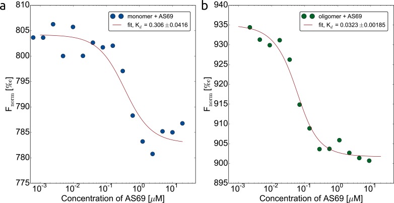
Figure 6—figure supplement 3. AS69 interacts with two distinct α-synuclein species.

Figure 6—figure supplement 3.

Binding of AS69 to monomeric (a) and oligomeric (Lorenzen et al., 2014) (bα-synuclein was quantified by microscale thermophoresis (MST) measurements (Wolff et al., 2016) in 20 mM phosphate buffer pH 7.4 and 50 mM NaCl. The oligomers used in this assay are kinetically stable and can be purified and studied in isolation (Lorenzen et al., 2014; Wolff et al., 2016). The preparation of labelled monomeric and oligomeric α-synuclein is described in detail in the Materials and methods section.