ABSTRACT
Merkel cell carcinoma (MCC) is a neuroendocrine skin cancer that typically presents as a painless erythematous nodule on body surfaces visible to the sun. Metastatic disease is typical to the lymph nodes, liver, and lungs. There are previous case reports of patients with metastases to the gastrointestinal tract including the stomach, small intestine, and pancreas. To our knowledge, there are only rare occurrences of metastases to the colon. We report a patient with a history of MCC treated with chemotherapy who presented with hematochezia and underwent a colonoscopy that showed a partially obstructing, edematous, friable 7-cm circumferential mass in the transverse colon. Biopsy confirmed the diagnosis of MCC that metastasized to the transverse colon.
INTRODUCTION
Merkel cell carcinoma (MCC) is a neuroendocrine skin cancer that is commonly found in whites who spend time outdoors, are immunocompromised, exposed to Merkel cell polyomavirus, or of older age; MCC is known for its poor prognosis.1 MCC typically appears as a painless erythematous or violaceous nodule on body surfaces visible to the sun.1 Metastatic disease is typical to the lymph nodes, liver, and lungs.1 There are few cases reporting patients with metastases to the gastrointestinal (GI) tract, and to our knowledge, MCC only rarely metastasizes to the colon. We present a patient with MCC that metastasized to the transverse colon.
CASE REPORT
A 71-year-old Hispanic woman with a medical history significant for MCC diagnosed in 2014 and asthma was admitted to our facility for acute asthma exacerbation and found to have systolic heart failure. She subsequently decompensated and was diagnosed with a non-ST segment elevation myocardial infarction. Left heart catheterization showed an ejection fraction of 25%, likely related to stress cardiomyopathy; no significant coronary artery disease was found. The gastroenterology service was consulted for hematochezia and diarrhea that developed while the patient was in the hospital. Her physical examination was normal, and her laboratory values were white blood cell count of 8.9/μL, hemoglobin 10.8 g/dL, hematocrit 33.8%, platelets 240/μL, bilirubin 0.4 mg/dL, aspartate aminotransferase 24 U/L, alanine aminotransferase 31 U/L, and alkaline phosphatase 81 U/L.
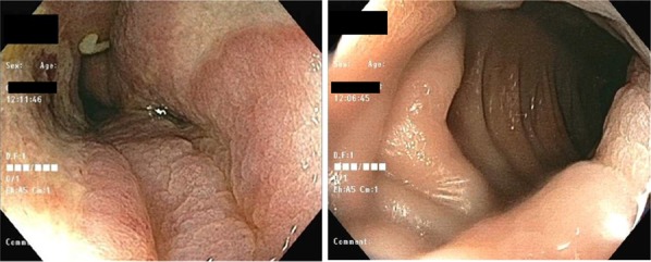
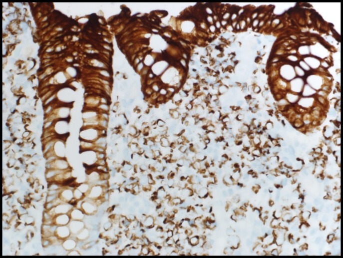
The patient presented with a small bowel obstruction in 2014 and was diagnosed with MCC that metastasized to the jejunum, which was resected after receiving neoadjuvant chemotherapy with etoposide and carboplatin. There was no evidence that the patient had active MCC in any other part of her GI tract at that time, including a negative computed tomography of the chest, abdomen, and pelvis. Subsequent colonoscopy performed within 6 months of partial enterectomy did not show any findings of active disease in the colon. She followed up with her oncologist for 2 years after the diagnosis of jejunal metastasis and was subsequently graduated back to her primary care physician. A colonoscopy was completed during the admission in 2018, where a partially obstructing, edematous, and friable 7-cm circumferential mass was found in the transverse colon, without any ulceration being appreciated (Figure 1). Biopsy showed invasive neoplasm with features consistent with metastatic MCC. The malignant cells showed positive staining with Synaptophysin, chromogranin, blush with CD56, and negative staining with CK7 and TTF1 (Figure 2). The lymphoid cells were positive for CD45. The patient followed up with her oncologist but decided to pursue hospice without palliative chemotherapy. The patient died approximately 6 months after colonoscopy showing newly found MCC of the transverse colon.
Figure 1.

Colonoscopy showed a partially obstructing, edematous, and friable circumferential mass without any ulceration in the transverse colon.
Figure 2.

Biopsy showed invasive neoplasm with features consistent with metastatic Merkel cell carcinoma.
DISCUSSION
MCC is a rare cancer that typically metastasizes to distant lymph nodes, liver, lungs, and bone.1 There have been few cases reported of MCC metastasizing to other areas of the GI tract apart from the colon. These include the stomach and duodenum; stomach; ileocecal valve; small bowel mesentery; jejunum; stomach, pancreas, and distal duodenum; stomach, jejunum, and ileum; and pancreas.1–16
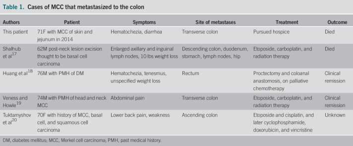
To our knowledge, there are only 4 case reports regarding MCC metastasizing to the colon (Table 1). Our patient is the fifth patient with MCC metastases to the colon, and the second specifically to the transverse colon.
Table 1.
Cases of MCC that metastasized to the colon
This case report is an example of the rarity of MCC metastasizing to the GI tract, particularly the colon, and serves as a reminder that a patient's history is an important marker of prediction of a mass. Biopsy is always warranted in these cases to confirm the diagnosis and allow prompt treatment and follow-up with oncology.
DISCLOSURES
Author contributions: All authors contributed equally to this manuscript. S. Mehta is the article guarantor.
Financial disclosure: None to report.
Informed consent was obtained for this case report.
REFERENCES
- 1.Matkowskyj KA, Hosseini A, Linn JG, Yang GY, Kuzel TM, Wayne JD. Merkel cell carcinoma metastatic to the small bowel mesentery. Rare Tumors. 2011;3:4–6. [DOI] [PMC free article] [PubMed] [Google Scholar]
- 2.Canales LI, Parker A, Kadakia S. Upper gastrointestinal bleeding from Merkel cell carcinoma. Am J Gastroenterol. 1992;87:1464–6. [PubMed] [Google Scholar]
- 3.Li M, Liu C. Cytokeratin 20 confirms Merkel cell metastasis to stomach. Appl Immunohistochem Mol Morphol. 2004;12:346–9. [DOI] [PubMed] [Google Scholar]
- 4.Idowu MO, Contos M, Gill S, Powers C. Merkel cell carcinoma: A report of gastrointestinal metastasis and review of the literature. Arch Pathol Lab Med. 2003;127:367–9. [DOI] [PubMed] [Google Scholar]
- 5.Wolov K, Tully O, Mercogliano G. Gastric metastasis of Merkel cell carcinoma. Clin Gastroenterol Hepatol. 2009;7:xxvi. [DOI] [PubMed] [Google Scholar]
- 6.Kini JR, Tapadia R, Shenoy S. Merkel cell carcinoma metastasis to stomach: An infrequent culmination of a rare neoplasm. J Gastrointest Canc. 2018;49:78–80. [DOI] [PubMed] [Google Scholar]
- 7.Hu ZI, Schuster JA, Kudelka AP, Huston TL. Merkel cell carcinoma with gastric metastasis and review of literature. J Cutan Med Surg. 2016;20:255–8. [DOI] [PubMed] [Google Scholar]
- 8.Cheung M, Lee H, Purkayastha S, Goldin R, Ziprin P. Ileocaecal recurrence of Merkel cell carcinoma of the skin: A case report. J Med Case Rep. 2010;4:43–7. [DOI] [PMC free article] [PubMed] [Google Scholar]
- 9.Foster R, Stevens G, Egan M. An unusual pattern of metastases from Merkel cell carcinoma. Australas Radiol. 1994;38:231–2. [DOI] [PubMed] [Google Scholar]
- 10.Naunton Morgan TC, Henderon RG. Small bowel metastases from a Merkel cell tumour. Br J Radiol. 1985;58:1212–3. [DOI] [PubMed] [Google Scholar]
- 11.Hizawa K, Kurihara S, Nakamori M, Nakahara T, Matsumoto T, Iida M. An autopsy case of Merkel cell carcinoma presenting aggressive intraabdominal metastasis and duodenal obstruction. Nihon Shokakibyo Gakkai Zasshi. 2007;104:1383–6. [PubMed] [Google Scholar]
- 12.Poskus E, Platkevicius G, Simanskaite V, Rimkeviciute E, Petrulionis M, Strupas K. Multiple gastrointestinal metastases of Merkel cell carcinoma. Medicine. 52;2016:321–4. [DOI] [PubMed] [Google Scholar]
- 13.Adsay NV, Andea A, Basturk O, Kiline N, Nassar H, Cheng JD. Secondary tumors of the pancreas: An analysis of a surgical and autopsy database and review of the literature. Virchows Arch. 2004;444:527–35. [DOI] [PubMed] [Google Scholar]
- 14.Bachmann J, Kleeff J, Bergmann F, et al. Pancreatic metastasis of Merkel cell carcinoma and concomitant insulinoma: Case report and literature review. World J Surg Oncol. 2005;3:58–64. [DOI] [PMC free article] [PubMed] [Google Scholar]
- 15.Dim DC, Nugent SL, Darwin P, Peng HQ. Metastatic Merkel cell carcinoma of the pancreas mimicking primary pancreatic endocrine tumor diagnosed by endoscopic ultrasound-guided fine needle aspiration cytology: A case report. Acta Cytol. 2008;53:223–8. [DOI] [PubMed] [Google Scholar]
- 16.Ouellette JR, Woodyard L, Toth L, Termuhlen PM. Merkel cell carcinoma metastatic to the head of the pancreas. J Pancreas. 2004;5:92–6. [PubMed] [Google Scholar]
- 17.Shalhub S, Clarke L, Morgan MB. Metastatic Merkel cell carcinoma masquerading as colon cancer. Gastrointest Endosc. 2004;60(5):856–8. [DOI] [PubMed] [Google Scholar]
- 18.Huang W, Lin PY, Lee I, Chin C, Wang J, Yang W. Metastatic Merkel cell carcinoma in the rectum: Report of a case. Dis Colon Rectum. 2007;50:1992–5. [DOI] [PubMed] [Google Scholar]
- 19.Veness MJ, Howle JR. Merkel cell carcinoma metastatic to the transverse colon: Disease free after six years—cure or just prolonged remission? Australas J Dermatol. 2011;52(4):295–7. [DOI] [PubMed] [Google Scholar]
- 20.Tuktamyshov R, Jain D, Ginsburg P. Recurrence of Merkel cell carcinoma in the gastrointestinal tract: A case report. BMC Res Notes. 2015;8:188–91. [DOI] [PMC free article] [PubMed] [Google Scholar]


