Abstract
Background
As one of the most common cancers, breast carcinoma is the most common disease in women. Intersectin 1 (ITSN1) contributes to the actin cytoskeleton reconstruction in breast cancer.
Purpose
The objective of this study to explore the functions of ITSN1 in breast carcinoma.
Methods
We downloaded microarray datasets GSE8087, GSE50697, and GSE98238 from the Gene Expression Omnibus database. Differentially expressed genes (DEGs) were used to construct a protein–protein interaction (PPI) network using STRING database, and the modules from PPI network were verified by Cytoscape software. Gene ontology terms and Kyoto Encyclopedia of Gene and Genome pathway were used to analyze the biological functions using the DAVID database. ONCOMINE, GEPIA, UALCAN, and Human Protein Atlas databases were used to investigate the characteristics of ITSN1 for the prognosis of breast carcinoma. Cell counting kit-8, flow cytometry, and colony formation assays were used to detect cell viability, cell apoptosis, and cell proliferation. RT-PCR and Western blot assays were used to detect ITSN1, Ki67, and cleaved caspase-3 expressions.
Results
Low expressions of ITSN1 were significantly associated with clinical cancer stages. RT-PCR and Western blot analysis showed low expression of ITSN1 in breast cancer tissues and cell lines. ITSN1 inhibition could promote cell proliferation and inhibit cell apoptosis, while ITSN1 overexpression could inhibit cell proliferation and increase cell apoptosis by regulating the levels of expression of Ki67 and cleaved-caspase-3.
Conclusion
The results indicated that ITSN1 could be a prognostic biomarker for survivals of breast cancer patients.
Keywords: ITSN1, bioinformatic analysis, proliferation, apoptosis, breast cancer
Introduction
Breast cancer, also known as primary breast cancer, originates from breast ducts or lobules and is a type of malignant tumor with the highest incidence in women worldwide.1 The factors influencing the incidence of breast cancer are very complex, mainly including age of menopause, familial inheritance, diet, obesity, and excessive intake of exogenous estrogen.1,2 At present, surgery is used as a main treatment method for breast cancer, with some combined treatment modes, such as chemoradiotherapy, molecular targeted therapy, and endocrinotherapy.3 Therefore, currently, it is important to use preventive and treatment measures to actively carry out an early screening and comprehensive diagnosis for breast cancer. It is widely believed that the imbalance in intracellular anti-oncogene and proto-oncogene is the main reason for tumor. Breast cancer is a highly heterogeneous disease in terms of gene.1,2 Tumor heterogeneity refers to the biological characteristics of tumor cells gradually changing from a monoclonal state to a polyclonal state in the process of tumor development, which can have an impact on the tumor in various aspects, such as growth rate, invasion ability, drug susceptibility, and prognosis.4
Correct early diagnosis, treatment, and prognosis assessment of BC are very difficult, even though many cancer-related genes and cellular pathways are related to the emergence and evolution of BC.5 Therefore, it is important to explore the molecular mechanisms of cell proliferation and apoptosis and to formulate valid diagnosis and treatment tactics.
With the fast expansion of microarray technology and bioinformatic analysis, the Gene Expression Omnibus (GEO) database (https://www.ncbi.nlm.nih.gov/) provides new clues to discover differentially expressed genes (DEGs) and the passageways of the initiation and evolution of BC.6 Thus, three RNA microarray datasets from the GEO database were downloaded and analyzed to identify DEGs in BC cells and BC cells treatment with paclitaxel/miR-203/RhoGDI beta-specific siRNA. Subsequently, we used protein–protein interaction (PPI) network analyses to identify significant modules from the GEO database (GSE8087, GSE50697, and GSE98238).7 We used gene ontology (GO) and Kyoto Encyclopedia of Gene and Genome (KEGG) analyses7,8 to comprehend the molecular mechanism involved in carcinogenesis and progression of BC. We screened 20 key genes as potential, candidate biomarkers for BC. ONCOMINE, GEPIA, UALCAN, and Human Protein Atlas databases were used to investigate the roles of 20 key genes in the prognosis of BC. Finally, we evaluated intersectin 1 (ITSN1) effects on proliferation and apoptosis in BC cell lines. The purpose of this study was to identify proliferation-apoptosis-related mRNAs and their potential molecular mechanisms based on integrated bioinformatic analyses and empirical tests.
Materials and methods
Microarray data
GEO is an open functional genomics data warehouse, which stores a large number of gene expression data, chips, and microarrays. We can download the three genetic expression datasets (GSE8087, GSE50697 and GSE98238; Affymetrix GPL96 platform). Based on the annotation on the platform, the probes are transformed into corresponding gene symbols. The GSE8087 dataset contained three samples of BC cells transfected with siLuc and three samples of BC cells transfected with si RhoGDIbeta. GSE50697 contained three samples of BC cells transfected with control and three samples of BC cells transfection miR-203. GSE98238 contained three samples of BC cells treated with DMSO and three samples of BC cells treated with paclitaxel.
Identification of DEGs
People used GEO2R to compare DEGs in control and laboratory specimens (http://www.ncbi.nlm.nih.gov/geo/geo2r). “GEO2R” is an interactive web tool, users are permitted to compare GEGs in high percentage of datasets in the series of GEO2R with respect to the experimental condition. The p-value balanced the statistical limitations of significant gene discovery and false positive by using false detection rates. The probes with or without equivalent gene symbols were deleted, as well as the genes with multiple probes. logFC (foldchange) >1.5 or P-value <0.05 were believed meaningful statistically.
PPI network construction and analysis of modules
Search Tool for the Retrieval of Reciprocity Genes (STRING) database (http://string-db.org/) was used to evaluate PPI information, containing direct (physical) and indirect (functional) correlation. In this study, the DEGs were selected utilizing STRING with a cutoff greater than 0.4 times that of the standard to assess the information of PPI. Cytoscape 3.7.1 was used to visualize the PPI network. In order to screen the genes, a node degree (>3) was chosen as the threshold, and screen modules of key genes from the PPI network with degree a threshold=3 were screened utilizing the Molecular Complex Detection (MCODE) plug-into. The selection criteria were as follows: degree cutoff value =2, node score cutoff value =0.2, maximum depth =100, and K score =2. KEGG and GO assessments were performed for the key genes from this module using DAVID (http://david.ncifcrf.gov).
Functional annotation and pathway enrichment
GO is a bioinformatics tool and used to annotate genes and analyze biological processes (BPs) of DEGs or key genes. In addition, GO offers three categories of defined terms, which include BP, cellular component (CC), and molecular function (MF). KEGG, as a sophisticated database resource for the systematic analysis of gene functions, links genomic information and high-order functional Information. DAVID was used to perform the GO and KEGG analysis. p<0.05 was set as the cutoff criterion.
ONCOMINE database
ONCOMINE database (www.oncomine.org), as an integrated online cancer microarray database, was used to analyze the sequences of DNA or RNA for gene expression analyses. In this study, 20 key genes in different cancer tissues and their corresponding adjacent normal control samples were obtained form ONCOMINE database. Differences in transcriptional expression were compared using Students’ t-test. The cutoff of p-value and fold change were as follows: p-value: 0.01, fold change: 1.5, gene rank: 10%, and data type: mRNA.
Human Protein Atlas
Human Protein Atlas (https://www.proteinatlas.org) is a website that contains immunohistochemistry-based expression data, which collects approximately 20 common types of cancers, and each tumor type includes 12 individual tumors. Users can identify tumor-type specific proteins expression patterns that are differentially expressed in a given tumor. In this study, a direct comparison of protein expression of ITSN1 in human normal and BC tissues was performed by immunohistochemistry.
UALCAN
As an interactive web resource, UALCAN database (http://ualcan.path.uab.edu) is based on level 3 RNA-seq and clinical data of 31 cancer types from TCGA database. It can be used to analyze the relative transcriptional expression of potential genes of interest comparing the transcriptional expression of tumors and normal samples with relative clinicopathological parameters. In this research, we used UALCAN to analyze mRNA expression of ITSN1 in primary BC tissues and its relation of clinicopathological parameters. Differences in transcriptional expression were compared by Students’ t test and p<0.01 was considered statically significant.
GEPIA
Gene Expression Profiling Interactive Analysis (GEPIA, http://gepia.cancer-pku.cn/) is an online tool that is fast and customizability and is based on TCGA and Genotype-Tissue Expression databases data. In this study, we use GEPIA to analyze mRNA expression of ITSN1in BC tissues and normal tissues.
Patients and tissue samples
BC tissues and matched adjacent normal tissues were obtained from 24 patients (mean age, 49.29±7.26) who underwent surgical resection in the Jiangxi Cancer hospital. The patients’ clinical information is listed in Table 1. Tissue samples were stored at −80°C, which were used to perform the following studies. All the patients gave their written informed consent. All samples were obtained with written informed consent and analyzed anonymously. All the patients did not receive radiotherapy and immunotherapy. The study was approved by the Ethics Committee of Jiangxi Cancer hospital. The study was conducted in accordance with the Declaration of Helsinki.
Table 1.
Association between ITSN1 and the clinicopathological characteristics of BC
| Variables | Cases (n=24) | ITSN1 | P-value | |
|---|---|---|---|---|
| Low (n=18) | High (n=6) | |||
| Age (years) | 0.098 | |||
| <50 | 13 | 8 | 5 | |
| ≥50 | 11 | 10 | 1 | |
| Menopause | 0.384 | |||
| No | 19 | 15 | 4 | |
| Yes | 5 | 3 | 2 | |
| Tumor size | 0.059 | |||
| ≤2.0 cm | 12 | 7 | 5 | |
| >2.0 cm | 12 | 11 | 1 | |
| Lymph node metastasis | 0.006* | |||
| No | 6 | 2 | 4 | |
| Yes | 18 | 16 | 2 | |
| TNM stage | 0.046* | |||
| I–II | 8 | 4 | 4 | |
| III–IV | 16 | 14 | 2 | |
Note: *P<0.05, statistically significant.
Cell culture
Human BC cell line (MCF-7, MDA-MB-231, T47D, and BT-549) and one normal human mammary epithelial cell line MCF-10A were purchased from Shanghai Cell Bank, Chinese Academy of Sciences (Shanghai, China). The cells were cultured in DMEM supplemented with 10% fetal serum and incubated at 37°C with 5% CO2. In further experiments, the cells at logarithmic phase growth.
Production and transfection of ITSN1 overexpression vectors
ITSN1-overexpressing vectors and negative controls were purchased from Thermo Scientific Co. LTD (USA). The primers ITSN1gene were 5 ′-ATGGCTCAGTTTCCAACACCTT -3′ (forward) and 5′- CTACGGCTCATCAAACAACTGCA -3′ (reverse), cloned into pLVX-Puro vector (Clontech). Plasmids (pLVX-Puro-ITSN1) were transfected into MCF-7 cells using Lipofectamine™ 2000 (Invitrogen, CA, USA).
RNA interference
A siRNA targeting human ITSN1 mRNA was designed by Sbo Biomedical Technologies Inc (Shanghai, China). siRNA-ITSN1 sequences were as follows: 5ʹ-GCAUGAUCAGCAGUUCCAUAGUUUA-3ʹ. The siRNAs (20 μmol/L) were transfected into BT-549 cells by utilizing Lipofectamine 2000 Reagent (Invitrogen). In addition, nonspecific siRNA (siRNA-NC) was used as a negative control.
Cell counting kit-8 (CCK-8) assay
The viability of BT-549 and MCF-7 cells transfected with ITSN1-overexpressing vectors or siRNA-ITSN1 was detected by using CCK-8 kit (Beyotime Biotechnology, Shanghai, China) according to the manufacturer’s instructions. Briefly, after the cells were transfected with ITSN1 overexpression vectors or siRNA-ITSN1 for 0, 24, 48, and 72 hrs, 3×10³ cells/well in cultured medium (90 μL/well) were immediately incubated with CCK-8 working solution (10 μL/well) at 37°C for 1 hr. The OD value was recorded using DNM-9602 microplate reader (PerLong, Beijing, China) at 450 nm wavelength.
RT-PCR analysis
Total of RNA from tissue samples and cells was extracted using the TRizol reagent (Thermo Fisher Scientific, Waltham, MA, USA). Agilent Bioanalyzer 2100 with RNA 6000 Nano kit (Agilent Technologies, Santa Clara, CA, USA) was used to detect the integrity of the isolated RNA. High-capacity cDNA reverse transcription kits (Thermo Fisher Scientific) was used to synthesize single-stranded cDNA from RNA. Real-time quantification was performed using the SYBR Green PCR kit (Thermo Fisher Scientific). Each gene’s cycle threshold (Ct) was then recorded.
ITSN1 has become normalization by GAPDH and quantified using the 2−∆∆Ct method (ΔCt = Cttarget gene – Ctinternal control). Amplification condition was as follows: 95°C, 10 mins (95°C, 15 s; 60°C, 45 s) × 40; 95°C, 15 s; 60°C, 1 min; 95°C, 15 s; 60°C, 15 s.
The forward primer of ITSN1 is TATCCTGGCAATGCACCTCA. The reverse primer of ITSN1 is AACTGGTTCCTCTGGTAGCC. The forward primer of GAPDH is ACCCAGAAGACTGTGGATGG. The reverse primer of GAPDH is TCAGCTCAGGGATGACCTTG.
Western blot analysis
RIPA lysis buffer (Beyotime Institute of Biotechnology Jiangsu, China) was used to extract whole cell protein from cell lines and BC tissues. The protein concentration of cell lysates was quantified using BCA Kit (Beyotime Institute of Biotechnology Jiangsu, China), and 20–50 μg of each protein was separated by SDS-PAGE on 10% gels and transferred to a polyvinylidene fluoride membrane (Millipore, USA). The membranes were blocked in 5% non-fat milk diluted with Tris buffered saline Tween-20 at 37°C for 1 hr and incubated overnight at 4°C with primary antibody: anti-ITSN1 (1:500; ab118262; Abcam, Cambridge, UK), anti-ki67 (1:1000; ab92742; Abcam, Cambridge, UK), and anti-cleaved-caspase-3 (1:1000; ab49822., Abcam, Cambridge, UK). The membranes were then incubated with goat anti-rabbit or anti-mouse IgG conjugated to horseradish peroxidase secondary antibody (1:1000; Cell Signaling Technology Inc., MA, USA) for 2 hrs. The proteins were visualized using ECL-plus reagents (Amersham Biosciences Corp., USA). The density of the bands was measured using the Image J software (USA), and values were normalized to the densitometric values of GAPDH (1:5000; ab181602; Abcam, Cambridge, UK) in each sample.
Colony formation assay
MCF-7 and BT-549 cells transfected with ITSN1-overexpressing vectors or siRNA-ITSN1 and their negative control were inoculated in RPMI 1640 medium containing 10% FCS and 0.5% agar (bottom layer). A total 5×10³ cells/well suspended in 20% FCS/0.7% agarose (top layer) were plated and incubated at 37°C for 14 days. The plates were stained with 0.005% crystal violet (Bogoo, Shanghai, Bogoo Biotechnology Co., Ltd) for more than 2 hrs, and colonies were counted in two colony grids per well by using a microscope.
Apoptosis assay
Annexin V-FITC/PI Kit (Beijing Biosea Biotechnology, Beijing, China) was used to perform cell apoptosis analysis, according to the instructions. Cells (1×10⁵ cells/well) were seeded in a 6-well plate. MCF-7 and BT-549 cells transfected with ITSN1-overexpressing vectors or siRNA-ITSN1and their negative control were washed twice with cold PBSds and resuspended in buffer and measured using a flow cytometer (Beckman Coulter, USA) to differentiate apoptotic cells (annexin V-positive and PI-negative) from necrotic cells (Annexin V-and PI-positive).
Statistical analyses
SPSS 16.0 (SPSS Inc., Chicago, IL, USA) was used for statistical analysis. One-way ANOVA was utilized for multiple group comparisons. p < 0.05 was considered statistically significant. The relative ITSN1 expression >1.3 as high expression and 1.3 is derived from the mean values of 24 samples. The correlations between ITSN1 expression and clinicopathological characteristics were examined by χ2 test. Survival curves were constructed using the Kaplan–Meier method and analyzed by the log-rank test. The experiments were performed three times.
Results
Bioinformatics identifies key genes in BC
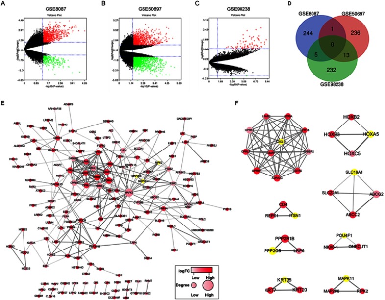
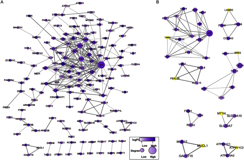
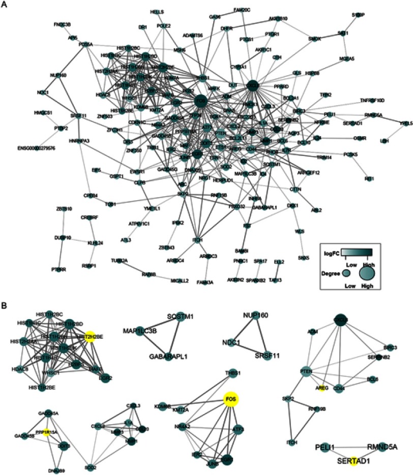
Three gene expression datasets about BC from the GEO database, including GSE8087, GSE50697 and GSE98238, were used to identify DEGs in BC cell lines (Figure 1A–C). The top 250 DEGs (P<0.05) from GSE8087, GSE50697, and GSE98238 datasets, respectively, were used to perform the follow-up bioinformatics exploration. The intersecting part of the three sets of DEGs consisted of 0 genes (Figure 1D). Therefore, Cytoscape was used to construct PPI network with 174 nodes and 326 edges from GSE8087 datasets, PPI network with 148 nodes and 264 edges from GSE50697 datasets, and PPI network with 162 nodes and 470 edges from GSE98238 datasets using STRING with a score of >0.4. MCODE plug-in of Cytoscape was used to screen key genes of significant modules from three PPI networks (Figure 1E and F, 2 and 3). A total of 20 keygenes (AREG, ATP6V1C2, CXCL11, EREG, FBXL20, FOS, HIST1H2BE, HOXA5, ITSN1, KRT35, LAMB3, MAPK11, MTIH, MUCL1, POPU4F1, PPP1R15A, PPP2CB, SERTAD1, SLC19A1, and TIMP3) were used to perform enrichment analysis (Table 2). Six key genes from the PPI network of GSE8087 dataset, including HOXA5, ITSN1, PPP2CB, POU4F1, KRT35, and MAPK11, were significantly upregulated (Table 2; logFC>1.5, P<0.05), and CXCL11 from the PPI network of GSE8087 dataset was significantly downregulated (Table 2; logFC<-1.5, P<0.05). Two key genes from the PPI network of GSE50679 dataset, including LAMB and EREG, were significantly upregulated (Table 2; logFC>1.5, P<0.05) and 4 key genes from the PPI network of GSE50679 dataset, including TIMP3, FBXL20, MUCL1, and ATP6V1C2, were significantly downregulated (Table 2; logFC<-1.5, P<0.05). FOS from the PPI network of GSE98238 dataset was significantly upregulated (Table 2; logFC>1.5, P<0.05).
Figure 1.
Volcano plot, Venn diagram, PPI network, and the significant module of DEGs. (A–C) Volcano plot of the DE-mRNAs from GSE8087, GSE50697, and GSE98238 datasets, the red dots and green dots represent the upregulated and downregulated (logFC>1.5 or <-1.5). (D) The top of 250 DEGs were selected with a P-value <0.05 among the mRNA expression profiling sets GSE8087, GSE50697, and GSE98238. The 3 datasets showed an overlap of 0 gene. (E) The PPI network of DEGs from GSE8087 set was constructed using Cytoscape. (F) The significant module was obtained from PPI network. The color of a node in the PPI network reflects the log (FC) value of the Z score of gene expression, and the size of the node indicates the number of interacting proteins with the designated protein.
Figure 2.
PPI network and the significant module of DEGs from GSE50697 dataset. (A) The PPI network of DEGs from GSE50697 set was constructed using Cytoscape. (B) The significant module was obtained from PPI network. The color of a node in the PPI network reflects the log (FC) value of the Z score of gene expression, and the size of the node indicates the number of interacting proteins with the designated protein.
Figure 3.
PPI network and the significant module of DEGs from GSE98238 dataset. (A) The PPI network of DEGs from GSE98238 dataset was constructed using Cytoscape. (B) The significant module was obtained from PPI network. The color of a node in the PPI network reflects the log (FC) value of the Z score of gene expression, and the size of the node indicates the number of interacting proteins with the designated protein.
Table 2.
| No. | Gene name | logFC | P-value | |
|---|---|---|---|---|
| GSE8087 | 1 | CXCL11 (C-X-C motif chemokine ligand 11) | −1.5823 | 0.0096 |
| 2 | HOXA5 (homeobox A5) | 3.0745 | 0.0086 | |
| 3 | SLC19A1 (solute carrier family 19 member 1) | 1.4501 | 0.0051 | |
| 4 | ITSN1 (intersectin 1) | 2.5274 | 0.0021 | |
| 5 | PPP2CB (protein phosphatase 2 catalytic subunit beta) | 3.6406 | 0.0018 | |
| 6 | POU4F1 (POU class 4 homeobox 1) | 3.0246 | 0.0055 | |
| 7 | KRT35 (keratin 35) | 2.7854 | 0.0020 | |
| 8 | MAPK11 (mitogen-activated protein kinase 11) | 2.7090 | 0.0034 | |
| GSE50679 | 1 | LAMB3 (laminin subunit beta 3) | 1.5865 | 0.0005 |
| 2 | TIMP3 (TIMP metallopeptidase inhibitor 3) | −2.2309 | 0.0002 | |
| 3 | FBXL20 (F-box and leucine rich repeat protein 20) | −2.2775 | 0.0004 | |
| 4 | EREG (epiregulin) | 1.7253 | 0.0001 | |
| 5 | MT1H (metallothionein 1H) | 1.0028 | 0.0008 | |
| 6 | MUCL1 (mucin like 1) | −4.2828 | 0.0003 | |
| 7 | ATP6V1C2 (ATPase H + transporting V1 subunit C2) | −2.3263 | 0.0009 | |
| GSE98238 | 1 | HIST1H2BE (H2B clustered histone 6) | 0.9740 | 8.84E-07 |
| 2 | PPP1R15A (protein phosphatase 1 regulatory subunit 15A) | 0.6819 | 6.08E-06 | |
| 3 | FOS (Fos proto-oncogene, AP-1 transcription factor subunit) | 2.0977 | 3.61E-09 | |
| 4 | AREG (amphiregulin) | 0.8058 | 2.59E-06 | |
| 5 | SERTAD1 (SERTA domain containing 1) | 0.8414 | 2.13E-07 |
KEGG and GO enrichment analyses of DEGs
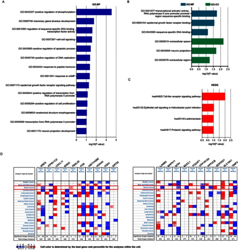
Twenty key genes were mapped using GO terms and KEGG pathways, and potential functions and related mechanisms in BC were analyzed by DAVID. Twenty key genes mainly enriched in the GO terms, including BP, CC, and MF (Figure 4A and B). GO analysis suggested that changes in BP term of 20 key genes were significantly enriched in “positive regulation of phosphorylation”, “mammary alveolar development”, “sequential-specific DNA-binding transcription factor activity”, “cell signal transduction and apoptotic process”, and “intranscriptional activator activity”. Changes in MF term of the 20 key genes were significantly enriched in “RNA polymerase II core facilitator proximate area sequence-specific binding” and “epidermal growth factor receptor binding”. Changes in CC term of the 20 key genes were significantly enriched in “extracellular space”. KEGG pathway analysis revealed that the 20 key genes involved in abundant in Toll-like receptor signaling pathway (Figure 4C). More importantly, ITSN1, HOXA5, and POPU4F1 were significantly correlated with cell apoptosis.
Figure 4.
GO/KEGG analysis and ONCOMINE database analysis of key genes. (A and B) A total of 20 key genes from PPI network of the GSE8087, GSE50697, and GSE98238 datasets. GO, Gene Ontology; MF, molecular function; CC, cellular component; BP, biological process. (C) A total 20 key genes from PPI network of the GSE8087, GSE50697, and GSE98238 data sets were performed KEGG pathway enrichment analysis. KEGG, Kyoto Encyclopedia of Genes and Genomes. (D) Transcriptional expression of 20 key genes in 20 different types of cancer diseases (ONCOMINE database), difference of transcriptional expression was compared by Students’ t-test. Cutoff of p-value and fold change were as follows: P-value: 0.01, fold change: 1.5, gene rank: 10%, data type: mRNA.
Low expression of ITSN1and association of mRNA expression of ITSN1 with clinicopathological parameters in patients with BC
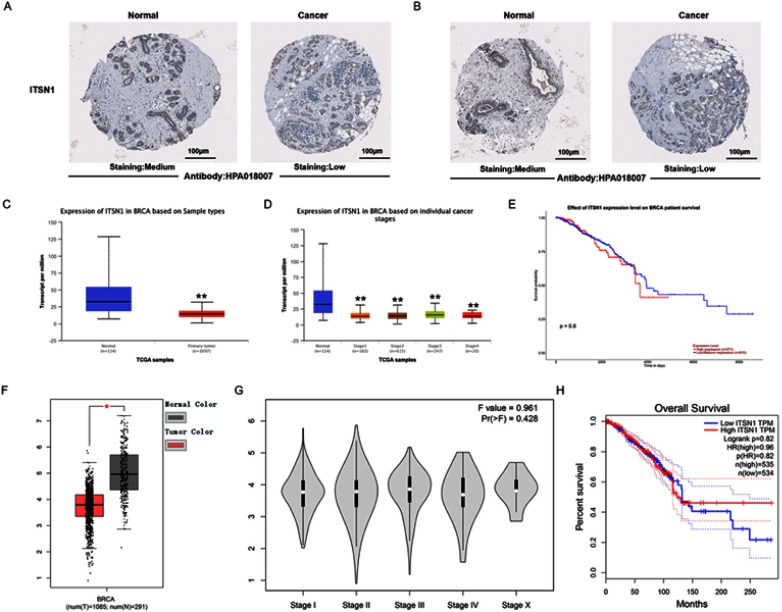
To research the distinct prognostic and potential therapeutic value of ITSN1 in BC patients, mRNA expression and protein expression were analyzed by ONCOMINE database (www.oncomine.org), Human Protein Atlas (https://www.proteinatlas.org), UALCAN (http://ualcan.path.uab.edu), and GEPIA (http://gepia.cancer-pku.cn). As shown in Figures 4D and 5, mRNA expressive abundance of ITSN1 in 20 types of cancers was first measured and compared to normal tissues by ONCOMINE database. ITSN1 mRNA levels were significantly downregulated in BC tissues. Next, the mRNA expression patterns of ITSN1 were further measured by UALCAN, whose resources were based on level three RNA-seq and clinical data from 31 cancer types of TCGA database, which was different from other databases. As shown in Figure 5A, ITSN1 protein level was found to be significantly downregulated in primary BC tissues in contrast to normal samples (P<0.05). The mRNA expressions of ITSN1 were remarkably correlated with patients’ individual cancer stages, and patients who were in more advanced cancer stages tended to express lower mRNA expression of ITSN1. Similarly, as shown in Figure 5B, ITSN1 protein level was found to be significantly downregulated in BC tissues in contrast to regular samples by GEPIA database. Moreover, mRNA expressions of ITSN1 had nothing to do with the patient’s overall survival time. After examining the mRNA expression patterns of ITSN1 in BC, we tried to explore the protein expression patterns of ITSN1 in BC by the Human Protein Atlas. As shown in Figure 5C, ITSN1 protein was low expressed in BC tissues, whereas medium expressions of ITSN1 were observed in normal tissues.
Figure 5.
Human Protein Atlas, UALCAN, and GEPIA database analyses of ITSN1. (A and B) Representative immunohistochemistry images of ITSN1 in BC tissues and normal breast tissues (Human Protein Atlas). ITSN1 protein’ medium expressions were observed in normal breast tissues, whereas their low expressions were observed in BC tissues; (C) mRNA expression of ITSN1 in BC tissues and adjacent normal liver tissues (UALCAN). mRNA expressions of ITSN1 were found to be downexpressed in primary BC tissues compared to normal samples, (D) ITSN1 is associated with clinical stages (UALCAN) in BC; (E) ITSN1 is not associated with poor survival rate in BC tissues (UALCAN); (F) mRNA expression of ITSN1 in BC tissues and adjacent normal liver tissues (GEPIA). mRNA expressions of ITSN1 were found to be downxpressed in primary BC tissues compared to normal samples; (G) ITSN1 is not associated with clinical stages in BC tissues (GEPIA); (H) ITSN1 is not associated with poor survival rate in BC tissues (GEPIA). **P<0.01 and *P<0.05.
ITSN1 expression is downregulated in BC tissues and cell lines
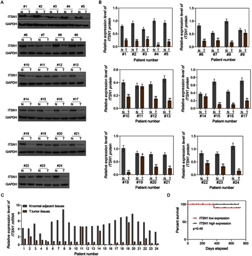
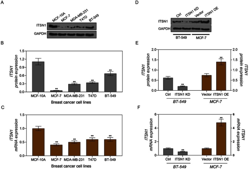
To further identify the mRNA and protein levels of ITSN1 in BC, we examined the ITSN1expression level in BC tissues and cell lines by using RT-PCR and Western blot analysis. Similar to the above results, low expression of ITSN1 was discovered in tumor tissues of 24 patients with BC (Figure 6A–C). In addition, our analysis showed that ITSN1 was positively associated with lymph node metastasis and TNM stage (Table 1) and ITSN1 had no connection with poor survival (Figure 6D). The protein and mRNA levels of ITSN1 were significantly downregulated in BC cell lines (Figure 7A–C). Our results showed that ITSN1 expression level in MCF-7 cells was lower than that of other groups. ITSN1 expression level in BT-549 cells was higher than that of MCF-7, MDA-MB-231, and T47D groups and lower than that of MCF-10A group. It showed that the change of ITSN1 in MDA-MB-231 and T47D cells was lower than the change of ITSN1 in BT-549 and MCF-7 cells. Therefore, siRNA-ITSN1 was transfected into BT-549 cells and the plasmids expressing ITSN1 were transfected into MCF-7 cells. The results of Western blot showed that ITSN1 protein level of ITSN1 knock-down (KD) group in BT-549 cells was significantly decreased, compared to the ctrl group and ITSN1 level of ITSN1 protein overexpression (OE) group in MCF-7 cells was significantly increased, compared to vector group (Figure 7D and E). The results of RT-PCR showed that the change tendency of ITSN1 mRNA level in ITSN1 KD group and ITSN1 OE group was similar to ITSN1 protein level (Figure 7F).
Figure 6.
ITSN1expression is downregulated in BC tissues. (A) ITSN1 protein was expressed less in BC tissues by Western blot assay. (B) Statistical graph of ITSN1 protein levels in BC tissues. (C) ITSN1 mRNA was expressed less in BC tissues by RT-PCR assay. (D) Kaplan–Meier survival analysis is shown. GAPDH was used as a load control. Data are presented as the mean ± standard deviation. **P<0.01 versus MCF-10A group/or Ctrl group/or Vector group.
Figure 7.
ITSN1 expression is downregulated in BC cell lines. (A and B) ITSN1 protein was expressed less in BC cell lines by Western blot assay; (C) ITSN1 mRNA was expressed less in BC cell lines by RT-PCR assay; (D and E) ITSN1 was expressed less in BT-549 cells transfected with siRNA-ITSN1 and highly expressed in MCF-7 cells transfected with ITSN1-overexpressing plasmid by Western blot analysis; (F) ITSN1 was expressed less in BT-549 cells transfected with siRNA-ITSN1 and highly expressed in MCF-7 cells transfected with ITSN1-overexpressing plasmid by RT-PCR assay. GAPDH was used as a load control. Data are presented as the mean ± standard deviation. **P<0.01 versus MCF-10A group/or Ctrl group/or Vector group.
ITSN1 regulated proliferation and apoptosis of BC cells by affecting the expression of Ki67 and caspase-3 proteins
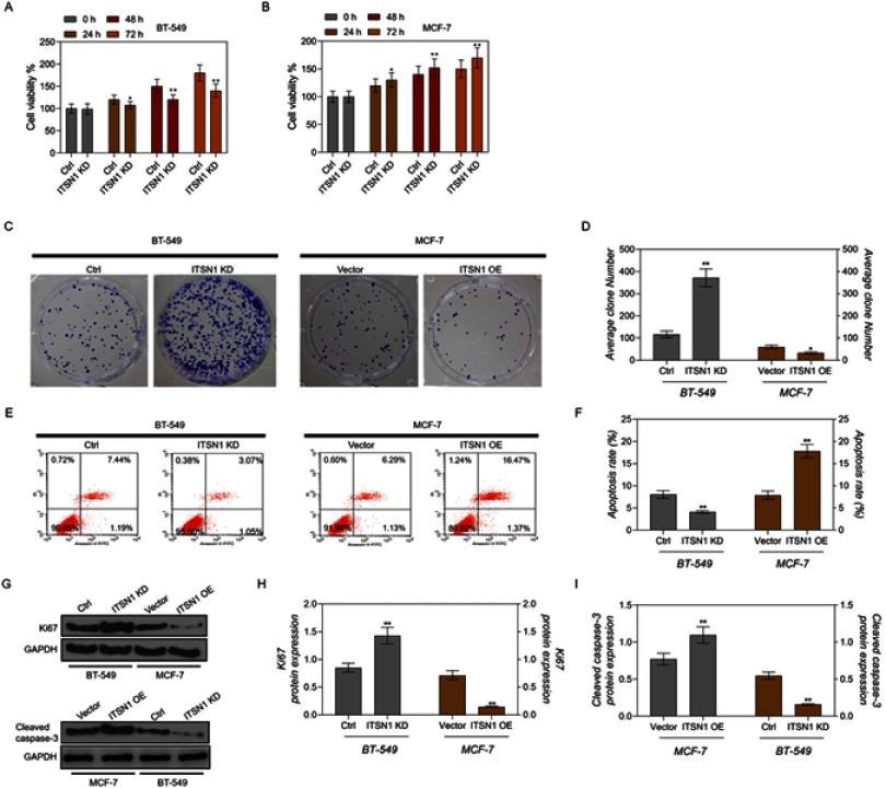
To investigate the role of ITSN1 in BC progression, CCK-8, flow cytometry, and colony formation assays were used to detect the functions of ITSN1 in cell proliferation and apoptosis. As shown in Figure 8A and B, the results of CCK-8 assay showed that downregulated ITSN1 promoted cell viability and upregulated ITSN1 inhibited cell viability. Colony formation ability was significantly increased by ITSN1 inhibition and was significantly decreased by ITSN1 overexpression (Figure 8C and D). ITSN1 inhibition inhibited apoptosis of BT-549 cells, whereas, upregulated ITSN1 promoted apoptosis of MCF-7 cells (Figure 8E and F). ITSN1 inhibition could increase Ki67 protein level of BT549 cells and decrease cleaved caspase-3 protein level of BT459 cells, while, ITSN1 overexpression could decrease Ki67 protein level of MCF-7 cells and increase cleaved caspase-3 protein level of MCF-7 cells (Figure 8G–I).
Figure 8.
ITSN1 regulated proliferation and apoptosis of BC cells by affecting the expression of Ki67 and caspase-3 protein. (A and B) CCK-8 assay was used to detect the viability of BT-549 cells transfected with siRNA-ITSN1 or MCF-7 cells transfected with ITSN1-overexpressing plasmid. (C and D) Colony formation was used to detect the proliferation of BT-549 cells transfected with siRNA-ITSN1 or MCF-7 cells transfected with ITSN1-overexpressing plasmid. (E and F) Flow cytometry assay was used to detect the apoptosis level of BT-549 cells transfected with siRNA-ITSN1 or MCF-7 cells transfected with ITSN1-overexpressing plasmid. (G–I) Western blot assay was used to detect the protein expression level of Ki67 and cleaved caspase-3 in BT-549 cells transfected with siRNA-ITSN1 or MCF-7 cells transfected with ITSN1-overexpressing plasmid. GAPDH was used as a load control. Data are presented as the mean ± standard deviation. **P<0.01 and *P<0.05 versus Ctrl group/or Vector group.
Discussion
The rapid development of high-throughput technologies, including DNA chips, and second-generation sequencing results in massive data,9 and researchers need to process useful information with the help of bioinformatics methods. Bioinformatics is a interdiscipline that combines computer science and life science, and it calculates and analyzes the underlying meaning in massive biological data by comprehensively using the theories of computer science, statistics, and biological science.10–12 Many research institutions have provided open platforms and analysis software, and their data contain genomic information and functional integration that greatly facilitates researchers to integrate and analyze biological data and effectively reduces experimental expenses and time costs. GEO is an open international dataset containing high-throughput chip and second-generation sequencing data that can be downloaded for free. In addition, GEO provides a type of webpage interactive analysis tool (GEO2R), to screen DEGs by using GEO query and Limma package from Bioconductor.
In this study, data were extracted from three datasets. From GSE8087 dataset, 761 upregulated and 250 downregulated DEGs were identified in the control siRNA group and RhoGDI beta-specific siRNA group. From GSE50697 dataset, 681 upregulated and 960 downregulated DEGs were identified in the control group and paclitaxel group. From GSE98238 dataset, 74 upregulated and 2 downregulated DEGs were identified in the control group and miR-203 expressing group (Figure 1A–C). The top 250 genes from each dataset were used for further analysis. STRING database was used to construct PPI networks and Cytoscape was used to identify significant module and key genes. Our results showed that 20 key genes were screened from 23 modules of PPI network (Figures 1–3). GO analysis results indicated that changes in BP of the ITSN1, HOXA5, and POPU4F1 genes mainly enriched in positive regulation of apoptosis process. KEGG pathway analysis showed that the 20 key genes were mostly replenished in Toll-like receptor signaling pathway (Figure 4A–C).
In order to further identify ITSN1 expression in protein and mRNA levels in BC tissues, ONCOMINE, GEPIA, UALCAN, and Human Protein Atlas databases were used to investigate the expression of ITSN1 in BC. ONCOMINE is a large tumor gene chip database used for analyzing gene differential expression, searching an outlier, and predicting co-expression genes that can be classified according to tumor staging, tumor grading, and tumor histologic type. Based on this database, we found that, except CXCL11 and SERTAD1, the remaining 18 genes were downregulated in breast cancer. FOS, HIST1H2BE, HOXA5, ITSN1, MUCL1, PPP2CB, and TIMP3 are significantly downregulated in breast cancer. We found that ITSN1 and HOXA5 were closely related to apoptosis, and TSN1 and HOXA5 were used in subsequent bioinformatics analysis.13,14 There are many studies on HOXA5 in breast cancer,15–17 but few data are reported on ITSN1 in breast cancer. The data in UALCAN and GEPIA showed that ITSN1 was expressed less in breast cancer tissues, and it was significantly correlated with cancer staging. Similarly, the data in Human Protein Atlas also indicate that ITSN1 was expressed less in breast cancer tissues (Figure 5). Thus, we choose ITSN1 to conduct follow-up experiment.
Cell proliferation, cell differentiation, and cell apoptosis are the three basic life activities of cells in every multicellular organism during the process of development, and they are interdependent and indispensable.18 Millions of new cells are produced in a normal organism per second to compensate for aging and dead cells. Once the dynamic balance of proliferation and apoptosis is broken, many types of diseases can be induced easily. Malignant tumor cells show excessive proliferation and abnormal apoptosis.19,20 Therefore, it is of great importance to seek targeted genes related to proliferation and apoptosis. As a large multi-domain scaffold protein, ITSN1 has many functions in many types of endocytosis, mainly as a scaffold at the initiation period of cell fission. Because of its multi-modular structure, ITSN1 participates in many PPIs and plays a key role in cell growth, apoptosis, cell cycle regulation, DNA damage repair, and innate immune response.21 The lack of intracellular DNA caused by ITSN1 knockout alters the signal balance of Smad2/3-ERK1/2 downstream of transferring growth factor receptor I, which causes the proliferation and neovascularization of pulmonary endothelial cells.21,22 Thus, we decided to investigate the role of ITSN in breast carcinoma. Our results showed that ITSN1 had no connection with poor survival (Figure 6), which could be caused by small sample size. The results of RT-PCR and Western blotting showed that the expression levels of ITSN1 were low in breast carcinoma tissues and cell lines (Figures 6 and 7). After BT-549 cell transfection with siRNA-ITSN1 or MCF-7 cells transfection with ITSN1-overexpressing plasmid, CCK-8 and clone formation assays were used to determine the cell proliferation ability. Our results indicate that the silencing of ITSN1 promoted the viability and clone formation ability of BT-549 cells, and upregulation of ITSN1 inhibited the viability and clone formation ability of MCF-7 cells (Figure 8A–D). In addition, ITSN1 inhibition increased the apoptosis level of BT-549 cells, and the overexpression of ITSN1 inhibited the apoptosis of MCF-7 cells (Figure 8E and F). These results suggested that ITSN1 was expressed less in breast carcinoma and affected the proliferation and apoptosis of breast cancer cells. Ki67 is a protein closely associated with proliferation, and caspase-3, as an executive protein of apoptosis, is expressed in breast carcinoma.23 To further study the molecular mechanism of ITSN1 in breast carcinoma cells, we used Western blot assay to verify Ki67 and cleaved caspase-3 protein levels in breast cancer cells transfected with siRNA-ITSN1 or the ITSN1-overexpressing plasmid. The results showed that the silencing of ITSN1 upregulated the expression of Ki67 and downregulated the expression of cleaved caspase-3, while the overexpression of ITSN1 had an opposite effect on the expression of Ki67 and cleaved caspase-3 (Figure 8G–I), suggesting that ITSN1 influences protein expression of Ki67 and cleaved caspase-3.
In conclusion, we successfully identified a proliferation-apoptosis-associated mRNA (ITSN1) based on bioinformatic analysis and experimental verification. This study shows that ITSN1 plays a major role in the proliferation and apoptosis of BC and has broad application potential.
Data statement
The datasets used and/or analyzed during the present study are available from the corresponding author on reasonable request.
Ethics statement
All samples were obtained with written informed consent and analyzed anonymously. The study was approved by the Ethics Committee of Jiangxi Cancer hospital. The study was conducted in accordance with the Declaration of Helsinki.
Author contributions
All authors contributed to data analysis, drafting or revising the article, gave final approval of the version to be published, and agree to be accountable for all aspects of the work.
Disclosure
The authors report no conflicts of interest in this work.
References
- 1.Nones K, Johnson J, Newell F, et al. Whole-genome sequencing reveals clinically relevant insights into the aetiology of familial breast cancers. Ann Oncol. 2019; 30(7): 1071-1079. [DOI] [PMC free article] [PubMed] [Google Scholar]
- 2.Wang B, Xu SY, Liu JX, et al. [Total alkaloids of Coptidis Rhizoma combined with exercise inhibits tumor growth of orthotopically transplanted 4T1 breast cancer mice by blocking cell cycle G_1/S transformation]. Zhongguo Zhong Yao Za Zhi. 2019;44(8):1635–1641. doi: 10.19540/j.cnki.cjcmm.20190102.002 [DOI] [PubMed] [Google Scholar]
- 3.Wang S, Lin H, Cong W. Chinese medicines improve perimenopausal symptoms induced by surgery, chemoradiotherapy, or endocrine treatment for breast cancer. Front Pharmacol. 2019; 5(10): 174. [DOI] [PMC free article] [PubMed] [Google Scholar]
- 4.Cros J, Raffenne J, Couvelard A, Poté N. Tumor heterogeneity in pancreatic adenocarcinoma. Pathobiology. 2018;85(1–2):64–71. doi: 10.1159/000477773 [DOI] [PubMed] [Google Scholar]
- 5.Wang B, Yang Y, Jiang Z, et al. Clinicopathological characteristics, diagnosis, and prognosis of pregnancy-associated breast cancer. Thoracic Cancer. 2019;10(5):1060–1068. doi: 10.1111/1759-7714.13045 [DOI] [PMC free article] [PubMed] [Google Scholar]
- 6.Liu F, Wu Y, Mi Y, Gu L, Sang M, Geng C. Identification of core genes and potential molecular mechanisms in breast cancer using bioinformatics analysis. Pathol Res Pract. 2019;215:152436. doi: 10.1016/j.prp.2019.152436 [DOI] [PubMed] [Google Scholar]
- 7.Li L, Lei Q, Zhang S, Kong L, Qin B. Screening and identification of key biomarkers in hepatocellular carcinoma: evidence from bioinformatic analysis. Oncol Rep. 2017;38(5):2607–2618. doi: 10.3892/or.2017.5946 [DOI] [PMC free article] [PubMed] [Google Scholar]
- 8.Yan M, Jing X, Liu Y, Cui X. Screening and identification of key biomarkers in bladder carcinoma: Evidence from bioinformatics analysis. Oncol Lett. 2018;16(3):3092–3100. doi: 10.3892/ol.2018.9002 [DOI] [PMC free article] [PubMed] [Google Scholar]
- 9.Penzo M, de Las Heras-Duena L, Mata-Cantero L, et al. High-throughput screening of the plasmodium falciparum cGMP-dependent protein kinase identified a thiazole scaffold which kills erythrocytic and sexual stage parasites. Sci Rep. 2019;9(1):7005. doi: 10.1038/s41598-019-42801-x [DOI] [PMC free article] [PubMed] [Google Scholar]
- 10.Do Valle IF, Menichetti G, Simonetti G, et al. Network integration of multi-tumour omics data suggests novel targeting strategies. Nat Commun. 2018;9(1):4514. [DOI] [PMC free article] [PubMed] [Google Scholar]
- 11.Hashemifar S, Neyshabur B, Khan AA, Xu J. Predicting protein-protein interactions through sequence-based deep learning. Bioinformatics. 2018;34(17):i802–i810. doi: 10.1093/bioinformatics/bty573 [DOI] [PMC free article] [PubMed] [Google Scholar]
- 12.Zhong Z, Wu H, Zhang Q, et al. Characteristics of T cell receptor repertoires of patients with acute myocardial infarction through high-throughput sequencing. J Transl Med. 2019;17(1):21. doi: 10.1186/s12967-019-1973-5 [DOI] [PMC free article] [PubMed] [Google Scholar]
- 13.Ma Y, Wang B, Li W, et al. Reduction of intersectin1-s induced apoptosis of human glioblastoma cells. Brain Res. 2010; 10(1351): 222-8. [DOI] [PubMed] [Google Scholar]
- 14.Wu Y, Zhou T, Tang Q, et al. HOXA5 inhibits tumor growth of gastric cancer under the regulation of microRNA-196a. Gene. 2019; 10(681): 62-68. [DOI] [PubMed] [Google Scholar]
- 15.Bagadi SA, Prasad CP, Kaur J, et al. Clinical significance of promoter hypermethylation of RASSF1A, RARbeta2, BRCA1 and HOXA5 in breast cancers of Indian patients. Life Sci. 2008;82(25–26):1288–1292. doi: 10.1016/j.lfs.2008.04.020 [DOI] [PubMed] [Google Scholar]
- 16.Henderson GS, van Diest PJ, Burger H, et al. Expression pattern of a homeotic gene, HOXA5, in normal breast and in breast tumors. Cell Oncol. 2006;28(5–6):305–313. [DOI] [PMC free article] [PubMed] [Google Scholar]
- 17.Teo WW, Merino VF, Cho S, et al. HOXA5 determines cell fate transition and impedes tumor initiation and progression in breast cancer through regulation of E-cadherin and CD24. Oncogene. 2016;35(42):5539–5551. doi: 10.1038/onc.2016.95 [DOI] [PMC free article] [PubMed] [Google Scholar]
- 18.Ahmed AA, Mohamed AD, Gener M, Li W, Taboada E. YAP and the Hippo pathway in pediatric cancer. Mol Cell Oncol. 2017;4(3):e1295127. doi: 10.1080/23723556.2017.1295127 [DOI] [PMC free article] [PubMed] [Google Scholar]
- 19.Yao R, Xu L, Wei B, et al. miR-142-5p regulates pancreatic cancer cell proliferation and apoptosis by regulation of RAP1A. Pathol Res Pract. 2019;215:152416. doi: 10.1016/j.prp.2019.04.008 [DOI] [PubMed] [Google Scholar]
- 20.Zhang C, Wang W, Lin J, et al. lncRNA CCAT1 promotes bladder cancer cell proliferation, migration and invasion. Int Braz J Urol. 2019; 45(3): 549-559. [DOI] [PMC free article] [PubMed] [Google Scholar]
- 21.Jeganathan N, Predescu D, Zhang J, et al. Rac1-mediated cytoskeleton rearrangements induced by intersectin-1s deficiency promotes lung cancer cell proliferation, migration and metastasis. Mol Cancer. 2016;15(1):59. doi: 10.1186/s12943-016-0543-1 [DOI] [PMC free article] [PubMed] [Google Scholar]
- 22.Gryaznova T, Gubar O, Burdyniuk M, et al. WIP/ITSN1 complex is involved in cellular vesicle trafficking and formation of filopodia-like protrusions. Gene. 2018; 20 (674): 49-56. [DOI] [PubMed] [Google Scholar]
- 23.Abdel-Salam IM, Ashmawy AM, Hilal AM, Eldahshan OA, Ashour M. Chemical composition of aqueous ethanol extract of luffa cylindrica leaves and its effect on representation of caspase-8, caspase-3, and the proliferation marker Ki67 in intrinsic molecular subtypes of breast cancer in vitro. Chem Biodivers. 2018;15(8):e1800045. doi: 10.1002/cbdv.v15.8 [DOI] [PubMed] [Google Scholar]


