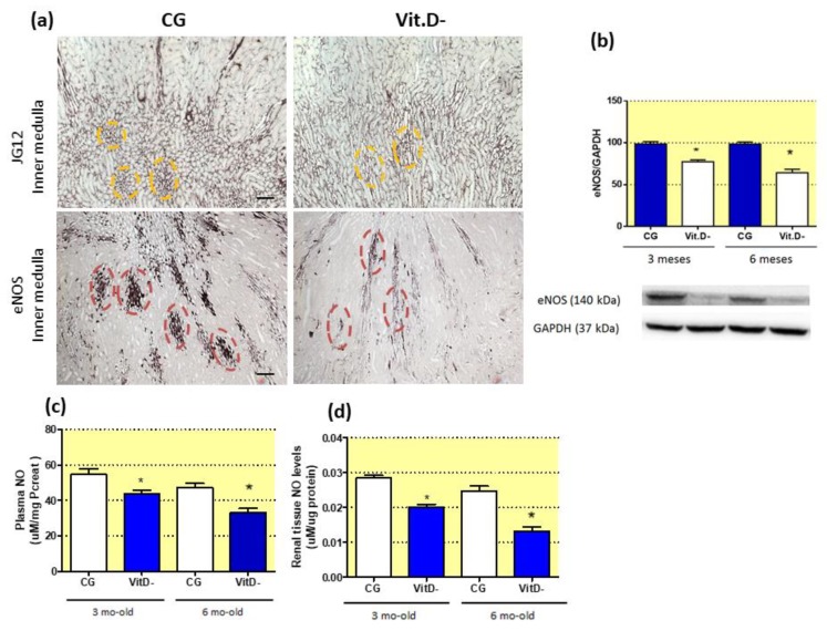
Figure 4.
Effect of Vitamin D (Vit.D) deficiency on the differentiation of the renal microvasculature. (a) Immunolocalization of aminopeptidase P (JG12) and endothelial nitric oxide synthase (eNOS) in the inner medullae of the control group (CG) and Vit.D- group at 6 months of age (CG n = 6, Vit.D- n = 8). Note the yellow circles showing the morphological arrangements of the vessels in the CG; in the Vit.D- group, these morphological arrangements are dispersed and fewer in number. Red circles demonstrate the immunolocalization of eNOS, showing the vessel integrity in the CG and reduced, severely dispersed expression in the Vit.D- group, which reflects impaired eNOS synthesis in these renal vessels in the Vit.D- group. Western blot analysis of renal tissues from all groups for eNOS expression (CG n = 4, Vit.D- n = 4) (b). The densitometric ratio between the densitometries of eNOS and glyceraldehyde 3-phosphate dehydrogenase (GAPDH) was calculated, and the data are expressed relative to the control, with the mean (±SEM) control value designated as 100%. Blots are representative images from independent experiments. NO levels in plasma (c), renal tissue (d), and urine (e) (CG n = 7, Vit.D- n = 8). Bar = 50 μm. * p < 0.05 compared with controls of the same age.

