Abstract
Electrolytic lesions to the amygdala, a limbic structure implicated in stress-related behaviors and memory modulation, have been shown to prevent stress-induced impairments of hippocampal long-term potentiation (LTP) and spatial memory in rats. The present study investigated the role of intrinsic amygdalar neurons in mediating stress effects on the hippocampus by microinfusing the GABAA receptor agonist muscimol into the amygdala and examining stress effects on Schaffer collateral/commissural-CA1 LTP and spatial memory. The critical period of the amygdalar contribution to stress effects on hippocampal functions was determined by applying muscimol either before stress or immediately after stress. Our results indicate that intra-amygdalar muscimol infusions before uncontrollable restraint-tailshock stress effectively blocked stress-induced physiological and behavioral effects. Specifically, hippocampal slices prepared from vehicle-infused stressed animals exhibited markedly impaired LTP, whereas slices obtained from muscimol-infused stressed animals demonstrated robust LTP comparable with that of unstressed animals. Correspondingly, vehicle-infused stressed animals displayed impaired spatial memory (on a hidden platform version of the Morris water maze task), whereas muscimol-infused stressed animals revealed unimpaired spatial memory. In contrast to prestress muscimol effects, however, immediate poststress infusions of muscimol into the amygdala failed to interfere with stress impairments of LTP and spatial memory. Together, these results suggest that the amygdalar neuronal activity during stress, but not shortly after stress, is essential for the emergence of stress-induced alterations in hippocampal LTP and memory.
Keywords: amygdala, hippocampus, GABA, glucocorticoids, corticosterone, long-term potentiation
Introduction
The hippocampus is considered to be particularly susceptible to uncontrollable stress effects because it is heavily enriched with corticosteroid receptors and it participates in the termination of stress responses via the glucocorticoid-mediated negative feedback of the hypothalamus-pituitary-adrenal (HPA) axis (Reul and de Kloet, 1985; McEwen and Sapolsky, 1995). Because this medial temporal lobe structure is crucial for declarative-explicit memory in humans and spatial-relational memory in rodents (Scoville and Milner, 1957; Squire and Zola-Morgan, 1996; Eichenbaum et al., 1992), evidence is emerging that stress generally impairs hippocampal-dependent memory tasks in both humans and rats (Sapolsky, 1992; McEwen, 2000; Kim and Diamond, 2002).
Analogous to behavioral findings, in vitro and in vivo electrophysiological studies indicate that stress impairs long-term potentiation (LTP) induction in the hippocampus. Hippocampal slices from rats that experienced uncontrollable restraint-tailshock stress, for example, exhibit subnormal LTP in CA1 and dentate gyrus (DG) (Foy et al., 1987; Shors et al., 1989; Shors and Dryver, 1994; Kim et al., 1996). Other stress paradigms, such as forced exposure to a brightly lit novel chamber or to a cat, likewise impair LTP and/or primed-burst potentiation (PBP) in awake, behaving rats (Diamond and Rose, 1994; Xu et al., 1997; Mesches et al., 1999). Because LTP is a candidate synaptic mnemonic mechanism (Bliss and Lomo, 1973; Malenka and Nicoll, 1999; Pittenger and Kandel, 2003), with qualities congruent to Bain's (1873) and Hebb's (1949) postulates (Wilkes and Wade, 1997), it has been hypothesized that stress-induced changes in hippocampal plasticity might be the neural basis of stress impairments in hippocampal memory (Kim and Yoon, 1998; Kim and Diamond, 2002).
Several lines of evidence indicate that the amygdala might be involved in mediating stress effects on LTP and memory. Amygdalar lesions have been shown to block/attenuate stress-induced gastric erosions (Henke, 1981; Grijalva et al., 1986), analgesia (Helmstetter, 1992), and anxiety-like behaviors (Adamec et al., 1999). In a series of experiments, McGaugh and colleagues (Packard et al., 1994; McGaugh, 2000; Roozendaal et al., 2003) found that pharmacological manipulations in the amygdala (that alter GABA, opioid, norepinephrine, and acetylcholine neurotransmissions) can enhance or impair the formation of memory that relies on the hippocampus. Recently, amygdalar lesions, stimulations, and drug infusions have been reported to modulate the magnitude of DG LTP (Abe, 2001). These findings suggest that the amygdala, via its projection to the hippocampus (Krettek and Price, 1977; Pikkarainen et al., 1999), might be involved in mediating stress effects on hippocampal functioning. In support of this notion, electrolytic lesions of the amygdala before stress have been found to prevent stress effects on LTP and spatial memory in rats (Kim et al., 2001); however, because electrolytic lesions damage both cells and fibers of passage, it is unclear whether the lesion effects were resulting from damaging amygdalar neurons or fibers that course through the amygdala. Therefore, the present study used the GABAA receptor agonist muscimol (MUSC) to inactivate amygdalar neurons and investigated stress effects on LTP and spatial memory. Moreover, muscimol was applied either before or immediately after stress to pinpoint the time course of amygdalar involvement in mediating stress effects on the hippocampus.
Materials and Methods
Experiment 1: intra-amygdalar muscimol infusions and stress effects on hippocampal LTP
The goal of experiment 1 was to determine whether intrinsic neurons in the amygdala are crucial for mediating stress effects on hippocampal LTP. To test this, the GABAA receptor agonist muscimol was used to inhibit the amygdalar neurons before stress. Previous studies have shown that rats emit 22 kHz ultrasonic vocalizations (USVs) (a distress call) in various situations of significant survival value (Anderson, 1954; Kaltwasser, 1990; Blanchard et al., 1991; Van der Poel and Miczek, 1991; Brudzynski and Chiu, 1995) and that the amygdala is necessary for rats to emit aversively induced USVs (Goldstein et al., 1996; Lee et al., 2001). Therefore, during stress, USVs were monitored to assess the efficacy of muscimol in the amygdala. Drug infusions and stress administration, histological verification, slice recording, and corticosterone assays were performed by different experimenters in a “blind” manner.
Subjects. Twenty-eight experimentally naive male Charles River (Wilmington, MA) Sprague Dawley rats (initially weighing 270-300 g) were housed individually in our Association for Assessment and Accreditation of Laboratory Animal Care accredited animal care facility and maintained on a reverse 12 h light/dark cycle (lights on at 7:00 P.M.) with ad libitum access to food and water. All experiments were conducted during the dark phase of the cycle and in strict compliance with the Yale Animal Resource Center guidelines.
Surgery. Animals were anesthetized via intraperitoneal injection of a 30 mg/kg ketamine and 2.5 mg/kg xylazine mixture, with supplemental injections given as needed. Under aseptic conditions, a stereotaxic instrument with nonpuncture ear bars (Stoelting, Wood Dale, IL) was used to implant 26 gauge guide cannulas (Plastics One, Roanoke, VA) bilaterally into the amygdala (from bregma: anteroposterior, -2.3 mm; mediolateral, ±5 mm; dorsoventral, -7.7 to 8.0 mm). Implanted cannulas were cemented to four anchoring screws on the skull. Dummy cannulas were inserted into the implanted cannulas to maintain patency of the guide cannulas. During 10-15 d of postoperative recovery, the rats were adapted to handling, and each dummy cannula was removed and replaced with a clean one.
Drugs and infusion. Muscimol free base (Sigma-Aldrich, St. Louis, MO), dissolved in artificial CSF (ACSF) (10 mm, pH ∼7.4) was micro-infused into the amygdala (bilaterally) by backloading the drug up a 33 gauge infusion cannula into polyethylene (PE 20) tubing connected to 10 μl Hamilton microsyringes (Hamilton Company, Reno, NV). The infusion cannula protruded 1 mm beyond the guide cannula. An infusion volume of 0.3 μl (per side) was delivered using a Harvard PHD2000 syringe pump (Harvard Apparatus, South Natick, MA) over the course of 3 min (at a rate of 0.1 μl/min). The infusion cannula remained in place for at least 30 s after the infusions before being pulled out.
Because our intra-amygdalar muscimol infusion parameter is similar to those used in fear-conditioning studies (Helmstetter and Bellgowan, 1994; Muller et al., 1997; Wilensky et al., 1999; Maren et al., 2001), the extent of muscimol diffusion in the amygdala should be reasonably comparable. Based on studies that examined 3H-muscimol spreading (Krupa et al., 1996; Arikan et al., 2002) in the cerebellum in which a 1 μl volume infusion diffused a radius of 1.6-2.0 mm, it was estimated that 0.3 μl of muscimol used in the present study would spread within a radius of ∼0.5-0.7 mm from the infusion needle tip. Hence, it is likely that infused muscimol would have diffused to the central, lateral, and basal nuclei of the amygdala and possibly to portions of adjacent neighboring structures.
Stress paradigm and USV data collection/analysis. Approximately 30 min after intra-amygdalar infusions, half of the animals from the MUSC- and ACSF-treated groups were restrained in a Plexiglas tube and exposed to 60 tailshocks (1 mA intensity; 1 s duration; 60 s intershock interval), whereas the remaining animals were left undisturbed (a 2 × 2 factorial design): ACSF-CONTROL (n = 6), ACSF-STRESS (n = 8), MUSC-CONTROL (n = 6), MUSC-STRESS (n = 8). The restraint-tailshock stress procedure, adapted from the “learned helplessness” paradigm (in which animals are exposed to unpredictable and uncontrollable aversive stimulus) (Seligman and Maier, 1967; Maier and Seligman, 1976), has been shown previously to reliably impair LTP in the hippocampus (Foy et al., 1987; Shors et al., 1989; Kim et al., 1996).
During stress, USV behavior was monitored to assess the efficacy of muscimol in the amygdala using a heterodyne bat detector (Mini-3, Noldus Information Technology, Wageninge, The Netherlands) that transformed high-frequency (22 ± 5 kHz) ultrasonic vocalization calls into the audible range (Lee et al., 2001). The output of the bat detector was fed through an audio amplitude filter (Noldus), which filtered out signals falling below an amplitude range that was individually adjusted for each animal. The resulting signal was then sent to an IBM-PC computer equipped with Noldus UltraVox vocalization analysis software. The software converted the signal into vocalization onset and offset times according to the following specifications: an onset was recorded if its duration was ≥500 ms, and the offset was recorded if the interval between episodes was ≥40 ms. If the interval was <40 ms, then the two bouts were counted as a single episode. The parameters for USV analysis were adapted from Brudzynski (1994). A custom-written analysis program (Labview 6i) was used to generate a raster plot representing the distribution of USV during the stress session.
In vitro electrophysiology procedure. Promptly after stress (within 10 min), animals were decapitated under halothane anesthesia, and the cannula-dental cement assembly was pulled from the skull. The removed brain was cut coronal-horizontally at an angle of ∼45° (from rostral-caudal direction) to partition the amygdala-containing brain portion (later used for verification of cannula tip) from the hippocampus-containing brain portion. From the latter part, hippocampal slices were prepared in a standard manner (Teyler, 1980). In brief, transverse hippocampal slices (400 μm) were maintained in an interface recording chamber (Fine Science Tools, Foster City, CA) that was perfused continuously (∼2 ml/min) with 95% O2/5% CO2-saturated ACSF containing (in mm): 124 NaCl, 3 KCl, 1.25 NaH2PO4, 1 MgSO2, 26 NaHCO3, 3 CaCl2 and 10 glucose, at 32°C. After at least 1 h of incubation, a concentric bipolar electrode (25 μm inner contact diameter) delivering 100 μs pulses stimulated the Schaffer collateral/commissural fibers. A glass electrode filled with 2 m NaCl (1.5-2.5 MΩ) was placed in the stratum radiatum in CA1 under a microscope to record field EPSPs (f-EPSPs). From a standard I-O curve (generated by averaging five f-EPSP slopes at each of different stimulation intensities), the test stimulus intensity was adjusted to produce a response that was ∼50% of the maximum evoked responses (in the absence of population spikes) (Kim et al., 1996). Baseline synaptic transmission was monitored for 20 min (every 20 s) before a tetanus was delivered (five trains of 100 Hz, each lasting 200 ms at an intertrain interval of 10 s). The f-EPSPs (amplified in the band of 0.1-5000 Hz) were monitored up to 90 min after the tetanus. During the tetanus, f-EPSPs evoked by the first pulse in each of the five trains were recorded to assess the development of potentiation. Data were collected and analyzed on-line using a computer program written in AXOBASIC/QUICK-BASIC (Axon Instruments, Union City, CA). The initial (negative) slope of f-EPSPs was used in statistical analyses (Kim et al., 1996). Only those slices that exhibited a stable baseline for 20 min were included in the analysis. The change in f-EPSPs after tetanus was averaged across slices for each rat (two hippocampal slices per rat). The magnitude of LTP was measured between 70 and 90 min after the tetanus and analyzed by means of two-way ANOVA (muscimol or ACSF infusions × stress or no stress conditions).
Corticosterone radioimmunoassay. During decapitation, trunk blood was collected for corticosterone radioimmunoassay. Blood serum was separated by centrifugation (5000 rpm, 20 min) and stored at -80°C until the time of assay. Serum corticosterone was calculated using the radioimmunoassay kit of ICN Biomedicals (Carson, CA) with 125I-corticosterone as a tracer.
Histology. The portion of the brain containing the amygdala was stored in 10% formalin for at least 2 weeks before slicing. Transverse sections (50 μm) were taken through the extent of the placement of cannulas, mounted on gelatinized slides, and stained with cresyl violet dye. An observer unaware of the data examined each section microscopically to determine the locations of the tips of the infusion cannulas, and subjects with inaccurate cannula placements (i.e., one or more cannula tips misplaced) were excluded from the statistical analyses.
Experiment 2: prestress and poststress intra-amygdalar muscimol infusions and stress effects on hippocampal LTP
Experiment 1 found that intra-amygdalar muscimol infusions before stress effectively prevent stress effects on hippocampal LTP. Several studies have shown that immediate posttraining inactivation of the amygdala interferes with the formation of hippocampal-dependent memory (McGaugh, 2000). Thus, the goal of experiment 2 was to test whether intra-amygdalar infusions of muscimol after stress can likewise prevent stress effects on hippocampal LTP.
Twenty-two experimentally naive Sprague Dawley male rats underwent bilateral cannula implantation, stress, and intra-amygdalar drug infusions as described above.
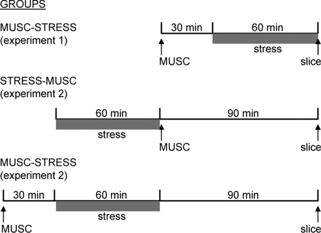
Subjects were assigned to one of three conditions. The first group of animals received intra-amygdalar muscimol infusions followed by restraint-tailshock stress (MUSC-STRESS; n = 8). The second group experienced stress first and then promptly (<5 min) received muscimol infusions (STRESS-MUSC; n = 8). The third group consisted of animals that underwent stress followed by ACSF infusions (STRESS-ACSF; n = 6). After stress, all animals were placed back in their home cage undisturbed. After ∼90 min, hippocampal slices were prepared and tested for LTP as in experiment 1. Serum corticosterone was measured in all groups.
It should be mentioned that slices were prepared ∼90 min after the termination of stress in experiment 2 (for both MUSC-STRESS and STRESS-MUSC groups), whereas slices were prepared promptly after stress in experiment 1 (for MUSC-STRESS group). This 90 min stress-to-slice preparation period was selected to time-match the muscimol infusion-to-slice preparation interval between the experiment 1 MUSC-STRESS group and the experiment 2 STRESS-MUSC group and to time-match the stress-to-slice preparation interval between MUSC-STRESS and STRESS-MUSC groups, both in experiment 2 (Fig. 1). Because previous studies have demonstrated that stress-induced LTP impairments last at least 48 h in rats (Shors et al., 1997) and 24 h in mice (Garcia et al., 1997), any potential differences in LTP magnitudes between prestress and poststress muscimol groups in experiment 2 can be attributed to whether the amygdala was inactivated during stress or immediately after stress (by comparing the experiment 1 MUSC-STRESS group with the experiment 2 STRESS-MUSC group) rather than to the temporal difference from muscimol infusions to hippocampal slice preparation (by comparing the STRESS-MUSC and MUSC-STRESS groups, both in experiment 2).
Figure 1.
Time intervals from intra-amygdalar MUSC infusions to stress presentation to hippocampal slice preparation in experiments 1 and 2.
Experiment 3: prestress and poststress intra-amygdalar muscimol infusions and stress effects on spatial memory
We found previously that electrolytic lesions of the amygdala, which damage both intrinsic cells and fibers of passage, blocked stress-induced impairments in spatial memory (Kim et al., 2001). To ascertain whether neuronal activities in the amygdala contribute to stress effects on spatial memory, experiment 3 investigated the effects of prestress and immediate poststress intra-amygdalar muscimol infusions on a hippocampal-dependent hidden platform version of the Morris water maze task.
Subjects, drug infusions, and stress. Fifty experimentally naive Sprague Dawley male rats implanted with bilateral cannulas underwent similar drug infusion and stress procedures as described previously.
Animals were assigned to one of five groups: ACSF-CONTROL (n = 10), ACSF-STRESS (n = 10), MUSC-CONTROL (n = 10), MUSC-STRESS (n = 10), and STRESS-MUSC (n = 10). Half of the animals in the ACSF-STRESS group received ACSF infusions before stress, and the remaining half were infused with ACSF immediately after stress. Because prestress and poststress ACSF-infused rats did not differ, the data were pooled.
Hidden platform water maze task. All animals underwent water maze training between 4 and 5.5 h after intra-amygdalar infusions. This relatively long infusion-to-training lag time was necessary because we observed noticeable motoric effects during water maze training (i.e., decreased swim speed, difficulty in platform climbing, decreased exploratory-rearing behavior on the platform) when animals were trained up to 90 min after muscimol infusions.
The training and testing procedures were adapted from those described previously and have been shown to be hippocampal based (Packard and McGaugh, 1994). Animals were subjected to eight training trials (with a 15 min intertrial interval) to find a fixed submerged platform (6″ diameter) and escape from a circular water maze (72″; diameter, 24.5″ height; 22-24°C water temperature). The starting point was distributed randomly across the four quadrants (two starting points per each quadrant; the animal always faced the wall when placed in the water). If escape did not occur within 60 s, the animal was manually guided to the platform. After climbing the platform, the animal stayed on it for 60 s and then was placed in a holding cage until the next trial. After the last trial, the animals were returned to their home cages. The next day, a retention test (a 60 s probe trial) was given in which the platform was removed from the pool. Animals' movements and the time taken to reach the position at which the platform had been located in training were monitored automatically using a computerized HVS 2020 Tracking System (HVS Image, Buckingham, UK).
Histology. At the completion of behavioral testing, the subjects were overdosed with ketamine HCl and xylazine and perfused intracardially with 0.9% saline followed by 10% buffered formalin. The brains were removed and stored in 10% formalin for at least 2 weeks before slicing. Transverse sections (50 μm) were taken through the extent of the cannula tract, mounted on gelatinized slides, and stained with cresyl violet.
Results
Experiment 1
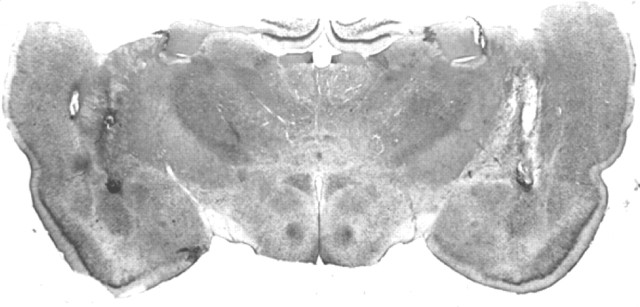
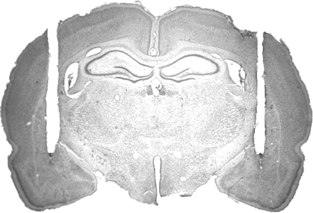
Figure 2 shows a photomicrograph of a transverse brain section stained with cresyl violet from a typical rat (from slice experiments 1 and 2) with infusion cannula tips located bilaterally in the amygdala. The frontal and parietal cortices as well as the rostral-most portion of the dorsal hippocampus are absent in this section because, as described previously, the brain was cut at an angle of ∼45° (from rostralcaudal direction) to separate the hippocampus (to prepare slices) from the amygdala (to confirm cannula tips). Four rats were excluded from the data analysis on the basis of histological results.
Figure 2.
Photomicrograph showing a transverse brain section stained with cresyl violet from a rat infused with muscimol bilaterally into the amygdala and used in the hippocampal slice experiment. Arrowheads indicate infusion cannula tip positions.
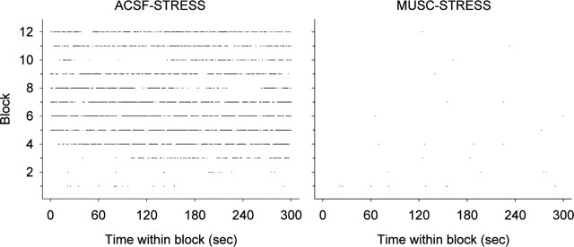
Table 1 shows the mean duration of USV emitted by ACSF-STRESS and MUSC-STRESS groups during the 60 min of stress session. As can be seen, intra-amygdalar infusions of muscimol before stress nearly eliminated stress-induced USVs compared with ACSF infusions (t(12) = 13.37; p < 0.01). To appreciate better the effect of intra-amygdalar muscimol on stress-induced USV across time, Figure 3 depicts USVs as event raster plots that show the distribution of USV episodes (raw data) from a typical ACSF and MUSC animal, respectively, experiencing stress. Each point on the raster plot represents an episode of USV. It is apparent that muscimol infusions into the amygdala robustly abolished stress-induced USV during the entire 60 min of stress session. It should be mentioned that USV was elicited during the intershock periods and not in response to the shock. In contrast, the reflexive audible “squeal” vocalization in response to the shock was observed in both the ACSF and MUSC groups (data not shown). Thus, intra-amygdalar muscimol does not interfere with the animal's perception of tailshock that elevates corticosterone (see below).
Table 1.
Stress-induced USV in experiment 1
|
Groups |
USV duration (mean ± SE) |
|---|---|
| ACSF-STRESS (n = 6) | 1787 ± 102 s |
| MUSC-STRESS (n = 7) |
2.8 ± 1 s |
Figure 3.
Examples of the USV raster plots; each dot represents a time-stamped episode of vocalization. USV emissions are shown from a typical ACSF- and muscimol-infused animal during 60 min of stress. The y-axis represent block of time; each block represent 5 min (12 blocks × 5 min = 60 min of stress).
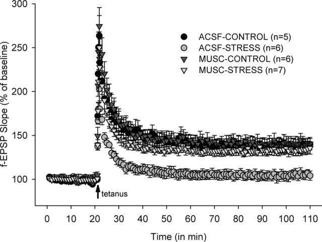
As shown in Figure 4, hippocampal slices from ACSF-STRESS animals exhibited impaired LTP (normalized f-EPSP slopes measured 70-90 min after the tetanus: 104.5 ± 5.2%), whereas LTP was reliable in slices from ACSF-CONTROL (138.6 ± 7.3%), MUSC-CONTROL (139.8 ± 7.9%), and MUSC-STRESS (133.6 ± 6.0%) animals. A significant drug × stress interaction (two-way ANOVA; F(1,23) = 4.58; p < 0.05) indicates that the intra-amygdalar muscimol infusions did not decrease or increase the magnitude of LTP per se, but did prevent stress-induced LTP impairments in the hippocampus. The I-O curve revealed that neither stress nor intra-amygdalar muscimol (in experiments 1 and 2) affected the baseline Schaffer collateral/commissural-CA1 synaptic transmission (data not shown).
Figure 4.
Effects of intra-amygdalar infusions of muscimol and stress on Schaffer collateral/commissural-CA1 LTP. Synaptic strength in the CA1 area of the hippocampus from ACSF-CONTROL, ACSF-STRESS, MUSC-CONTROL, and MUSC-STRESS animals is expressed as a percentage of the average pretetanus f-EPSP over time (in minutes).
Analysis of trunk blood showed significantly higher levels of serum corticosterone in rats exposed to stress than those that did not, regardless of whether the animals received ACSF or muscimol infusions into their amygdala (ACSF-STRESS = 66.8 ± 7.7, MUSC-STRESS = 51.2 ± 9.3, ACSF-CONTROL = 5.4 ± 4.9, MUSC-CONTROL = 8.9 ± 5.0 μg/dl; two-way ANOVA, main effect of stress: F(1,23) = 165.12; p < 0.01). Although there was no main effect of drug (F(1,23) = 2.23; p > 0.05), there was a statistically reliable drug × stress interaction (F(1,23) = 5.68; p < 0.05), which is likely attributable to the corticosterone level showing an increasing trend from ACSF-CONTROL to MUSC-CONTROL animals and a decreasing trend from ACSF-STRESS to MUSC-STRESS animals; however, post hoc tests indicated that corticosterone levels were not statistically different between ACSF-CONTROL and MUSC-CONTROL groups or between ACSF-STRESS and MUSC-STRESS groups [all p values > 0.05; Tukey honestly significant difference (HSD)].
Experiment 2
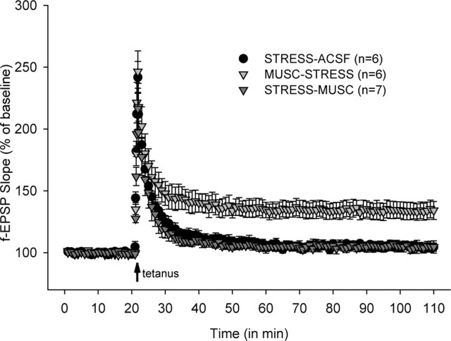
Based on histological results, three rats were excluded from the data analysis. A one-way ANOVA indicated a significant group difference in the magnitude of LTP (F(2, 18) = 13.67; p < 0.01) (Fig. 5). As in experiment 1, intra-amygdalar infusions of muscimol before stress (MUSC-STRESS) reliably prevented stress effects on hippocampal LTP (MUSC-STRESS vs STRESS-ACSF; p < 0.01; Tukey HSD). In marked contrast, immediate poststress intra-amygdalar muscimol infusions (STRESS-MUSC) had virtually no effect on stress-associated LTP deficits; LTP impairments were comparable between STRESS-MUSC and STRESS-ACSF groups (p > 0.05; Tukey HSD).
Figure 5.
Prestress and immediate poststress intra-amygdalar infusions of muscimol and stress effects on Schaffer collateral/commissural-CA1 LTP. Synaptic strength in the CA1 area of the hippocampus from STRESS-ACSF, MUSC-STRESS, and STRESS-MUSC animals is expressed as a percentage of the average pretetanus f-EPSP over time (in minutes).
The corticosterone levels were considerably lower when trunk blood was collected 90 min after stress (ACSF-STRESS = 23.4 ± 4.7, MUSC-STRESS = 18.0 ± 10.9, STRESS-MUSC = 9.5 ± 5.9 μg/dl) compared with trunk blood collected promptly after stress (experiment 1). One-way ANOVA indicated that there is no reliable group difference (F(2,16) = 3.32; p > 0.05).
Experiment 3
Figure 6 shows a photomicrograph of a transverse brain section from a representative rat used in the water maze experiment. As can been seen, infusion cannula tracts are in the amygdala. Of 50 rats, 8 rats were excluded from the data analysis based on histological results.
Figure 6.
Photomicrograph showing a transverse brain section stained with cresyl violet from a rat with bilateral guide cannulas implanted in the amygdala and used in the behavioral experiment. Arrowheads indicate infusion cannula tip positions.
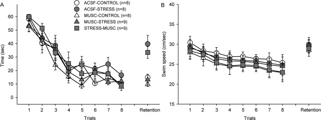
In a hidden platform version of the water maze task, all groups significantly decreased their latencies to find the hidden platform during the eight training trials (Fig. 7A). The rate of acquisition was comparable among ACSF-CONTROL, ACSF-STRESS, MUSC-CONTROL, MUSC-STRESS, and STRESS-MUSC groups (oneway ANOVA with trials as a repeated measure; main effect of group: F(4.37) = 1.79; p > 0.05; group × trials interaction: F < 1.0). This suggests that the sensory/motor systems (e.g., abilities to navigate using distal cues and swim) and the motivation to escape the water were unaffected in rats that earlier underwent stress and/or intraamygdala muscimol infusions. On the retention (probe) test 1 d later, however, there were significant group differences (F(4,37) = 8.19; p < 0.01). Specifically, animals in the ACSF-STRESS and STRESS-MUSC groups required a considerably longer time to find the original location of the platform compared with ACSF-CONTROL, MUSC-CONTROL, and MUSC-STRESS animals (all p values < 0.05; Tukey HSD). As shown in Figure 7B, the latency differences cannot be attributed to possible motoric effects because there were no reliable group differences in swim speed during either the acquisition or probe test (p > 0.05). Although a single training session consisting of eight trials did not yield a reliable quadrant bias or difference in number of quadrant entries per annulus crossing on the probe trial in any of the groups (data not shown) (Packard and McGaugh, 1994; Kim et al., 2001), the latency and distance measures indicate that intra-amygdalar infusions of muscimol before stress, but not immediately after stress, block stress effects on spatial memory.
Figure 7.
Effects of intra-amygdala infusions of muscimol and stress on spatial memory. A, Mean (±SE) latencies to find a submerged platform from ACSF-CONTROL, ACSF-STRESS, MUSC-CONTROL, MUSC-STRESS, and STRESS-MUSC animals during acquisition and a single retention test. B, Mean (±SE) swim speed of five groups during acquisition and a single retention test.
Discussion
The present findings indicate that amygdalar activity is crucially involved in the emergence of stress-induced impairments in hippocampal LTP and hippocampal-dependent spatial memory in rats. Specifically, we found that intra-amygdalar infusions of the GABAA receptor agonist muscimol before stress (MUSC-STRESS group) effectively prevented stress impairments of CA1 LTP in vitro (experiments 1 and 2) and spatial memory in a water maze task (experiment 3). These observations are congruent with previous findings that amygdalar lesions block or attenuate a range of stress-induced effects such as LTP and spatial memory (Kim et al., 2001), gastric erosion (Henke, 1981), and analgesia (Helmstetter, 1992). Anatomically, the amygdala sends direct (from the magnocellular and parvicellular divisions of the basolateral amygdala to the CA1 and subiculum) and indirect (via the entorhinal cortex) projections to the hippocampus (Krettek and Price, 1977; Aggleton, 1986; Pikkarainen et al., 1999), routes by which it can influence hippocampal functions (Kim and Diamond, 2002). Because muscimol increases Cl- ion conductance across cell membranes (Feldman et al., 1997), the drug effects presumably are caused by inhibition of amygdalo-hippocampal activity during stress.
In contrast to prestress muscimol effects, inactivation of the amygdala poststress failed to prevent stress effects on either LTP or spatial memory. Hippocampal slices from rats that received amygdalar infusions of muscimol immediately after experiencing stress (STRESS-MUSC group in experiment 2) demonstrated LTP impairments comparable with slices from vehicle-treated stressed animals. Similarly, the magnitude of spatial memory impairments was indistinguishable between the STRESS-MUSC and STRESS-ACSF groups (experiment 3). The lack of poststress amygdalar muscimol effects suggests that inhibiting amygdalar activity after stress does not impact stress effects on hippocampal functioning.
The dissimilar effects on LTP between animals that received muscimol infusions before stress (MUSC-STRESS) and after stress (STRESS-MUSC) cannot be accounted for by differences in muscimol-to-stress or stress-to-hippocampal slice preparation periods between the groups. As shown in Figure 1, the MUSC-STRESS group in experiment 1 and the STRESS-MUSC group in experiment 2 were time-matched in terms of muscimol infusions-to-slice preparation (90 min), whereas STRESS-MUSC and MUSC-STRESS groups, both in experiment 2, were time-matched in terms of stress termination-to-slice preparation (90 min). It appears that the critical time window of amygdalar activity in mediating stress effects on the hippocampus is during stress, not after stress.
Previous studies have shown that high levels of corticosterone can affect the intrinsic properties of hippocampal neurons (i.e., prolonging the afterhyperpolarization) (Joels and De Kloet, 1989; Kerr et al., 1989), impair LTP and/or PBP (Diamond et al., 1992), and interfere with performance in spatial memory tasks (Bodnoff et al., 1995; de Quervain et al., 1998). It is possible then that the prestress and poststress amygdalar muscimol differences observed in the present study might be caused by differences in corticosterone levels; however, this is unlikely for two reasons. First, amygdalar muscimol prevented stress-induced LTP impairments without significantly affecting the increase in corticosterone secretion caused by stress (MUSC-STRESS animals in experiment 1). Second, although the corticosterone levels were comparable between MUSC-STRESS and STRESS-MUSC groups 90 min after the termination of stress, LTP impairments were observed only in the latter group (experiment 2). These observations indicate that amygdalar muscimol can prevent stress effects on LTP whether the corticosterone level is high (MUSC-STRESS animals in experiment 1) or low (MUSC-STRESS animals in experiment 2) at the time of hippocampal slice preparation and that stress-induced LTP impairments occur even when the corticosterone level is low at the time of slice preparation (MUSC-CONTROL animals in experiment 2). The dissociation of corticosterone level and LTP inducibility suggests that the elevation in corticosterone is not a sufficient condition for mediating stress effects on the hippocampal. This view is further supported by other findings such as the following: (1) amygdalar lesions block stress effects on LTP and spatial memory without impeding the stress-induced increase in corticosterone secretion (Kim et al., 2001); (2) LTP is reduced in adrenalectomized rats after stress and is not restored by exogenous administration of corticosterone (Shors et al., 1990); and (3) in normal animals administered with dexamethasone (a synthetic glucocorticoid that blocks the HPA axis activity), stress-induced impairments in LTP still occurred (Foy et al., 1990).
Although stress impaired the retention of spatial memory in water maze, during the acquisition phase stressed animals displayed rates of improvement in locating the hidden platform similar to those of unstressed animals, suggesting that stress effects on LTP are correlated with retention (but not acquisition) of spatial memory. A similar observation in LTP and water maze performance has been reported previously. Specifically, Morris and colleagues (1986) found that rats infused with the NMDA receptor antagonist AP5, which completely blocked LTP in the hippocampus, exhibited mild (but not reliable) acquisition deficits in the water maze. They attributed this spared learning in the absence of LTP to the animal's use of “nonspecific instrumental learning that may have masked an impairment in true place-learning.” It is possible that LTP-impaired stressed animals used a similar nonspecific strategy to locate the hidden platform; however, the possible contribution of NMDA receptor-independent forms of plasticity (e.g., posttetanic potentiation) in the acquisition of water maze cannot be excluded (Morris, 2003).
Results from various posttraining drug infusion studies suggest that the amygdala is necessary for modulating the formation of memory that relies on the hippocampus (Packard et al., 1994; Cahill and McGaugh, 1998; Roozendaal et al., 1998). For instance, immediate posttraining pharmacological manipulations that alter noradrenergic, acetylcholinergic, GABAergic, and opioid activities in the amygdala can enhance or impair memory consolidation in hippocampal-dependent tasks (McGaugh, 2000). Amygdalar lesions, drug infusions, and stimulations have also been reported to influence DG LTP (Ikegaya et al., 1994, 1995, 1996; Akirav and Richter-Levin, 1999). Therefore, it has been suggested that posttraining amygdalar manipulations influence memory consolidation in the hippocampus by altering LTP or LTP-like changes (McGaugh, 2000). Consistent with this notion, a recent study found that the amygdala is necessary for glucocorticoid- or stress-induced impairments of spatial memory (Roozendaal et al., 2003).
Although the present results also implicate the amygdala in influencing hippocampal functioning, our findings suggest that amygdalar activity during stress, but not after stress, is critical for stress impairments of LTP and spatial memory. It is possible that the 60 min stress session used in the present study is too long to detect poststress amygdalar muscimol effects. For example, if stress effects on the hippocampus were to transpire within a few minutes of stress, then intra-amygdalar muscimol promptly after stress (60 min after stress initiation) might have missed the critical time window to effectively block stress effects. Indeed, there is evidence that infusing lidocaine (a voltage-dependent Na+ channel blocker) into the amygdala immediately after a brief aversive experience can impair contextual fear conditioning (Vazdarjanova and McGaugh, 1999); however, amygdalar muscimol immediately after one-trial fear conditioning failed to affect memory consolidation (Wilensky et al., 1999; Lee et al., 2001). Thus, it remains to be determined whether the present finding, that poststress amygdalar muscimol does not interfere with stress impairments in LTP and spatial memory, is the result of differences in stress versus learning. Additional studies are also necessary to examine whether other poststress drug manipulations (e.g., glucocorticoid receptor antagonists) can reverse stress effects on hippocampal plasticity and memory.
As mentioned previously, the amygdala seems to modulate the magnitude of LTP in the hippocampus. Ikegaya and colleagues (1994) have shown that electrolytic lesions to the basolateral (but not central) nuclei of the amygdala significantly attenuate the DG LTP in vivo, whereas high-frequency stimulation of the amygdala augment LTP (Ikegaya et al., 1996). Moreover, amygdalar infusions of NMDA receptor antagonist have been shown to decrease DG LTP without affecting the baseline synaptic response (Ikegaya et al., 1995), a finding which suggests that amygdalar NMDA receptors influence LTP in the hippocampus. In contrast, although both amygdalar muscimol infusions (present study) and lesions (Kim et al., 2001) blocked stress effects on CA1 LTP in vitro, neither manipulation affected the magnitude of LTP in unstressed animals. As reported previously (Shors et al., 1989; Xu et al., 1997), the I-O curve indicated that neither stress nor amygdalar inactivation altered the baseline synaptic transmission in CA1. Thus, the stress impairment of LTP is not likely caused by stress producing LTP (or LTP-like changes) that occludes subsequent LTP induction (Kim and Yoon, 1998). The differing effects observed with amygdalar inactivation on DG LTP (impairment) and CA1 LTP (no effect) might be caused by the amygdala differentially influencing synaptic plasticity in different regions of the hippocampus (because of differences in anatomical projections from amygdala to hippocampal subfields) or by in vitro and in vivo procedural differences. These issues will need to be examined in future studies.
At present, it is not known why (or whether) it might be evolutionarily advantageous to impair hippocampal memory functioning after stress. One possibility is that stress effects on subsequent learning might serve to reduce retroactive interference of the original memory of a stressful event, which might be essential to guide future behavior. An alternative possibility is that stress-induced memory impairments may regulate the strength (or generalizability) of traumatic memory such that reduced memory functions actually help the subject cope with the psychological impact of the stressful event. According to the latter view, the development of stress disorders (such as posttraumatic stress disorder) might be caused by deficiency in stress-induced memory impairments. Regardless of the precise evolutionary underpinning, the amygdala plays an integral role in the mediation of stress effects on hippocampal functioning.
In summary, amygdalar activity during stress, but not after stress, is critical for the emergence of stress impairments in hippocampal LTP and spatial memory. The present findings have clinical implications in that after traumatic stress experiences, drug manipulations targeting the amygdala activity may not be sufficient to counteract stress effects on the hippocampus (although there may be other beneficial effects). Perhaps other drugs or combined drug-behavioral manipulations may prove more therapeutically effective. As such, future studies should investigate whether poststress behavioral and pharmacological intervention can prevent stress effects on hippocampal functioning.
Footnotes
This work was supported by the Whitehall Foundation and National Institute of Mental Health Grant 1R01 MH64457 to J.J.K. We thank Drs. Karyn Frick and Sheri Mizumori for helpful comments on this manuscript, Min Jung Kim for the histology, and Taekwan Lee for the USV analysis program.
Correspondence should be addressed to Jeansok J. Kim, Department of Psychology, Guthrie Hall, Box 351525, University of Washington, Seattle, WA 98195-1525. E-mail: jeansokk@u.washington.edu.
Copyright © 2005 Society for Neuroscience 0270-6474/05/251532-08$15.00/0
References
- Abe K (2001) Modulation of hippocampal long-term potentiation by the amygdala: a synaptic mechanism linking emotion and memory. Jpn J Pharmacol 86: 18-22. [DOI] [PubMed] [Google Scholar]
- Adamec RE, Burton P, Shallow T, Budgell J (1999) Unilateral block of NMDA receptors in the amygdala prevents predator stress-induced lasting increases in anxiety-like behavior and unconditioned startle—effective hemisphere depends on the behavior. Physiol Behav 65: 739-751. [DOI] [PubMed] [Google Scholar]
- Aggleton JP (1986) A description of the amygdalo-hippocampal interconnections in the macaque monkey. Exp Brain Res 64: 515-526. [DOI] [PubMed] [Google Scholar]
- Akirav I, Richter-Levin G (1999) Biphasic modulation of hippocampal plasticity by behavioral stress and basolateral amygdala stimulation in the rat. J Neurosci 19: 10530-10535. [DOI] [PMC free article] [PubMed] [Google Scholar]
- Anderson JW (1954) The production of ultrasonic sounds by laboratory rats and other mammals. Science 119: 808-809. [DOI] [PubMed] [Google Scholar]
- Arikan R, Blake NM, Erinjeri JP, Woolsey TA, Giraud L, Highstein SM (2002) A method to measure the effective spread of focally injected muscimol into the central nervous system with electrophysiology and light microscopy. J Neurosci Methods 118: 51-57. [DOI] [PubMed] [Google Scholar]
- Bain A (1873) Mind and body. The theories of their relation. London: Henry King.
- Blanchard RJ, Blanchard DC, Agullana R, Weiss SM (1991) Twenty-two kHz alarm cries to presentation of predator, by laboratory rats living in visible burrow systems. Physiol Behav 50: 967-972. [DOI] [PubMed] [Google Scholar]
- Bliss TVP, Lomo T (1973) Long-lasting potentiation of synaptic transmission in the dentate area of the anaesthetized rabbit following stimulation of the perforant path. J Physiol (Lond) 232: 331-356. [DOI] [PMC free article] [PubMed] [Google Scholar]
- Bodnoff SR, Humphreys AG, Lehman JC, Diamond DM, Rose GM, Meaney MJ (1995) Enduring effects of chronic corticosterone treatment on spatial learning, synaptic plasticity, and hippocampal neuropathology in young and mid-aged rats. J Neurosci 15: 61-69. [DOI] [PMC free article] [PubMed] [Google Scholar]
- Brudzynski SM (1994) Ultrasonic vocalization induced by intracerebral carbachol in rats: localization and dose-response study. Behav Brain Res 63: 133-143. [DOI] [PubMed] [Google Scholar]
- Brudzynski SM, Chiu EMC (1995) Behavioural responses of laboratory rats to playback of 22 kHz ultrasonic calls. Physiol Behav 57: 1039-1044. [DOI] [PubMed] [Google Scholar]
- Cahill L, McGaugh JL (1998) Mechanisms of emotional arousal and lasting declarative memory. Trends Neurosci 21: 294-299. [DOI] [PubMed] [Google Scholar]
- de Quervain DJF, Roozendaal B, McGaugh JL (1998) Stress and glucocorticoids impair retrieval of long-term spatial memory. Nature 394: 787-790. [DOI] [PubMed] [Google Scholar]
- Diamond DM, Rose GM (1994) Stress impairs LTP and hippocampal-dependent memory. Ann NY Acad Sci 746: 411-414. [DOI] [PubMed] [Google Scholar]
- Diamond DM, Bennett MC, Fleshner M, Rose GM (1992) Inverted-U relationship between the level of peripheral corticosterone and magnitude of hippocampal primed burst potentiation. Hippocampus 2: 421-430. [DOI] [PubMed] [Google Scholar]
- Eichenbaum H, Otto T, Cohen NJ (1992) The hippocampus—what does it do? Behav Neural Biol 57: 2-36. [DOI] [PubMed] [Google Scholar]
- Feldman RS, Meyer J, Quenzer LF (1997) Principles of neuropsychopharmacology. Sunderland, MA: Sinauer.
- Foy MR, Stanton ME, Levine S, Thompson RF (1987) Behavioral stress impairs long-term potentiation in rodent hippocampus. Behav Neural Biol 48: 138-149. [DOI] [PubMed] [Google Scholar]
- Foy MR, Foy JG, Levine S, Thompson RF (1990) Manipulation of pituitaryadrenal activity affects neural plasticity in rodent hippocampus. Psychol Sci 3: 201-204. [Google Scholar]
- Garcia R, Musleh W, Tocco G, Thompson RF, Baudry M (1997) Time-dependent blockade of STP and LTP in hippocampal slices following acute stress in mice. Neurosci Lett 233: 41-44. [DOI] [PubMed] [Google Scholar]
- Goldstein LE, Rasmusson AM, Bunney BS, Roth EH (1996) Role of the amygdala in the coordination of behavioral, neuroendocrine, and prefrontal cortical monoamine responses to psychological stress in the rat. J Neurosci 16: 4787-4798. [DOI] [PMC free article] [PubMed] [Google Scholar]
- Grijalva CV, Tache Y, Gunion MW, Walsh JH, Geiselman PJ (1986) Amygdaloid lesions attenuate neurogenic gastric mucosal erosions but do not alter gastric secretory changes induced by intracisternal bombesin. Brain Res Bull 16: 55-61. [DOI] [PubMed] [Google Scholar]
- Hebb DO (1949) The organization of behavior: a neuropsychological theory. New York: Wiley.
- Helmstetter FJ (1992) The amygdala is essential for the expression of conditional hypoalgesia. Behav Neurosci 106: 518-528. [DOI] [PubMed] [Google Scholar]
- Helmstetter FJ, Bellgowan PS (1994) Effects of muscimol applied to the basolateral amygdala on acquisition and expression of contextual fear conditioning in rats. Behav Neurosci 108: 1005-1009. [DOI] [PubMed] [Google Scholar]
- Henke PG (1981) Attenuation of shock-induced ulcers after lesion in the medial amygdala. Physiol Behav 27: 143-146. [DOI] [PubMed] [Google Scholar]
- Ikegaya Y, Saito H, Abe K (1994) Attenuated hippocampal long-term potentiation in basolateral amygdala-lesioned rats. Brain Res 656: 157-164. [DOI] [PubMed] [Google Scholar]
- Ikegaya Y, Saito H, Abe K (1995) Amygdala N-methyl-d-aspartate receptors participate in the induction of long-term potentiation in the dentate gyrus in vivo. Neurosci Lett 192: 193-196. [DOI] [PubMed] [Google Scholar]
- Ikegaya Y, Saito H, Abe K (1996) The basomedial and basolateral amygdaloid nuclei contribute to the induction of long-term potentiation in the dentate gyrus in vivo. Eur J Neurosci 8: 1833-1839. [DOI] [PubMed] [Google Scholar]
- Joels M, De Kloet R (1989) Effects of glucocorticoids and norepinephrine on the excitability in the hippocampus. Science 245: 1502-1505. [DOI] [PubMed] [Google Scholar]
- Kaltwasser MT (1990) Startle-inducing acoustic stimuli evoke ultrasonic vocalization in the rat. Physiol Behav 48: 13-17. [DOI] [PubMed] [Google Scholar]
- Kerr DS, Campbell LW, Hao SY, Landfield PW (1989) Corticosteroid modulation of hippocampal potentials: increased effect with aging. Science 245: 1505-1509. [DOI] [PubMed] [Google Scholar]
- Kim JJ, Diamond DM (2002) The stressed hippocampus, synaptic plasticity and lost memories. Nat Rev Neurosci 3: 453-462. [DOI] [PubMed] [Google Scholar]
- Kim JJ, Yoon KS (1998) Stress: metaplastic effects in the hippocampus. Trends Neurosci 21: 505-509. [DOI] [PubMed] [Google Scholar]
- Kim JJ, Foy MR, Thompson RF (1996) Behavioral stress modifies hippocampal plasticity through N-methyl-d-aspartate receptor activation. Proc Natl Acad Sci USA 93: 4750-4753. [DOI] [PMC free article] [PubMed] [Google Scholar]
- Kim JJ, Lee HJ, Han J-S, Packard MG (2001) Amygdala is critical for stress-induced modulation of hippocampal LTP and learning. J Neurosci 21: 5222-5228. [DOI] [PMC free article] [PubMed] [Google Scholar]
- Krettek JE, Price JL (1977) Projections from the amygdaloid complex and adjacent olfactory structures to the entorhinal cortex and subiculum in the rat and cat. J Comp Neurol 172: 723-752. [DOI] [PubMed] [Google Scholar]
- Krupa DJ, Thompson JK, Thompson RF (1996) Localization of a memory trace in the mammalian brain. Science 260: 989-991. [DOI] [PubMed] [Google Scholar]
- Lee HJ, Choi J-S, Brown TH, Kim JJ (2001) Amygdalar NMDA receptors are critical for the expression of multiple conditioned fear responses. J Neurosci 21: 4116-4124. [DOI] [PMC free article] [PubMed] [Google Scholar]
- Maier SF, Seligman MEP (1976) Learned helplessness: theory and evidence. J Exp Psychol Gen 105: 3-45. [Google Scholar]
- Malenka RC, Nicoll RA (1999) Long-term potentiation—a decade of progress? Science 285: 1870-1874. [DOI] [PubMed] [Google Scholar]
- Maren S, Yap SA, Goosens KA (2001) The amygdala is essential for the development of neuronal plasticity in the medial geniculate nucleus during auditory fear conditioning in rats. J Neurosci 21: RC135(1-5). [DOI] [PMC free article] [PubMed] [Google Scholar]
- McEwen BS (2000) Effects of adverse experiences for brain structure and function. Biol Psychiatry 48: 721-731. [DOI] [PubMed] [Google Scholar]
- McEwen BS, Sapolsky RM (1995) Stress and cognitive function. Curr Opin Neurobiol 5: 205-216. [DOI] [PubMed] [Google Scholar]
- McGaugh JL (2000) Memory: a century of consolidation. Science 287: 248-251. [DOI] [PubMed] [Google Scholar]
- Mesches MH, Fleshner M, Heman KL, Rose GM, Diamond DM (1999) Exposing rats to a predator blocks primed burst potentiation in the hippocampus in vitro J Neurosci 19: RC18(1-4). [DOI] [PMC free article] [PubMed] [Google Scholar]
- Morris RGM (2003) Long-term potentiation and memory. Philos Trans R Soc Lond B Biol Sci 358: 643-647. [DOI] [PMC free article] [PubMed] [Google Scholar]
- Morris RGM, Anderson E, Lynch GS, Baudry M (1986) Selective impairment of learning and blockade of long-term potentiation by an N-methyl-d-aspartate receptor antagonist, AP5. Nature 319: 774-776. [DOI] [PubMed] [Google Scholar]
- Muller J, Corodimas KP, Fridel Z, LeDoux JE (1997) Functional inactivation of the lateral and basal nuclei of the amygdala by muscimol infusion prevents fear conditioning to an explicit conditioned stimulus and to contextual stimuli. Behav Neurosci 111: 683-691. [DOI] [PubMed] [Google Scholar]
- Packard MG, McGaugh JL (1994) Quinpirole and d-amphetamine administration post-training enhances memory on spatial and cued discrimination in a water maze. Psychobiology 22: 54-60. [Google Scholar]
- Packard MG, Cahill L, McGaugh JL (1994) Amygdala modulation of hippocampal-dependent and caudate nucleus-dependent memory processes. Proc Natl Acad Sci USA 91: 8477-8481. [DOI] [PMC free article] [PubMed] [Google Scholar]
- Pikkarainen M, Ronkko S, Savander V, Insausti R, Pitkanen A (1999) Projections from the lateral, basal, and accessory basal nuclei of the amygdala to the hippocampal formation in rat. J Comp Neurol 403: 229-260. [PubMed] [Google Scholar]
- Pittenger C, Kandel ER (2003) In search of general mechanisms for longlasting plasticity: Aplysia and the hippocampus. Philos Trans R Soc Lond B Biol Sci 358: 757-763. [DOI] [PMC free article] [PubMed] [Google Scholar]
- Reul JM, de Kloet ER (1985) Two receptor systems for corticosterone in rat brain: microdistribution and differential occupation. Endocrinology 117: 2505-2511. [DOI] [PubMed] [Google Scholar]
- Roozendaal B, Sapolsky RM, McGaugh JL (1998) Basolateral amygdala lesions block the disruptive effects of long-term adrenalectomy on spatial memory. Neuroscience 84: 453-465. [DOI] [PubMed] [Google Scholar]
- Roozendaal B, Griffith QK, Buranday J, de Quervain DJ, McGaugh JL (2003) The hippocampus mediates glucocorticoid-induced impairment of spatial memory retrieval: dependence on the basolateral amygdala. Proc Natl Acad Sci USA 100: 1328-1333. [DOI] [PMC free article] [PubMed] [Google Scholar]
- Sapolsky RM (1992) Stress, the aging brain, and the mechanisms of neuron death. Cambridge, MA: MIT.
- Scoville WB, Milner B (1957) Loss of recent memory after bilateral hippocampal lesions. J Neurol Neurosurg Psychiatry 20: 11-21. [DOI] [PMC free article] [PubMed] [Google Scholar]
- Seligman MEP, Maier SF (1967) Failure to escape traumatic shock. J Exp Psychol 74: 1-9. [DOI] [PubMed] [Google Scholar]
- Shors TJ, Dryver E (1994) Effects of stress and long-term potentiation (LTP) on subsequent LTP and the theta burst response in the dentate gyrus. Brain Res 666: 232-238. [DOI] [PubMed] [Google Scholar]
- Shors TJ, Seib TB, Levine S, Thompson RF (1989) Inescapable versus escapable shock modulates long-term potentiation in the rat hippocampus. Science 244: 224-226. [DOI] [PubMed] [Google Scholar]
- Shors TJ, Levine S, Thompson RF (1990) Effect of adrenalectomy and demedullation on the stress-induced impairment of long-term potentiation. Neuroendocrinology 51: 70-75. [DOI] [PubMed] [Google Scholar]
- Shors TJ, Gallegos RA, Breindl A (1997) Transient and persistent consequences of acute stress on long-term potentiation (LTP), synaptic efficacy, theta rhythms and bursts in area CA1 of the hippocampus. Synapse 26: 209-217. [DOI] [PubMed] [Google Scholar]
- Squire LR, Zola SM (1996) Structure and function of declarative and nondeclarative memory systems. Proc Natl Acad Sci USA 93: 13515-13522. [DOI] [PMC free article] [PubMed] [Google Scholar]
- Teyler TJ (1980) Brain slice preparation: hippocampus. Brain Res Bull 5: 391-403. [DOI] [PubMed] [Google Scholar]
- Van der Poel AM, Miczek KA (1991) Long ultrasonic calls in male rats following mating, defeat and aversive stimulation: frequency modulation and bout structure. Behaviour 119: 127-142. [Google Scholar]
- Vazdarjanova A, McGaugh JL (1999) Basolateral amygdala is involved in modulating consolidation of memory for classical fear conditioning. J Neurosci 19: 6615-6622. [DOI] [PMC free article] [PubMed] [Google Scholar]
- Wilensky AE, Schafe GE, LeDoux JE (1999) Functional inactivation of the amygdala before but not after auditory fear conditioning prevents memory formation. J Neurosci 19: RC48(1-5). [DOI] [PMC free article] [PubMed] [Google Scholar]
- Wilkes AL, Wade NJ (1997) Bain on neural networks. Brain Cognit 33: 295-305. [DOI] [PubMed] [Google Scholar]
- Xu L, Anwyl R, Rowan MJ (1997) Behavioural stress facilitates the induction of long-term depression in the hippocampus. Nature 387: 497-500. [DOI] [PubMed] [Google Scholar]


