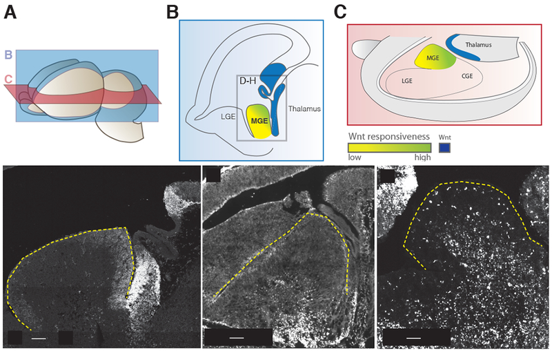
Figure 1. Expression of Wnt signaling factors present near the medial ganglionic eminence at embryonic day 13.5.

(A) Planes of section for (B) and (C) in relation to a whole e13.5 brain. (B) Parasagittal schematic of e13.5 caudal Wnt sources and Wnt responsiveness across the MGE. Box denotes cropped region for in situ hybridization images in D-H. (C) Schematic diagram in the transverse plane showing the same spatial relationship of caudal Wnt sources and Wnt responsiveness across the MGE. (D) In situ hybridization of Wnt7b, (E) TCF7L2 immuno staining for TCF7L2 protein, an early read-out for canonical Wnt signaling. TCF7L2 positive cells in the mantle are enriched in the caudal portion of the MGE. (F) Horizontal section of MGE in Tcf/Lef:H2B-dGFP reporter animals at 13.5, eGFP expression is enriched in the caudal MGE. The outline of the MGE is denoted by the yellow dashed line.
