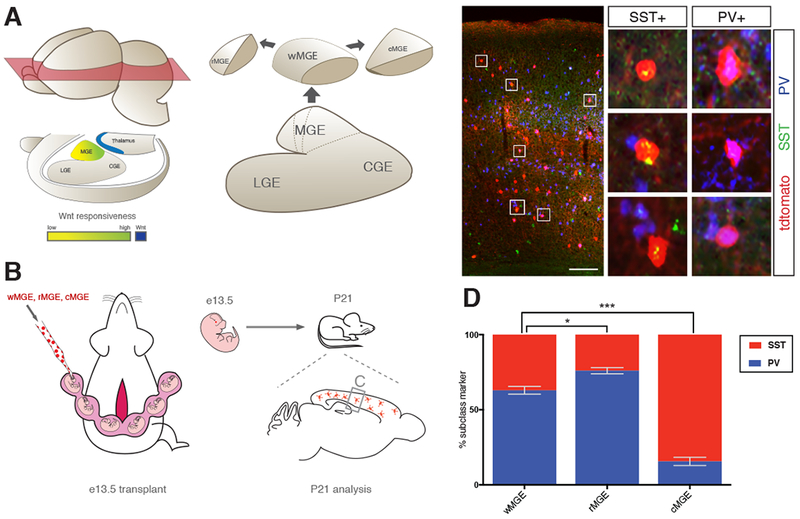
Figure 2. Ultrasound-guided in utero transplantation to determine the cortical interneuron output of MGE subdomains.

(A) Schematic diagram of spatial relationship between Wnt source (blue) and Wnt responsiveness within the MGE (green-to-yellow) suggests the presence of MGE subdomains along the rostral caudal axis. Whole (wMGE), rostral (rMGE) and caudal (cMGE) were dissected for transplantation studies. (B) Schematic of transplantation experiment. Pan-RFP expressing w-, r- and cMGE were transplanted into e13.5 mouse MGE in separate experiments by ultrasound backscatter microscopy. Host mice were sacrificed 27 days post transplant (postnatal day 21, P21) and donor cortical interneurons were analyzed for SST+ and PV+ expression. (C) Representative coronal section of P21 host forebrain showing RFP+ donor cells engrafted in cortex (scale bar 200um). Middle panel is higher magnification of box on left, boxes 1-6 show high power images of transplanted cells positive for RFP and either SST or PV. (D) P21 analysis of w-, r-, and cMGE transplantation studies for SST+ and PV+ expression in transplanted interneurons. Error bars standard error of the mean (* denotes p<0.05; ** denotes p<0.01; *** denotes p<0.001).
