Abstract
15N labeled amino acids are routinely used to label proteins or nucleic acids for study by NMR. However, NMR studies of 15N labeled amino acids in metabolite studies have not been pursued extensively, presumably due to line broadening present under standard experimental conditions. In this work, we show that lowering the temperature to −5 °C allows facile characterization of 15N-labeled amino acids. Further, we show that this technique can be exploited to measure 15NH3 produced in an enzyme catalyzed reaction and the transport and metabolism of individual amino acids in mammalian cell culture. With respect to 13C-labeled amino acids, 15N-labeled amino acids are less costly and enable direct characterization of nitrogen metabolism in complex biological systems by NMR. In summary, the present work significantly expands the metabolite pools and their reactions for study by NMR.
Subject terms: Biological techniques, NMR spectroscopy
Introduction
The use of 15N-labeled amino acids for the study of biomolecular structures, interactions and dynamics is very well established1. In these studies, 15N-labeled proteins, RNA or DNA are routinely labeled by preparation in Escherichia coli grown in media containing 15NH4+ 2,3. In contrast, 15N-labeled amino acids have not been used extensively for NMR-based metabolite studies. Presumably this stems from the extensive line-broadening of HN signals due to water exchange in aqueous samples at room temperatures (e.g. the exchange rates of NH3 groups with water are >~100/s4), which render the HN-15N group undetectable. On the other hand, 13C-labeled amino acids have been used extensively for NMR-based metabolite studies. For example, 13C-labeled glutamine is routinely used to characterize TCA cycle intermediates5. Recently, Beecher and Larive6 demonstrated that the HN-15N signal of glucosamine is observable at temperatures <0 °C with the addition of organic solvent to obviate freezing. In another study, Millard et al.7 observed the HN-15N signals of hydrolyzed amino acids prepared from 13C/15N-labeled algal and E. coli extracts. In the present work, we present NMR-based studies of commercially available 15N-labeled amino acids for biochemical analyses and characterization of amino acid transport and nitrogen metabolism in mammalian cells.
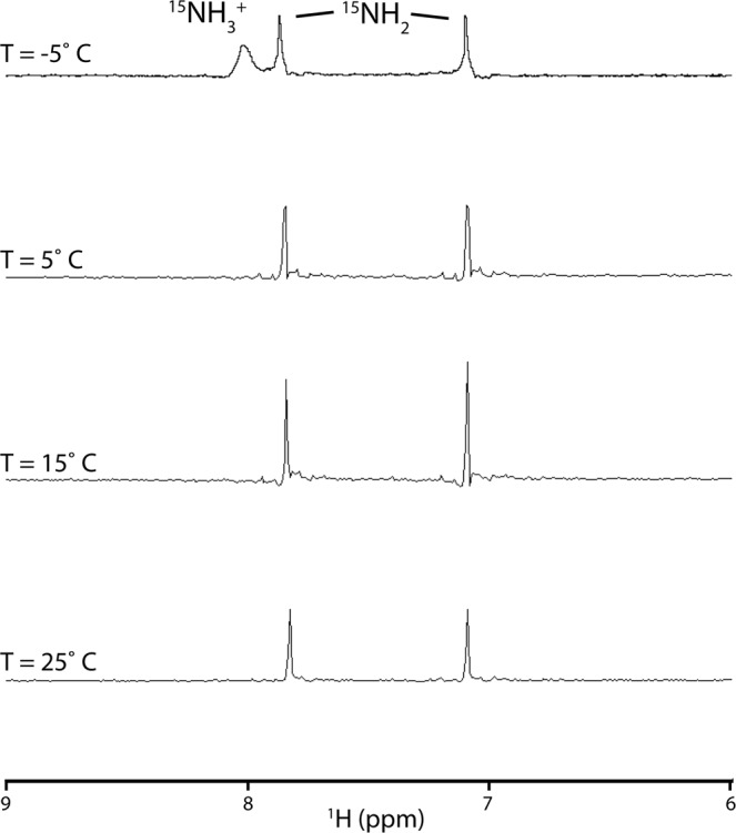
As a first step toward analyzing the potential use of 15N-labeled amino acids in NMR-based studies of metabolites, we used 15N-labeled Asn, which contains NH3+ and NH2 protons that are in fast and slow exchange, respectively. For these experiments we chose acidic conditions, for which HN exchange rate is minimized, and 20% acetone co-solvent, which enables studies below the freezing point of H2O. As shown in Fig. 1, only the NH2 groups are readily observed in the 15N-filtered NMR spectra at temperatures ranging from 5–25 °C. In contrast, the NH3+ group is observable at −5 °C, consistent with previous observations of the exchangeable NH3+ group in glucosamine under similar experimental conditions6. Consequently, subsequent studies of exchangeable HN groups were performed under conditions of low temperature and low pH.
Figure 1.

15N-filtered 1D spectrum of 15N-Asn as a function of temperature. Experimental conditions were 10 mM 15N-Asn in 100 mM 2H-acetate/pH 4.0, 10% 2H2O, 20% 2H-acetone.
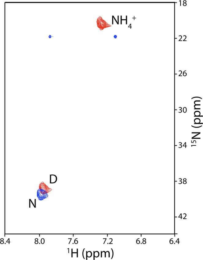
In the next step, we assayed the ability to detect amino acid metabolism in the media from cultured cells. L-Asparaginase (ASNase) is a FDA-approved therapy for the treatment of acute lymphoblastic leukemia8. Moreover, preclinical and clinical data have shown that ASNase is a promising therapy for other hematological cancers and select solid tumors9–11. ASNase treatment causes depletion of Asn in the blood by conversion to Asp plus NH3. Accordingly, we assayed the metabolism of 15N-Asn in cell cultures of LOUCY cells, a model leukemia cell line12, as a function of ASNase treatment. As shown by the blue correlations in Fig. 2, the 15NH3+ group of Asn is readily observed in media after 60 min in the absence of ASNase (the NH2 groups, which are normally observed at ~115 ppm are relatively weak and appear at an incorrect 15N chemical shift in this spectrum due to spectral aliasing). As shown by the red correlations in Fig. 2, treatment with ASNase for 60 min results in quantitative transformation of 15N-Asn to 15N-Asp and 15NH4+ and thus we are measuring the activity of the enzyme in the media (NH3, the product of ASNase catalysis of Asn, is in equilibrium with NH4+, which is the predominant species at acidic pH).
Figure 2.

15N-edited HSQC of 15N-Asn in the presence (red) and absence (blue) of ASNase. Amino acid labels correspond to the one letter code. Buffer conditions were 100 mM 2H-acetate/pH 4.0, 10% 2H2O, 20% 2H-acetone.
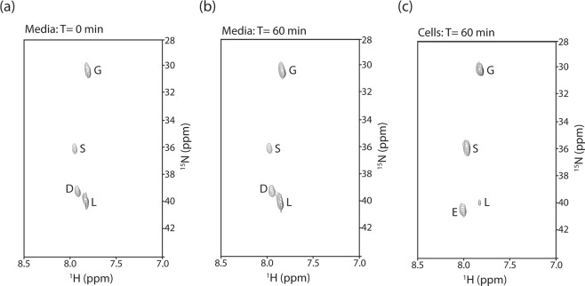
Lastly, we assayed the ability to characterize amino acid transport in cell culture by 15N NMR. For these experiments a mixture of 4 15N-labeled amino acids (Asp, Gly, Leu and Ser) was added to the media and the presence of 15N-labeled groups was assessed in the media and LOUCY cell extracts. Consequently, the media samples assay the amino acid stability and the cell extract samples assay the amino acid transport and stability. As shown in Fig. 3a, the 15NH3+ of the 4 amino acids are readily observed in the media at time = 0. As shown in Fig. 3b, the 4 amino acids are still observed after 60 min and there are no new correlations observed suggesting that they are not metabolized in the media. Interestingly, as shown in Fig. 3c, the 15N-metabolite profile in the cell extracts taken at time = 60 min was very different from that of the media. First note Gly and Ser are readily observed suggesting that they are being transported into the cells. In contrast Asp and Leu are observed at much lower concentrations, suggesting that they are transported at relatively low efficiency and/or being metabolized into other species. Surprisingly, the cell extracts contain a new 15N correlation corresponding to Glu, which is clearly absent from the media, suggesting a intracellular transaminase reaction. We note that aspartate transaminase, which catalyzes the interconversion of Asp + a-ketoglutarate ⇔ oxaloacetate + Glu and is present in a wide variety of tissues13, is one potential explanation. Alternatively, the branched amino acid transaminase, which catalyzes the interconversion of Leu + 2-oxoglutarate ⇔ 4-methyl-2-oxopentanoate + Glu and is upregulated in numerous cancers14,15, is an alternative explanation.
Figure 3.
Metabolite profiles of 15N-labeled Asp, Gly, Leu and Ser added to LOUCY cells, as assayed by 15N-edited HSQC of the media at time = 0 (a), the media at time = 60 min (b) and the cell extract at time = 60 min (c). Amino acid labels correspond to the one letter code. In all experiments, the buffer conditions were 100 mM 2H-acetate/pH 4.0, 10% 2H2O, 20% 2H-acetone.
In summary, we have demonstrated the ability to observe 15N-labeled amino acids and 15NH4+, a surrogate for NH3, in cell culture by NMR. Based of the results presented herein (Figs 2, 3 and the chemical shifts summarized in Table 1), at least 6 amino acids and NH4+ are sufficiently resolved in the HSQC spectra to be used in future NMR-based metabolite experiments. We note that signal intensity and linewidth may be expected to be further improved through the use of the HISQC sequence, which removes NyHz relaxation during nitrogen evolution, and the use of an external D2O standard for the NMR lock signal, which removes cross-peak asymmetry4. Nonetheless, we have exploited the HSQC technique at low pH and low temperature to characterize nitrogen metabolism and amino acid transport. Importantly, the observation of a novel transaminase activity in leukemia cells may suggest a potential metabolic vulnerability to be exploited. Previously 13C-labeled amino acids have been extensively used to study amino acid metabolism5; however, 15N-labeled amino acids are ~4X less expensive and enable direct studies of nitrogen metabolism. With respect to Mass Spectrometry (MS) studies, NMR offers a number of advantages including easy quantitation, the ability to re-use samples for other analyses, the ability to identify novel metabolites using other NMR experiments (e.g. COSY, ROESY), and the facile interpretation of data (e.g. a typical LC-MS study of cell extracts yields >10,000 signals versus the few signals observed in the 15N-edited HSQC spectrum). Nonetheless, MS is clearly much more sensitive and thus more desirable in sample limited situations (e.g. patient biopsy or the study of metabolites at very low concentrations). Finally, we note that the NMR-based technique could be easily extended to in vivo studies of amino acids and nitrogen metabolism. For example, non-toxic 15N-labeled compounds, which could include amino acids, nucleotides or ammonium, are readily introduced into animals either in the diet or by IV delivery, as previously shown by MS studies16.
Table 1.
Summary of chemical shifts.
| Compound | 1HN | 15N |
|---|---|---|
| Asn | 7.98 | 39.81 |
| Asp | 7.94 | 39.09 |
| Glu | 8.00 | 40.65 |
| Gly | 7.81 | 30.33 |
| Leu | 7.82 | 40.02 |
| Ser | 7.95 | 35.96 |
| NH4+ | 7.25 | 20.46 |
Experimental conditions were 20% d-acetone, 100 mM d-acetate/pH 4.0 and 10% 2H2O at −5 °C.
Methods
Isotope-labeled compounds
15N-labeled amino acids were purchased from CIL; 2H-acetate and 2H-acetone were purchased from Sigma.
Preparation of media and cells
For the analysis of media and cell extracts, the T-cell acute lymphoblastic leukemia suspension cell line LOUCY12 was used. Cells were harvested and re-suspended to 1*106 cells/mL of fresh RPMI 1640 media supplemented with 10% FBS. Cells were allowed to acclimate to new media for 1 hour in a humidified 37° incubator. In the enzyme treated flasks, 1 IU/mL of Era-TM ASNase12 was added. After 1 hour in the incubator, either 15N-Asn or a mixture of 15N- Asp, Gly, Ser and Leu was added to a final concentration of 500 µM of each. For media samples, 800 µL of media was spun for 5 minutes at 1k RCF at 4 °C. 500 µL of supernatant was transferred to a clean 1.5 mL Eppendorf tubes and stored at −20 °C. 1 hour after adding the 15N-amino acids mixture, each flask was harvested, chilled on ice and centrifuged for 5 minutes at 1k RCF. The cells were re-suspended in 1 mL of cold DPBS, centrifuged for 5 minutes at 1k RCF. The supernatant was discarded and the cell pellet and conditioned media were stored at −20 °C.
Preparation of NMR samples
Samples of media +/− ASNase were prepared directly be diluting media into buffer with final concentrations: 25% media, 20% d-acetone, 100 mM d-acetate/pH 4.0 and 10% 2H2O. Media with individual 15N amino acids was prepared by adding 1 ml of 100% ice cold methanol to 0.2 ml media, incubating on ice for 30 min, centrifugation (15 min at 10k), 4 hours of lyophilization, and suspension in buffer (20% d-acetone, 100 mM d-acetate/pH 4.0 and 10% 2H2O). Cell extracts were prepared by resuspension of cells in 80% ice cold methanol, incubating on ice for 30 min, centrifugation (15 min at 10 k), 4 hours of lyophilization, and suspension in buffer (20% d-acetone, 100 mM d-acetate/pH 4.0 and 10% 2H2O).
NMR experiments
NMR experiments were performed on a Bruker AVANCE 800 MHz spectrometer with a liquid nitrogen cooling unit to enable studies below 5 °C and using a TXI room temperature probe. For detection of HN-15N correlations, a standard HSQC sequence with water suppression by flip back pulses was employed on samples in 3 mm NMR tubes with a final volume of 200 µL. Total experimental times ranged from 0.5 to 2 hours. Chemical shifts were calibrated using DSS and indirect referencing of 15N17. The effects of low temperature and acetone on the chemical shifts were observed to be minimal (<0.04 ppm). Assignments for the 1HN-15N correlations were obtained from the analysis of individual reference compounds (15N-Asn, 15N-Asp, 15N-Glu, 15N-Gly, 15N-Ser, 15N-Leu and 15NH4Cl).
Author Contributions
B.R., A.L. and M.C. designed the research. M.D. prepared the enzyme and cells. B.R. and M.C. performed the NMR experiments. B.R. and M.C. wrote the manuscript and B.R., M.D., A.L. and M.C. discussed and improved the manuscript.
Competing Interests
The authors declare no competing interests.
Footnotes
Publisher’s note: Springer Nature remains neutral with regard to jurisdictional claims in published maps and institutional affiliations.
References
- 1.Foster MP, McElroy CA, Amero CD. Solution NMR of large molecules and assemblies. Biochemistry. 2007;46:331–340. doi: 10.1021/bi0621314. [DOI] [PMC free article] [PubMed] [Google Scholar]
- 2.Ohki S, Kainosho M. Stable isotope labeling methods for protein NMR spectroscopy. Prog. Nucl. Magn. Reson. Spectros. 2008;53:208–226. doi: 10.1016/j.pnmrs.2008.01.003. [DOI] [Google Scholar]
- 3.Nelissen FHT, Tessari M, Wijmenga SS, Heus HA. Stable isotope labeling methods for DNA. Prog. Nucl. Magn. Reson. Spectrosc. 2016;96:89–108. doi: 10.1016/j.pnmrs.2016.06.001. [DOI] [PubMed] [Google Scholar]
- 4.Iwahara J, Jung YS, Clore GM. Heteronuclear NMR spectroscopy for lysine NH(3) groups in proteins: unique effect of water exchange on (15)N transverse relaxation. J. Am. Chem. Soc. 2007;129:2971–2980. doi: 10.1021/ja0683436. [DOI] [PubMed] [Google Scholar]
- 5.Bruntz RC, Lane AN, Higashi RM, Fan TW. Exploring cancer metabolism using stable isotope-resolved metabolomics (SIRM) J. Biol. Chem. 2017;292:11601–11609. doi: 10.1074/jbc.R117.776054. [DOI] [PMC free article] [PubMed] [Google Scholar]
- 6.Beecher CN, Larive CK. (1)H and (15)N NMR Characterization of the Amine Groups of Heparan Sulfate Related Glucosamine Monosaccharides in Aqueous Solution. Anal. Chem. 2015;87:6842–6848. doi: 10.1021/acs.analchem.5b01181. [DOI] [PubMed] [Google Scholar]
- 7.Millard P, Cahoreau E, Heuillet M, Portais JC, Lippens G. 15)N-NMR-Based Approach for Amino Acids-Based (13)C-Metabolic Flux Analysis of Metabolism. Anal. Chem. 2017;289:2101–2106. doi: 10.1021/acs.analchem.6b04767. [DOI] [PubMed] [Google Scholar]
- 8.Curran E, Stock W. How I treat acute lymphoblastic leukemia in older adolescents and young adults. Blood. 2015;125:3702–3710. doi: 10.1182/blood-2014-11-551481. [DOI] [PMC free article] [PubMed] [Google Scholar]
- 9.Takahashi H, et al. Acute myeloid leukemia with mediastinal myeloid sarcoma refractory to acute myeloid leukemia therapy but responsive to L-asparaginase. Int. J. Hematol. 2012;96:136–140. doi: 10.1007/s12185-012-1111-0. [DOI] [PubMed] [Google Scholar]
- 10.Agrawal V, Woo JH, Borthakur G, Kantarjian H, Frankel AE. Red blood cell-encapsulated L-asparaginase: potential therapy of patients with asparagine synthetase deficient acute myeloid leukemia. Protein Pept. Lett. 2013;20:392–402. [PubMed] [Google Scholar]
- 11.Buaboonnam J, et al. Sequential administration of methotrexate and asparaginase in relapsed or refractory pediatric acute myeloid leukemia. Pediatr. Blood Cancer. 2013;60:1161–1164. doi: 10.1002/pbc.24470. [DOI] [PMC free article] [PubMed] [Google Scholar]
- 12.Nguyen HA, et al. A novel L-asparaginase with low L-glutaminase coactivity is highly efficacious against both T and B cell acute lymphoblastic leukemias in vivo. Cancer Res. 2018;78:1549–1560. doi: 10.1158/0008-5472.CAN-17-2106. [DOI] [PMC free article] [PubMed] [Google Scholar]
- 13.Hirotsu K, Goto M, Okamoto A, Miyahara I. Dual substrate recognition of aminotransferases. Chem. Rec. 2005;5:160–172. doi: 10.1002/tcr.20042. [DOI] [PubMed] [Google Scholar]
- 14.Hattori A, et al. Cancer progression by reprogrammed BCAA metabolism in myeloid leukaemia. Nature. 2017;545:500–504. doi: 10.1038/nature22314. [DOI] [PMC free article] [PubMed] [Google Scholar]
- 15.Raffel S, et al. BCAT1 restricts αKG levels in AML stem cells leading to IDHmut-like DNA hypermethylation. Nature. 2017;551:384–388. doi: 10.1038/nature24294. [DOI] [PubMed] [Google Scholar]
- 16.Patterson BW, Carraro F, Wolfe RR. Measurement of 15N enrichment in multiple amino acids and urea in a single analysis by gas chromatography/mass spectrometry. Biol. Mass Spectrom. 1993;22:518–523. doi: 10.1002/bms.1200220905. [DOI] [PubMed] [Google Scholar]
- 17.Wishart DS, et al. 1H, 13C and 15N chemical shift referencing in biomolecular NMR. J. Biomol. NMR. 1995;6:135–140. doi: 10.1007/BF00211777. [DOI] [PubMed] [Google Scholar]


