Abstract
Long-term potentiation (LTP) and long-term depression (LTD) of synaptic strength are ubiquitous mechanisms of synaptic plasticity, but their functional relevance in humans remains obscure. Here we report that a long-term increase in perceived pain to electrical test stimuli was induced by high-frequency electrical stimulation (HFS) (5 × 1 sec at 100 Hz) of peptidergic cutaneous afferents (27% above baseline, undiminished for >3 hr). In contrast, a long-term decrease in perceived pain (27% below baseline, undiminished for 1 hr) was induced by low-frequency stimulation (LFS) (17 min at 1 Hz). Pain testing with punctate mechanical probes (200 μm diameter) in skin adjacent to the HFS–LFS conditioning skin site revealed a marked twofold to threefold increase in pain sensitivity (secondary hyperalgesia, undiminished for >3 hr) after HFS but also a moderate secondary hyperalgesia (30% above baseline) after strong LFS. Additionally, HFS but not LFS caused pain to light tactile stimuli in adjacent skin (allodynia). In summary, HFS and LFS stimulus protocols that induce LTP or LTD in spinal nociceptive pathways in animal experiments led to similar LTP- and LTD-like changes in human pain perception (long-term hyperalgesia or hypoalgesia) mediated by the conditioned pathway. Additionally, secondary hyperalgesia and allodynia in adjacent skin induced by the HFS protocol and, to a minor extent, also by the LFS protocol, suggested that these perceptual changes encompassed an LTP-like heterosynaptic facilitation of adjacent nociceptive pathways by a hitherto unknown mechanism.
Keywords: pain, hyperalgesia, central sensitization, spinal cord, neuropathic pain, pain memory
Introduction
Since its discovery, use-dependent long-lasting modification of synaptic strength [long-term potentiation (LTP) and long-term depression (LTD)] is an intensively studied cellular model of learning and memory formation (Artola and Singer, 1993; Bliss and Collingridge, 1993). Much progress has been made in understanding the signal transduction pathways involved in LTP and LTD, ranging from serotonergic modulation of invertebrate reflex arcs to the plasticity of glutamatergic transmission in the brains of higher mammals (Bailey et al., 2000; Kemp and Bashir, 2001). Nevertheless, the role, if any, of LTP and LTD for perception, cognition, and behavior in humans remains obscure (Malinow, 1994), primarily because of a lack of adequate input stimuli for induction and of output parameters for measuring hippocampal or neocortical LTP or LTD in human subjects.
In this study, we took advantage of the availability of defined input stimuli and output measures of the nociceptive system in humans to demonstrate the existence of LTP- and LTD-like phenomena in pain perception. In contrast to the hippocampus or neocortex, input to the spinal cord consists of specialized classes of primary afferents that can be excited selectively by noninvasive stimuli in awake humans (Schmidt et al., 1995; Magerl et al., 2001), and pain ratings can serve as reliable output measures (Mayer et al., 1975; Willer, 1977). Both synaptic LTP and LTD have been identified in the dorsal horn of the spinal cord of anesthetized animals and in slice preparations of the spinal cord (Randic et al., 1993; Liu and Sandkühler, 1997; Sandkühler et al., 1997; Svendsen et al., 1998; Miletic and Miletic, 2000; Ikeda et al., 2003) and are believed to play major roles in use-dependent changes in nociception. Responses of spinal dorsal horn neurons to nociceptive input are enhanced after a strong painful stimulus (Simone et al., 1991; Dougherty et al., 1998). This central sensitization leads to an increase in pain sensitivity in a large area around the conditioned skin site (secondary hyperalgesia). These perceptual changes have also been termed “neurogenic hyperalgesia,” because they are brought about not by tissue damage itself but by the ensuing nociceptive input to the spinal cord (LaMotte et al., 1991; Simone et al., 1991). In neuropathic pain, a similar neurogenic hyperalgesia is caused by a lesion or dysfunction of the PNS or CNS (Treede et al., 1992; Fields et al., 1998; Woolf and Salter, 2000; Baumgärtner et al., 2002). It has been suggested that LTP in nociceptive pathways may underlie neurogenic hyperalgesia and chronic pain (Randic et al., 1993; Miletic and Miletic, 2000; Sandkühler, 2000a; Woolf and Salter, 2000). In turn, LTD in nociceptive pathways may play a role in afferent-induced forms of analgesia [e.g., after low-frequency, high-intensity transcutaneous electrical nerve stimulation (TENS) (acupuncture-like TENS) (Sandkühler, 2000b)].
The aim of this study was to assess long-lasting modulation of human pain perception after electrical stimulation protocols that induce LTP or LTD in spinal nociceptive pathways in animals. These stimuli were applied to peptidergic afferents in the skin via a specially designed surface electrode. Some of the data were presented at the 10th World Congress on Pain (Klein et al., 2002).
Materials and Methods
Subjects. After obtaining approval from the ethics committee, experiments were performed on eight healthy subjects (one female and seven males ranging in age from 22 to 55 years; mean age, 29.6 years), who participated in four sessions varying in electrical conditioning stimulation pattern. Each subject was familiarized with the experimental procedures beforehand, and each gave written, informed consent.
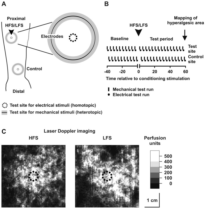
Conditioning stimuli. Conventional transcutaneous nerve stimulation with large surface electrodes recruits Aβ fibers at much lower current strengths than nociceptive Aδ and C fibers. Thus it is difficult to obtain sufficient nociceptive input by this electrode configuration. In the present study, we exploited the fact that the electrical threshold for nociceptive afferents decreases dramatically within their receptive field (Meyer et al., 1991; Schmidt et al., 1995) by applying all electrical stimuli through punctate electrodes (diameter, 200 μm) (see Fig. 1 A). Because of the small diameter of the electrodes, a high current density was achieved already at low stimulus intensities, which favors activation of superficial nociceptive Aδ fiber and C fiber afferents (Nilsson and Schouenborg, 1999; Inui et al., 2002). To achieve spatial summation within the receptive field of spinal cord neurons, 10 of these electrodes were mounted in a small circular plastic frame (attached to the skin by double-adhesive tape) and were stimulated simultaneously.
Figure 1.
Experimental procedure for testing the roles of LTP and LTD in human pain perception. A, Conditioning electrical HFS or LFS was applied to the proximal volar forearm via a circular array of electrodes. Homotopic effects of HFS and LFS on the conditioned pathway were tested by pain ratings to single pulses through the conditioning electrode. Heterotopic effects outside of the conditioned pathway were tested by pain ratings to mechanical stimuli at a distance of 15 mm from the electrode arrays. B, Testing of mechanical sensitivity consisted of stimulus–response functions for pricking pain with punctate probes and soft stroking. Testing was arranged in runs comprising all 10 mechanical stimuli in a randomized order (small black bars) followed by three electrical pulses at 10× the detection threshold (•) with 5–10 sec interstimulus intervals. Runs were alternated between the conditioned skin site and the control site for 40 min before and 60 min after HFS or LFS. At the end of the experiment, the area of hyperalgesia to mechanical stimuli was mapped using a 200 mN von Frey hair. C, Laser Doppler imaging revealed large areas of increased skin perfusion (flare) at sites of HFS and LFS (20 × T).
Cathodal electrical stimuli were applied on the forearm 5 cm distal to the cubital fossa via a constant current stimulator (DS7H; Digitimer, Welwyn Garden City, UK), and a large surface electrode on the ipsilateral upper arm served as the anode. Stimulus intensity was adjusted at 10 × and 20 × individual detection threshold (T) determined by applying single electrical pulses.
Two patterns of conditioning pulse trains were used: (1) high-frequency stimulation (HFS) trains of 100 Hz for 1 sec (pulse width, 2 msec), repeated five times at 10 sec intervals to induce LTP, and (2) low-frequency stimulation (LFS) at 1 Hz with stimulus trains of 1000 pulses to induce LTD. These protocols had been shown previously to induce LTP or LTD at primary afferent synapses with neurons of lamina II of the rat spinal cord (Randic et al., 1993; Liu and Sandkühler, 1997; Sandkühler et al., 1997; Svendsen et al., 1998). HFS corresponds to the burst mode of nociceptive afferents, which even in C fibers may reach discharge rates of up to 200 Hz for short periods of time (Weidner et al., 2002). LFS corresponds to the low-frequency discharge that nociceptive afferents can maintain for longer periods of time. Another circular array of punctate electrodes was placed ∼5 cm proximal to the wrist and served as the unconditioned control site.
Test stimuli. Pain perception was tested by two different approaches: (1) single electrical test pulses (at 10 × T) for testing modulation of human pain perception at the site of conditioning stimulation (homotopic hyperalgesia or hypoalgesia) and (2) two sets of mechanical test stimuli (punctate probes, light touch) for testing modulation of human pain perception adjacent to the site of conditioning stimulation (heterotopic hyperalgesia or hypoalgesia).
One set of mechanical stimuli was designed to activate nociceptive afferents (Greenspan and McGillis, 1994); these stimuli were shown previously to be suitable to detect secondary hyperalgesia surrounding an injury site and hypoalgesia in lesions of the nervous system (Ziegler et al., 1999; Magerl et al., 2001; Baumgärtner et al., 2002). They consisted of seven punctate probes (forces of 8, 16, 32, 64, 128, 256, and 512 mN) that were cylindrical stainless-steel wires (200 μm tip diameter) mounted on plastic rods that moved freely within a wider handheld tube.
Pain to light touch (allodynia) was tested with a second set of mechanical stimuli consisting of a short stroke (1 cm) of a soft cotton wisp (CW) (3 mN), a soft make-up brush (BR) (200–400 mN), and a Q-tip (QT) fixed alongside a flexible piece of cardboard, which allowed delivery of forces of ∼100 mN when just noticeably bent (Magerl et al., 2001). These three stimuli only activate low-threshold mechanoreceptors (Leem et al., 1993) and are not painful in normal skin. The mechanical stimuli were applied in a balanced order within a circular area 15 mm from the electrode array (Fig. 1 A).
Pain ratings. Subjects rated the magnitude of pain to mechanical and electrical test stimuli as well as to conditioning stimulation on a numerical rating scale ranging from 0 (nonpainful) to 100 (most intense pain imaginable). They were instructed to distinguish pain from the perception of touch or pressure by the presence of a sharp or slightly pricking or burning sensation.
Experimental protocol. Mechanical and electrical test stimuli were applied in runs alternating between conditioned and control skin sites during a time period of 40 min before (baseline) and 60 min after (test period) conditioning electrical stimulation (Fig. 1 B). In three additional subjects, the testing was further extended to 200 min. The order of the four conditioning stimulation patterns (HFS and LFS stimulation at 10 × Tor20 × T) was balanced across subjects. Mechanical and electrical testing were continued immediately after conditioning stimulation. The area of hyperalgesia was mapped at the end of the experiment using a calibrated von Frey hair (diameter, 0.45 mm; force, 200 mN).
Laser Doppler imaging. To check successful excitation of peptidergic afferents, we measured the increase in cutaneous blood flow after high-frequency and low-frequency electrical stimulation (20 × T) in an area surrounding the electrodes (Fig. 1C) using a laser Doppler perfusion imager (MoorLDI; 633 nm; Moor Instruments, Axminster, UK).
Data evaluation and statistics. Ratings were transformed into decadic logarithmic values to obtain a lognormal distribution. To avoid a loss of zero values, a small constant (0.1) was added to all of the ratings (Magerl et al., 1998). Except for touch-evoked pain, ratings were normalized to baseline by dividing postconditioning pain ratings by the mean value of the 40 min baseline period. Ratings to touch-evoked pain did not allow a normalization to baseline, because baseline ratings were zero by definition. Postconditioning rating data were included in a two-way ANOVA (test site × conditioning stimulus intensity) to determine differences between conditioned and control skin sites and of conditioning stimulus intensities. Post hoc paired t tests were performed to compare values at each observed time point.
Additionally, the shift of the stimulus–response function for both sets of mechanical test stimuli was analyzed by another two-way ANOVA (test stimulus intensity × preconditioning vs postconditioning). Here, post hoc paired t tests were performed to compare values at each mechanical test stimulus intensity. p values of <0.05 were considered significant.
Results
To study the perceptual correlates of LTP and LTD in the spinal cord, conditioning electrical pulse protocols were delivered to an array of punctate electrodes (Fig. 1A) attached to the skin of human subjects. Even single pulses at detection threshold intensity (T = 0.11 ± 0.06 mA; mean ± SD; n = 32) were already perceived as painful, indicating preferential activation of superficial nociceptors attributable to the high current density within the epidermis. Electrical stimulation did not lead to visible injuries in the skin. Compared with the threshold for conventional nerve stimulation with large surface electrodes (1.5 ± 0.3 mA; mean ± SD; n = 21) (Treede and Kunde, 1995), these stimuli would not be detectable if delivered through the skin to a nerve trunk. This is consistent with the observation that superficial endings of nociceptive afferents (Aδ and C fibers) have much lower thresholds for electrical stimuli than their axons within the nerve trunk (Meyer et al., 1991; Schmidt et al., 1995). Despite their low intensity, the stimuli induced substantial vasodilatation around the electrode array (Fig. 1C), indicating that the conditioning stimuli were sufficient to activate peptidergic afferents, most likely C fibers. Activation of this class of nociceptive afferents is a prerequisite for the induction of LTP and other forms of central sensitization in the spinal cord (Dougherty and Willis, 1991; Thompson et al., 1994; Liu and Sandkühler, 1997).
Perceptual correlates of the conditioning process
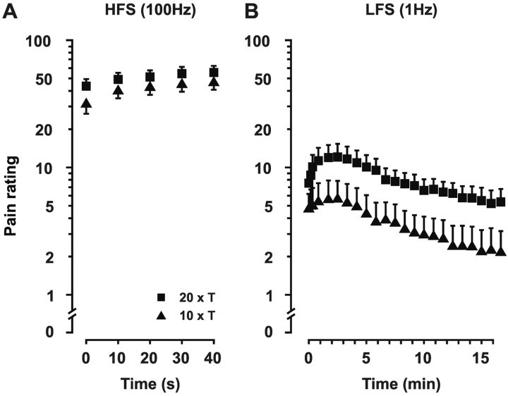
A single train of HFS evoked mild to moderate pain (mean pain rating, 32 of 100 at 10 × T and 44 of 100 at 20 × T), and repetition of such trains (five times at 10 sec intervals) resulted in a gradual increase in the perceived pain (Fig. 2A). Pain ratings for LFS were much lower than for HFS, because LFS consisted of single electrical pulses (first pulse: mean rating, 5 of 100 at 10 × T and 8 of 100 at 20 × T) (Fig. 2B). Pain to LFS initially increased over a period of ∼2.5 min (150 pulses) but then gradually declined until perceived pain was less than the initial pain ratings.
Figure 2.
Pain sensations evoked by conditioning electrical stimuli. A, HFS (5× 1 sec at 100 Hz; 10 sec intervals between bursts) elicited pain sensations that increased over the five bursts. B, During LFS (1000 pulses at 1 Hz), pain ratings increased over the first 150 pulses and then slowly declined until the end of the conditioning stimulation, when pain ratings were lower than the initial values. Mean ± SEM values across eight subjects are shown. Each filled triangle or filled square represents average pain ratings on a 100 point verbal rating scale. ▴, 10 × T; ▪, 20 × T.
Long-term changes in pain sensitivity at the site of conditioning stimulation
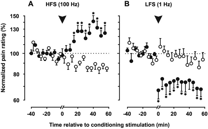
Conditioning HFS led to a significant increase in pain evoked by single electrical test stimuli of 10 × T delivered through the conditioning electrode (Fig. 3A). No differences were found between conditioning stimulus intensities of 10 × T and 20 × T (Table 1); thus data were pooled across intensities. The facilitation took ∼20 min to develop and stabilized at 27% above baseline (p < 0.01) (Table 1). The increase persisted until the end of the observation period (60 min). In two subjects, the observation period was extended to 200 min after conditioning stimulation (20 × T). Hyperalgesia to electrical stimuli stabilized at 36% above baseline after 60 min and remained undiminished until 200 min after conditioning stimulation (39% above baseline).
Figure 3.
Homotopic effects of conditioning electrical stimuli. A, After LFS, pain evoked by single electrical test pulses (intensity, 10 × T) through the HFS electrode increased to ∼30% above baseline (•). This facilitation lasted until the end of the observation period. Pain evoked by identical test pulses through a remote electrode exhibited a small decrease over time (○). B, After LFS, pain evoked by single electrical test pulses through the LFS electrode decreased to ∼30% below baseline (•). This inhibition was not reversible within the observation period. Mean ± SEM values across seven subjects are shown. Dotted lines indicate mean level of baseline period. Each circle represents normalized pain ratings averaged over a 5 min time window across stimulation intensities (10 × T; 20 × T). Asterisks indicate post hoc paired t tests; conditioned versus control site; p < 0.05.
Table 1.
ANOVA for pain ratings after conditioning stimulation
|
|
HFS |
LFS |
||||||||
|---|---|---|---|---|---|---|---|---|---|---|
|
|
df |
F
|
p
|
df |
F
|
p
|
||||
| Homotopic effects (electrical; n = 7) | ||||||||||
| Conditioned versus control | 1,6 | 25.97 | p < 0.01 | 1,6 | 25.86 | p < 0.01 | ||||
| Conditioning stimulus intensity | 1,6 | 0.19 | p = 0.68 | 1,6 | 2.15 | p = 0.19 | ||||
| Interaction | 1,6 | 0.28 | p = 0.62 | 1,6 | 1.67 | p = 0.24 | ||||
| Heterotopic effects (pin prick; n = 8) | ||||||||||
| Conditioned versus control | 1,7 | 54.73 | p < 0.001 | 1,7 | 8.34 | p < 0.05 | ||||
| Conditioning stimulus intensity | 1,7 | 1.18 | p = 0.31 | 1,7 | 11.85 | p < 0.05 | ||||
| Interaction | 1,7 | 1.8 | p = 0.22 | 1,7 | 7.07 | p < 0.05 | ||||
| Heterotopic effects (light touch; n = 8) | ||||||||||
| Conditioned versus control | 1,7 | 8.65 | p < 0.05 | |||||||
| Conditioning stimulus intensity | 1,7 | 2.49 | p = 0.16 | |||||||
| Interaction |
1,7 |
2.49 |
p = 0.16 |
|
|
|
||||
Allodynia after LFS was observed only in one subject at 20 × T; thus the statistical analysis appeared inadequate.
Conditioning LFS led to a significant decrease in pain (hypoalgesia) evoked by electrical test stimuli that developed continuously during LFS (see above) and persisted at 27% below baseline (p < 0.01) (Table 1) (Fig. 3B). Neither HFS nor LFS changed the pain ratings for stimulation of the remote control site. These findings indicate that HFS and LFS protocols induced LTP- or LTD-like long-term changes in human pain perception mediated by the conditioned pathway (homotopic hyperalgesia and hypoalgesia).
Long-term changes in pain sensitivity adjacent to the site of conditioning stimulation
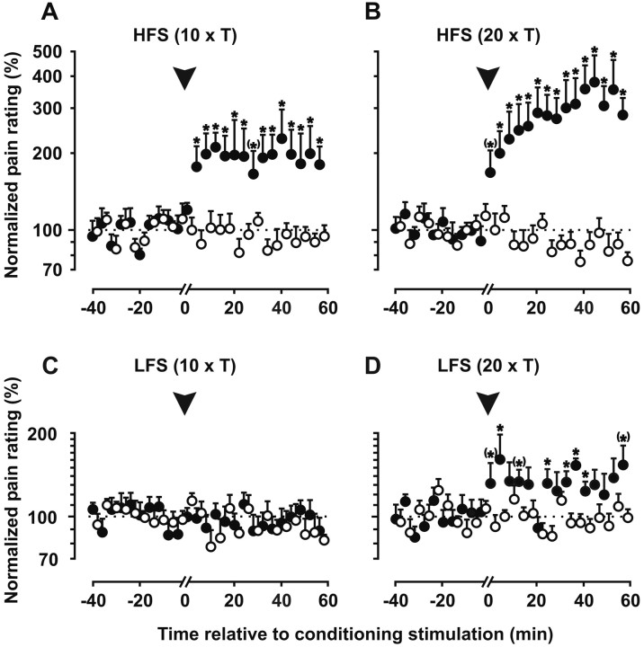
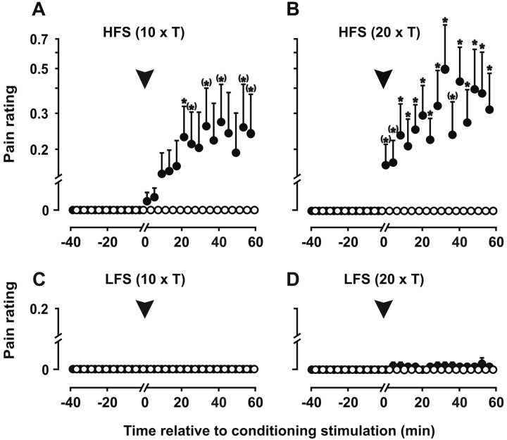
To test for perceptual correlates of heterotopic LTP and LTD outside of the conditioned pathway, mechanical test stimuli were applied around the conditioned site and around the unconditioned control site. As shown in Figure 4, A and B, HFS induced a significant enhancement of pin prick-evoked pain around the conditioning electrode (secondary hyperalgesia; p < 0.01) (Table 1), which persisted until the end of the observation period (60 min). Pain perception stabilized at a plateau phase of 90% above baseline after 10 × T and of 210% after 20 × T. One hour after conditioning stimulation, the mean area of hyperalgesia was 38 cm2 (10 × T) and 56 cm2 (20 × T). In three subjects, secondary hyperalgesia was assessed for 200 min after conditioning stimulation (20 × T) when pain perception was still enhanced to ∼220% above baseline (compared with 296% at 60 min). There was no mechanical hyperalgesia around the unconditioned control site. Because the neural pathway for these mechanical test stimuli was spatially segregated from that of the conditioning stimuli, the findings suggest a spread of facilitation to unstimulated inputs of the spinal cord (i.e., heterosynaptic facilitation). No such spread was seen for the inhibitory effect of LFS (Fig. 4C,D). In contrast, LFS caused a small but significant increase in mechanically evoked pain (p < 0.05) (Table 1). The significant effect of the stimulus intensity and significant interaction term reflect the finding that this secondary hyperalgesia occurred only at 20 × T (Fig. 4D) (to 30% above baseline) but not at 10 × T (Fig. 4C) (to 4% below baseline).
Figure 4.
Heterotopic effects of conditioning electrical stimuli on pin prick-evoked pain. A, B, Conditioning HFS induced a significant enhancement of pin prick-evoked pain adjacent to the conditioning electrode (•) but not adjacent to the control electrode (○). This secondary hyperalgesia occurred after conditioning stimulus intensities of 10 × T and 20 × T. Pain perception was significantly increased already at 5 min and remained potentiated throughout the 60 min observation period (10 × T) or even increased further (20 × T). C, Conditioning LFS at 10 × T induced no changes in pin prick-evoked pain at any test site. D, For conditioning LFS at 20 × T, a small but significant increase in pin prick-evoked pain was found near the conditioning electrode but not the control electrode. Mean ± SEM values across eight subjects are shown. Dotted lines indicate mean level of baseline period. Each circle represents the normalized average of pain ratings across all seven stimulus intensities over a 5 min time window. Asterisks indicate post hoc paired t tests; conditioned versus control site; p < 0.05.
HFS also induced touch-evoked pain (allodynia) around the HFS electrode but not around the remote unconditioned control site (Fig. 5A,B). Light tactile stimuli elicited mildly painful sensations in 9 of 16 experiments after HFS (p < 0.05) (Table 1). LFS at 10 × T did not elicit allodynia (Fig. 5C), but one subject reported touch-evoked pain after 20 × T (Fig. 5D).
Figure 5.
Heterotopic effects of conditioning electrical stimuli on touch-evoked pain. A, B, Conditioning HFS induced a state in which these tactile, normally non-noxious stimuli became painful adjacent to the conditioning electrode (•) (allodynia) but not adjacent to the control electrode (○). Allodynia gradually developed after HFS and persisted throughout the 60 min observation period. C, Conditioning LFS at 10 × T elicited no allodynia. D, For conditioning LFS at 20 × T, one subject developed allodynia near the conditioning electrode but not the control electrode. Mean ± SEM values across eight subjects are shown. Each circle represents the normalized average of pain ratings across all three stimulus intensities over a 5 min time window. Asterisks indicate post hoc paired t tests; conditioned versus control site; p < 0.05.
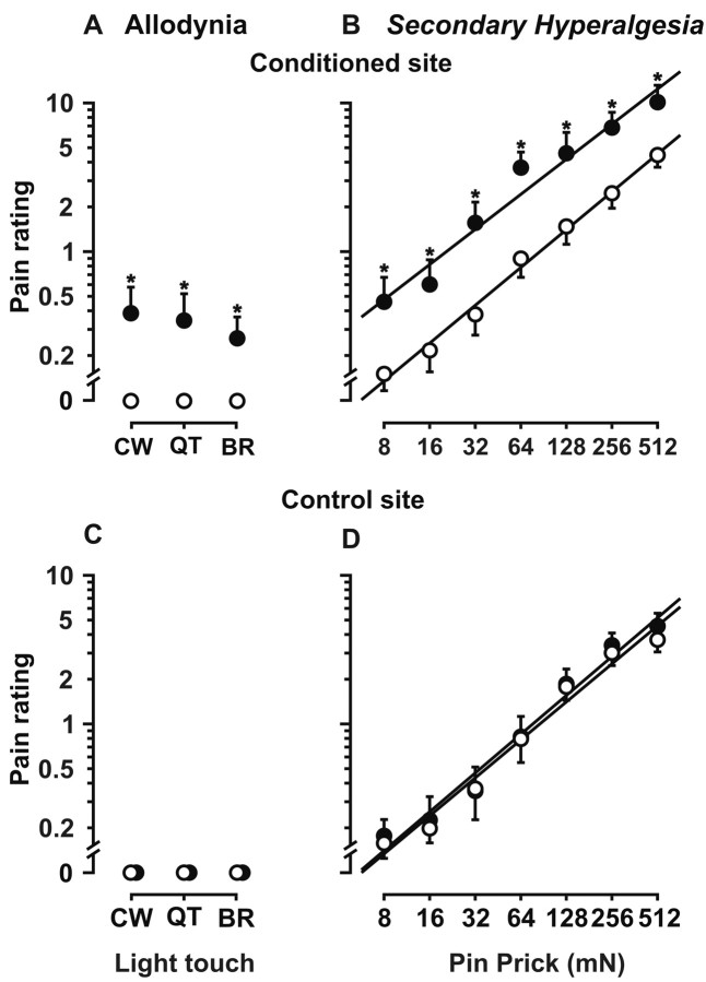
The stimulus–response functions of the two different mechanosensitive inputs are shown in detail in Figure 6. The change in perceived quality of light tactile stimuli toward a burning and painful sensation is represented by a significant upward shift (F(1,7) = 10.97; p < 0.05) (Fig. 6A) that was homogenous across all three test stimuli (F(2,14) = 1.34; NS). This effect was absent at the remote control site (Fig. 6C). Punctate probes of graded intensities induced graded pain sensations both before and after conditioning stimulation (F(6,42) = 79.1; p < 0.001) (Fig. 6B). The effect of conditioning HFS was a parallel leftward shift of the stimulus response function (F(1,7) = 33.21; p < 0.001) (Fig. 6B), indicating both a decrease in pain thresholds and an increase in pain ratings elicited by suprathreshold stimuli. This effect was confined to skin areas adjacent to the conditioning electrode, as shown by the complete absence of a shift in the stimulus–response function at the control site (Fig. 6D).
Figure 6.
Stimulus–response functions for punctate probes and light tactile stimuli after HFS at 20 × T. A, Allodynia. Light tactile stimuli were not painful when moved across normal skin before conditioning stimulation (○). Adjacent to the conditioning electrode, the same stimuli were mildly painful after HFS at 20 × T (•). CW, 3 mN; QT, 100 mN; BR, 400 mN. B, Secondary hyperalgesia. The stimulus–response function for punctate probes was approximately linear in log–log coordinates in normal skin (○). After HFS, there was a parallel leftward shift of this function adjacent to the conditioning electrode (•). C, D, There was neither allodynia nor hyperalgesia adjacent to the unstimulated control site. Mean ± SEM values across eight subjects are shown. Each circle represents average pain ratings on a 100 point verbal rating scale. Asterisks indicate post hoc paired t tests; preconditioning versus postconditioning stimulation; p < 0.05.
Discussion
This study has shown that the conditioning electrical pulse protocol that elicits LTP of synaptic efficacy in the hippocampus, neocortex, and spinal cord leads to an enhanced pain perception (hyperalgesia) in humans when applied to cutaneous nociceptive afferents. This enhanced pain perception lasted at least 3 hr when tested with the same electrical stimuli that activated the conditioning pathway. When tested adjacent to the conditioned skin site with punctate probes (secondary hyperalgesia) and light tactile stimuli (allodynia), pain perception was also found to be enhanced for at least 3 hr. The conditioning electrical pulse protocol that elicits LTD in spinal cord slices led to a diminished pain perception (hypoalgesia) for >1 hr when tested with the same electrical stimuli that activated the conditioning pathway. In contrast, this protocol led to secondary hyperalgesia in the surrounding skin. These findings suggest that LTP and LTD of the conditioned pathway in the spinal cord (homosynaptic LTP–LTD) lead to equivalent changes in human pain perception, and that LTP but not LTD may spread to unstimulated adjacent input (heterosynaptic LTP).
Perceptual correlate of homosynaptic LTP
Conditioning stimulation of one specific input pathway to the human spinal cord was sufficient to induce a long-term change in human pain perception mediated by this pathway. As demonstrated by spreading vasodilatation (Fig. 1C), the input pathway encompassed peptidergic cutaneous afferents containing substance P and calcitonin gene-related peptide (Brain and Williams, 1988), the central terminals of which preferentially synapse in lamina II (outer part) of the spinal cord dorsal horn (Hunt and Mantyh, 2001). LTP has been described after conditioning HFS of the synaptic connection between peptidergic C-fiber afferents and the nociceptive lamina I spinoparabrachial neurons (Ikeda et al., 2003) that mediate hyperalgesia in behaving animals (Khasabov et al., 2002). HFS also induced LTP in lamina II (Randic et al., 1993; Liu and Sandkühler, 1997) and in deep dorsal horn neurons (Svendsen et al., 1998).
In addition to the spinal cord, synaptic LTP in other parts of the CNS (e.g., the thalamus or neocortex) or altered descending inhibitory or facilitating control may have contributed to the observed long-term changes in pain perception. However, human models of cortical LTP involve the motor rather than the sensory cortex, and they require either simultaneous activation of two input pathways (Bütefisch et al., 2000) or direct depolarization of cortical neurons (Nitsche and Paulus, 2001). Moreover, high intensities of electrical stimulation, such as with the HFS protocol, activate the inhibitory rather than the facilitating descending pathways (Porreca et al., 2002; Suzuki et al., 2002). Therefore, spinal LTP is the most likely mechanism of the long-term enhancement of pain perception observed at the conditioned skin site.
Perceptual correlate of homosynaptic LTD
Conditioning pulse trains at 1 Hz (LFS) resulted in a long-term decrease in pain perception (hypoalgesia) mediated by the conditioned pathway throughout the observation period. This LTD-like reduction of human pain perception was similar to LTD at the synapses between nociceptive afferents and superficial dorsal horn neurons in rats, both in magnitude and in its restriction to the conditioned pathway (Chen and Sandkühler, 2000). Pain perception during conditioning LFS exhibited a biphasic time course. The initial increase in pain is attributable to the action potential “wind-up” in dorsal horn neurons. However, wind-up in the dorsal horn in animals reaches a plateau after 10–100 electrical pulses and outlasts conditioning stimulation only for seconds (Woolf, 1996; Herrero et al., 2000). The subsequent decline in pain ratings in our study is compatible with a delayed onset of LTD (Mizuno et al., 2001).
The LTD-like reduction of human pain perception observed in this study is unlikely to be attributable to descending inhibitory mechanisms such as diffuse noxious inhibitory control, which would have affected all sites outside of the conditioned site, including the unstimulated control site (Gozariu et al., 1997). However, the reduction of pain perception was confined strictly to the conditioned pathway.
Some clinical methods of stimulation-induced analgesia may at least partly be attributable to LTD in the spinal cord, such as spinal cord stimulation (North et al., 1993), cutaneous field stimulation (Nilsson et al., 2003), and the low-frequency, high-intensity mode of transcutaneous electrical nerve stimulation (acupuncture-like TENS) (Sandkühler, 2000b). In contrast, the short-lasting effect of nonpainful, high-frequency, low-intensity TENS (conventional TENS) is commonly attributed to segmental spinal inhibition induced by activation of Aβ fibers (Garrison and Foreman, 1996; Sandkühler, 2000b), as suggested in the gate control theory.
Perceptual correlates of heterosynaptic LTP
LTP in the spinal cord has been proposed as the underlying mechanism of central sensitization (Randic et al., 1993; Miletic and Miletic, 2000; Sandkühler, 2000a; Willis, 2002). Sensitization in the rat or monkey spinal cord was specific for mechanically evoked input, whereas heat-evoked input was not enhanced (Simone et al., 1991; Dougherty et al., 1998; Pertovaara, 1998). Likewise, central sensitization in humans led to an increase in pain sensitivity to mechanical but not to heat stimuli (secondary hyperalgesia and allodynia) in a large area around the injured skin site (LaMotte et al., 1991; Treede et al., 1992; Ali et al., 1996). We conclude that secondary hyperalgesia and allodynia to mechanical stimuli after the HFS protocol also represent nociceptive spinal LTP. However, they differ from homosynaptic LTP on several accounts. First, the facilitated input comes from adjacent skin sites and thus is spatially remote from the conditioning input (heterotopic). Second, both secondary hyperalgesia and allodynia involve the interaction of two input pathways: the conditioning input is carried by C fibers, whereas the facilitated input is carried by Aδ fibers and Aβ fibers, respectively (LaMotte et al., 1991; Kilo et al., 1994; Ziegler et al., 1999; Magerl et al., 2001). Third, whereas the conditioning pathway expresses the capsaicin-sensitive receptor TRPV1, both facilitated pathways do not (Magerl et al., 2001). These lines of evidence suggest that the central sensitization underlying secondary hyperalgesia is not restricted to the activated synapse (homosynaptic LTP) but also spreads to adjacent synapses (heterosynaptic LTP) (cf. Woolf and Salter, 2000). Because hyperalgesia in neuropathic pain involves facilitation of the same input pathways as secondary hyperalgesia (Treede et al., 1992; Fields et al., 1998; Baumgärtner et al., 2002), it may also be related to heterosynaptic LTP in the spinal cord.
Convergence of A- and C-fiber nociceptors on nociceptive spinal neurons could provide the anatomical basis for heterosynaptic LTP. Alternatively, mechanosensitive inputs may be facilitated at adjacent neurons by a diffusable factor that reaches extrasynaptic receptors (Liu et al., 1994). Nociceptive neuropeptides are likely candidates for this type of volume transmission, because substance P may spread over considerable distances (Duggan et al., 1995). The action of substance P on neurokinin 1 (NK1) receptors was essential both for the induction of LTP in spinal cord neurons (Liu and Sandkühler, 1997; Ikeda et al., 2003) and for the induction of secondary hyperalgesia (Laird et al., 2001). Moreover, NK1 receptor-bearing spinal cord neurons were shown to be essential for the development of hyperalgesia (Mantyh et al., 1997; Khasabov et al., 2002) and for the induction of LTP (Ikeda et al., 2003). For behaviorally relevant consequences, homosynaptic LTP at glutamatergic synapses in the rat hippocampus needs to be supplemented by heterosynaptic facilitation (Bailey et al., 2000). Thus the behaviorally relevant consequences of LTP in the spinal cord may also require heterosynaptic facilitation.
Heterosynaptic LTP appears to be induced not only by HFS but also by LFS protocols given at C-fiber intensity, as shown bythe induction of secondary hyperalgesia in our data (Fig. 4D). Secondary hyperalgesia also developed during prolonged LFS (5 Hz for 120 min) by an intracutaneous needle electrode (Koppert et al., 2001). Recent electrophysiological data indicate that some spinal neurons (those projecting to the periaqueductal gray) may develop LTP after LFS at 2 Hz (Ikeda and Sandkühler, 2003). These phenomena, however, deserve further study.
Conclusions
The present findings provide the missing link between clinical observations in humans (neurogenic hyperalgesia after tissue damage or lesions of the nervous system) and a basic neurobiological mechanism (synaptic LTP) that was proposed to turn acute pain into chronic pain. It may not be surprising that long-term memory in the human injury-detection system is found to be related to the highly conserved mechanism of learning (LTP), considering the fact that during evolution, strong injury-related selection pressures have shaped the nervous systems of all species (Walters, 1994). Research in many areas of neuroscience uses experimental paradigms that simulate aspects of actual or threatened injury to study mechanisms of learning and memory (e.g., conditioning of the gill withdrawal reflex in Aplysia) (Illich and Walters, 1997; Kandel, 2001). Our data show that neurogenic hyperalgesia, which is an aspect of injury-related behavior in humans, shares characteristics with both homosynaptic and heterosynaptic types of LTP.
Footnotes
This work was supported by the Bundesministerium für Bildung und Forschung (German Research Network Neuropathic Pain, 01EM0107), the Deutsche Forschungsgemeinschaft (Tr 236/16-1), and the Pain Research Programme of the Medical Faculty Heidelberg.
Correspondence should be addressed to Dr. Rolf-Detlef Treede, Institute of Physiology and Pathophysiology, Johannes Gutenberg University, Saarstrasse 21, D-55099 Mainz, Germany. E-mail: treede@uni-mainz.de.
DOI:10.1523/JNEUROSCI.1222-03.2004
Copyright © 2004 Society for Neuroscience 0270-6474/04/230964-08$15.00/0
T.K. and W.M. contributed equally to this work.
References
- Ali Z, Meyer RA, Campbell JN (1996) Secondary hyperalgesia to mechanical but not heat stimuli following a capsaicin injection in hairy skin. Pain 68: 401–411. [DOI] [PubMed] [Google Scholar]
- Artola A, Singer W (1993) Long-term depression of excitatory synaptic transmission and its relationship to long-term potentiation. Trends Neurosci 16: 480–487. [DOI] [PubMed] [Google Scholar]
- Bailey CH, Giustetto M, Huang YY, Hawkins RD, Kandel ER (2000) Is heterosynaptic modulation essential for stabilizing Hebbian plasticity and memory? Nat Rev Neurosci 1: 11–20. [DOI] [PubMed] [Google Scholar]
- Baumgärtner U, Magerl W, Klein T, Hopf HC, Treede R-D (2002) Neurogenic hyperalgesia versus painful hypoalgesia: two distinct mechanisms of neuropathic pain. Pain 96: 141–151. [DOI] [PubMed] [Google Scholar]
- Bliss TVP, Collingridge GL (1993) A synaptic model of memory: long-term potentiation in the hippocampus. Nature 361: 31–39. [DOI] [PubMed] [Google Scholar]
- Brain SD, Williams TJ (1988) Substance P regulates the vasodilator activity of calcitonin gene-related peptide. Nature 335: 73–75. [DOI] [PubMed] [Google Scholar]
- Bütefisch CM, Davis BC, Sawaki L, Kopylev L, Classen J, Cohen LG (2000) Mechanisms of use-dependent plasticity in the human motor cortex. Proc Natl Acad Sci USA 97: 3661–3665. [DOI] [PMC free article] [PubMed] [Google Scholar]
- Chen J, Sandkühler J (2000) Induction of homosynaptic long-term depression at spinal synapses of sensory Aδ-fibers requires activation of metabotropic glutamate receptors. Neuroscience 98: 141–148. [DOI] [PubMed] [Google Scholar]
- Dougherty PM, Willis WD (1991) Enhancement of spinothalamic neuron responses to chemical and mechanical stimuli following combined micro-iontophoretic application of N-methyl-d-aspartic acid and substance P. Pain 47: 85–93. [DOI] [PubMed] [Google Scholar]
- Dougherty PM, Willis WD, Lenz FA (1998) Transient inhibition of responses to thermal stimuli of spinal sensory tract neurons in monkeys during sensitization by intradermal capsaicin. Pain 77: 129–136. [DOI] [PubMed] [Google Scholar]
- Duggan AW, Riley RC, Mark MA, MacMillan SJ, Schaible HG (1995) Afferent volley patterns and the spinal release of immunoreactive substance P in the dorsal horn of the anaesthetized spinal cat. Neuroscience 65: 849–858. [DOI] [PubMed] [Google Scholar]
- Fields HL, Rowbotham M, Baron R (1998) Postherpetic neuralgia: irritable nociceptors and deafferentation. Neurobiol Dis 5: 209–227. [DOI] [PubMed] [Google Scholar]
- Garrison DW, Foreman RD (1996) Effects of transcutaneous nerve stimulation (TENS) on spontaneous and noxiously evoked dorsal horn cell activity in cats with transacted spinal cords. Neurosci Lett 216: 125–128. [DOI] [PubMed] [Google Scholar]
- Gozariu M, Bragard D, Willer JC, Le Bars D (1997) Temporal summation of C-fiber afferent inputs: competition between facilitatory and inhibitory effects on C-fiber reflex in the rat. J Neurophysiol 78: 3165–3179. [DOI] [PubMed] [Google Scholar]
- Greenspan JD, McGillis SL (1994) Thresholds for the perception of pressure, sharpness, and mechanically evoked cutaneous pain: effects of laterality and repeated testing. Somatosens Mot Res 11: 311–317. [DOI] [PubMed] [Google Scholar]
- Herrero JF, Laird JMA, Lopez-Garcia JA (2000) Wind-up of spinal cord neurones and pain sensation: much ado about something? Prog Neurobiol 61: 169–203. [DOI] [PubMed] [Google Scholar]
- Hunt SP, Mantyh PW (2001) The molecular dynamics of pain control. Nat Rev Neurosci 2: 83–91. [DOI] [PubMed] [Google Scholar]
- Ikeda H, Sandkühler J (2003) Two forms of synaptic long-term potentiation in ascending pathways. Soc Neurosci Abstr 29: 131. [Google Scholar]
- Ikeda H, Heinke B, Ruscheweyh R, Sandkühler J (2003) Synaptic plasticity in spinal lamina I projection neurons that mediate hyperalgesia. Science 299: 1237–1240. [DOI] [PubMed] [Google Scholar]
- Illich PA, Walters ET (1997) Mechanosensory neurons innervating Aplysia siphon encode noxious stimuli and display nociceptive sensitization. J Neurosci 17: 459–469. [DOI] [PMC free article] [PubMed] [Google Scholar]
- Inui K, Tran TD, Hoshiyama M, Kakigi R (2002) Preferential stimulation of Adelta fibers by intra-epidermal needle electrode in humans. Pain 96: 247–252. [DOI] [PubMed] [Google Scholar]
- Kandel ER (2001) The molecular biology of memory storage: a dialogue between genes and synapses. Science 294: 1030–1038. [DOI] [PubMed] [Google Scholar]
- Kemp N, Bashir ZI (2001) Long-term depression: a cascade of induction and expression mechanisms. Prog Neurobiol 65: 339–365. [DOI] [PubMed] [Google Scholar]
- Khasabov SG, Rogers SD, Ghilardi JR, Peters CM, Mantyh PW, Simone DA (2002) Spinal neurons that possess the substance P receptor are required for the development of central sensitization. J Neurosci 22: 9086–9098. [DOI] [PMC free article] [PubMed] [Google Scholar]
- Kilo S, Schmelz M, Koltzenburg M, Handwerker HO (1994) Different patterns of hyperalgesia induced by experimental inflammation in human skin. Brain 117: 385–396. [DOI] [PubMed] [Google Scholar]
- Klein T, Magerl W, Hopf H-C, Sandkühler J, Mantzke U, Treede R-D (2002) Long-term potentiation of human pain perception. Tenth World Congress on Pain, San Diego, August.
- Koppert W, Dern SK, Sittl R, Albrecht S, Schuttler J, Schmelz M (2001) A new model of electrically evoked pain and hyperalgesia in human skin: the effects of intravenous alfentanil, S(+)-ketamine, and lidocaine. Anesthesiology 95: 395–402. [DOI] [PubMed] [Google Scholar]
- Laird JMA, Roza C, DeFelipe C, Hunt SP, Cervero F (2001) Role of central and peripheral tachykinin NK1 receptors in capsaicin-induced pain and hyperalgesia in mice. Pain 90: 97–103. [DOI] [PubMed] [Google Scholar]
- LaMotte RH, Shain CN, Simone DA, Tsai E-FP (1991) Neurogenic hyperalgesia: psychophysical studies of underlying mechanisms. J Neurophysiol 66: 190–211. [DOI] [PubMed] [Google Scholar]
- Leem JW, Willis WD, Weller SC, Chung JM (1993) Differential activation and classification of cutaneous afferents in the rat. J Neurophysiol 70: 2411–2424. [DOI] [PubMed] [Google Scholar]
- Liu H, Brown JL, Jasmin L, Maggio JE, Vigna SR, Mantyh PW, Basbaum AI (1994) Synaptic relationship between substance P and the substance P receptor: light and electron microscopic characterization of the mismatch between neuropeptides and their receptors. Proc Natl Acad Sci USA 91: 1009–1013. [DOI] [PMC free article] [PubMed] [Google Scholar]
- Liu X-G, Sandkühler J (1997) Characterization of long-term potentiation of C-fiber-evoked potentials in spinal dorsal horn of adult rat: essential role of NK1 and NK2 receptors. J Neurophysiol 78: 1973–1982. [DOI] [PubMed] [Google Scholar]
- Magerl W, Wilk SH, Treede R-D (1998) Secondary hyperalgesia and perceptual wind-up following intradermal injection of capsaicin in humans. Pain 74: 257–268. [DOI] [PubMed] [Google Scholar]
- Magerl W, Fuchs PN, Meyer RA, Treede R-D (2001) Roles of capsaicin-insensitive nociceptors in pain and secondary hyperalgesia. Brain 124: 1754–1764. [DOI] [PubMed] [Google Scholar]
- Malinow R (1994) LTP: desperately seeking resolution. Science 266: 1195–1196. [DOI] [PubMed] [Google Scholar]
- Mantyh PW, Rogers SD, Honore P, Allen BJ, Ghilardi JR, Li J, Daughters RS, Lappi DA, Wiley RG, Simone DA (1997) Inhibition of hyperalgesia by ablation of lamina I spinal neurons expressing the substance P receptor. Science 278: 275–279. [DOI] [PubMed] [Google Scholar]
- Mayer DJ, Price DD, Becker DP (1975) Neurophysiological characterization of the anterolateral spinal cord neurons contributing to pain perception in man. Pain 1: 51–58. [DOI] [PubMed] [Google Scholar]
- Meyer RA, Davis KD, Cohen RH, Treede R-D, Campbell JN (1991) Mechanically insensitive afferents (MIAs) in cutaneous nerves of monkey. Brain Res 561: 252–261. [DOI] [PubMed] [Google Scholar]
- Miletic G, Miletic V (2000) Long-term changes in sciatic-evoked A-fiber dorsal horn field potentials accompany loose ligation of the sciatic nerve in rats. Pain 84: 353–359. [DOI] [PubMed] [Google Scholar]
- Mizuno T, Kanazawa I, Sakurai M (2001) Differential induction of LTP and LTD is not determined solely by instantaneous calcium concentration: an essential involvement of a temporal factor. Eur J Neurosci 14: 701–708. [DOI] [PubMed] [Google Scholar]
- Nilsson H-J, Schouenborg J (1999) Differential inhibitory effect on human nociceptive skin senses induced by local stimulation of thin cutaneous fibers. Pain 80: 103–112. [DOI] [PubMed] [Google Scholar]
- Nitsche MA, Paulus W (2001) Sustained excitability elevations induced by transcranial DC motor cortex stimulation in humans. Neurology 57: 1899–1901. [DOI] [PubMed] [Google Scholar]
- North RB, Kidd DH, Zahurak M, James CS, Long DM (1993) Spinal cord stimulation for chronic, intractable pain: experience over two decades. Neurosurgery 32: 384–395. [DOI] [PubMed] [Google Scholar]
- Pertovaara A (1998) A neuronal correlate of secondary hyperalgesia in the rat spinal dorsal horn is submodality selective and facilitated by supraspinal influence. Exp Neurol 149: 193–202. [DOI] [PubMed] [Google Scholar]
- Porreca F, Ossipov MH, Gebhart GF (2002) Chronic pain and medullary descending facilitation. Trends Neurosci 25: 319–325. [DOI] [PubMed] [Google Scholar]
- Randic M, Jiang MC, Cerne R (1993) Long-term potentiation and long-term depression of primary afferent neurotransmission in the rat spinal cord. J Neurosci 13: 5228–5241. [DOI] [PMC free article] [PubMed] [Google Scholar]
- Sandkühler J (2000a) Learning and memory in pain pathways. Pain 88: 113–118. [DOI] [PubMed] [Google Scholar]
- Sandkühler J (2000b) Long-lasting analgesia following TENS and acupuncture: spinal mechanisms beyond gate control. In: Proceedings of the Ninth World Congress on Pain. Progress in Pain Research and Management, Vol 16 (Devor M, Rowbotham M, Wiesenfeld-Hallin Z, eds), pp 359–369. Seattle: International Association for the Study of Pain. [Google Scholar]
- Sandkühler J, Chen JG, Cheng G, Randic M (1997) Low-frequency stimulation of afferent Aδ-fibers induces long-term depression at primary afferent synapses with substantia gelatinosa neurons in the rat. J Neurosci 17: 6483–6491. [DOI] [PMC free article] [PubMed] [Google Scholar]
- Schmidt R, Schmelz M, Forster C, Ringkamp M, Torebjörk E, Handwerker H (1995) Novel classes of responsive and unresponsive C nociceptors in human skin. J Neurosci 15: 333–341. [DOI] [PMC free article] [PubMed] [Google Scholar]
- Simone DA, Sorkin LS, Oh U, Chung JM, Owens C, LaMotte RH, Willis WD (1991) Neurogenic hyperalgesia: central neural correlates in responses of spinothalamic tract neurons. J Neurophysiol 66: 228–246. [DOI] [PubMed] [Google Scholar]
- Suzuki R, Morcuende S, Webber M, Hunt SP, Dickenson AH (2002) Superficial NK1-expressing neurons control spinal excitability through activation of descending pathways. Nat Neurosci 5: 1319–1326. [DOI] [PubMed] [Google Scholar]
- Svendsen F, Tjolsen A, Hole K (1998) AMPA and NMDA receptor-dependent spinal LTP after nociceptive tetanic stimulation. NeuroReport 9: 1185–1190. [DOI] [PubMed] [Google Scholar]
- Thompson SW, Dray A, Urban L (1994) Injury-induced plasticity of spinal reflex activity: NK1 neurokinin receptor activation and enhanced A- and C-fiber mediated responses in the rat spinal cord in vitro J Neurosci 14: 3672–3687. [DOI] [PMC free article] [PubMed] [Google Scholar]
- Treede R-D, Kunde V (1995) Middle-latency somatosensory evoked potentials after stimulation of the radial and median nerves: component structure and scalp topography. J Clin Neurophysiol 12: 291–301. [DOI] [PubMed] [Google Scholar]
- Treede R-D, Meyer RA, Raja SN, Campbell JN (1992) Peripheral and central mechanisms of cutaneous hyperalgesia. Prog Neurobiol 38: 397–421. [DOI] [PubMed] [Google Scholar]
- Walters ET (1994) Injury-related behavior and neuronal plasticity: an evolutionary perspective on sensitization, hyperalgesia, and analgesia. Int Rev Neurobiol 36: 325–427. [DOI] [PubMed] [Google Scholar]
- Weidner C, Schmelz M, Schmidt R, Hammarberg B, Ørstavik K, Hilliges M, Torebjörk HE, Handwerker HO (2002) Neural signal processing: the underestimated contribution of peripheral human C-fibers. J Neurosci 22: 6704–6712. [DOI] [PMC free article] [PubMed] [Google Scholar]
- Willer JC (1977) Comparative study of perceived pain and nociceptive flexion reflex in man. Pain 3: 69–80. [DOI] [PubMed] [Google Scholar]
- Willis WD (2002) Long-term potentiation in spinothalamic neurons. Brain Res Brain Res Rev 40: 202–214. [DOI] [PubMed] [Google Scholar]
- Woolf CJ (1996) Windup and central sensitization are not equivalent. Pain 66: 105–108. [PubMed] [Google Scholar]
- Woolf CJ, Salter MW (2000) Neuronal plasticity: increasing the gain in pain. Science 288: 1765–1769. [DOI] [PubMed] [Google Scholar]
- Ziegler EA, Magerl W, Meyer RA, Treede R-D (1999) Secondary hyperalgesia to punctate mechanical stimuli: central sensitization to A-fiber nociceptor input. Brain 122: 2245–2257. [DOI] [PubMed] [Google Scholar]


