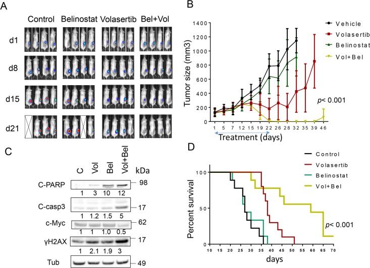
This article has been corrected: Due to errors in image assembly, the IVIS image for the control mouse (panel 3) was inadvertently duplicated as the volasertib-treated mouse (panel 2) at time 0 (d1) in Figure 6A. After reviewing the original files, the correct image for the volasertib-treated time 0 (d1) mouse (panel 2) was located. The corrected Figure 6A is shown below. The authors declare that these corrections do not change the results or conclusions of this paper.
Original article: Oncotarget. 2017; 8:31478–31493. 31478-31493. https://doi.org/10.18632/oncotarget.15649
Figure 6. Co-treatment with volasertib and belinostat suppresses tumor growth in a murine xenograft model and prolongs animal survival.
NOD/SCID-γ mice were subcutaneously inoculated in the right rear flank with 10 × 106 U2932/Luc cells which stably express luciferase. Studies involved 9-10 mice per group. Treatment was initiated after the tumor were visualized, measured, and randomly grouped 10 days after injection of tumor cells. Belinostat was administrated at a dose of 80 mg/kg by i.p 5 days a week. Volasertib was administered at a dose of 12 mg/kg i.p once a week. Control animals were administered equal volumes of vehicle. A. Tumor growth was monitored twice weekly by injection of luciferin and imaged by the IVIS 200 imaging system. d=day, empty boxes represent deceased mice. B. Tumor size was measured by caliper twice weekly. Tumor volumes were calculated (length × width2/2) and plotted against days of treatment. The combination group exhibited significantly smaller tumor size than either single-agent volasertib or belinostat treatment (one-way Anova, p < 0.001). C. Mice were treated for 14 days, and after tumors reached 1 cm in diameter, a representative mouse in each group was sacrificed. Tumors were resected, homogenized and subjected to Western blot analysis for c-PARP, cleaved caspase-3, p-histone H3 and γH2A.X expression. D. Kaplan–Meier analysis was performed to analyze survival of animals. The survival of mice treated with the combination was significantly prolonged compared to mice treated with single agents (p< 0.001). Treatment was discontinued after day 21.


