Neuroscientists believe that immediately after a mammal is born, brain cells called microglia spring into action, pruning away connections between neurons. This may be a way for the brain to refine its neural networks. Until recently, neurobiologists had assumed the microglia worked by phagocytosis, reaching out a cup-shaped tendril to swallow the neuron-to-neuron bridges whole. But in 2017 when Laetitia Weinhard, then a graduate student at EMBL Rome in Italy, observed pruning via a combination of light-sheet and electron microscopy, she saw something others had missed.
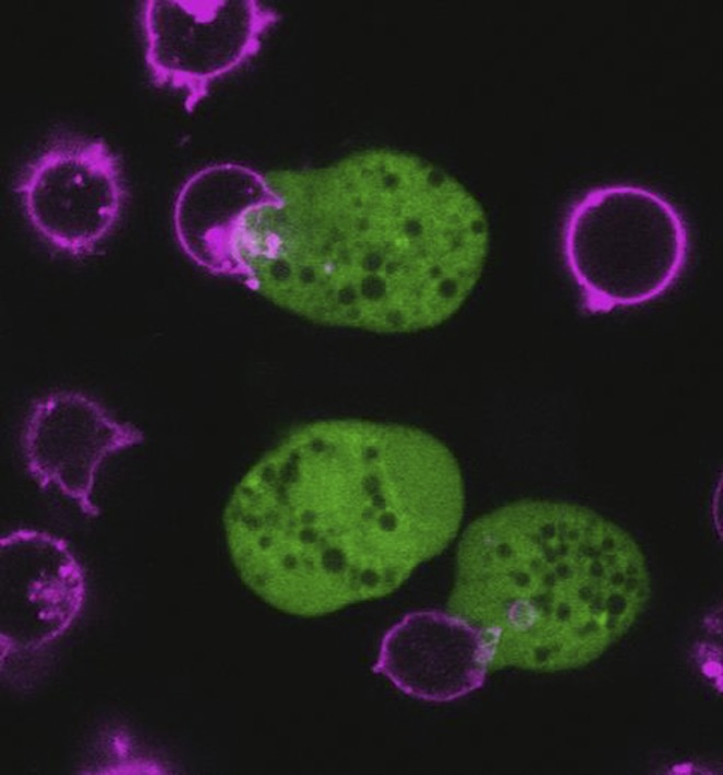
Live confocal video microscopy captures the amoebic parasite Entamoeba histolytica (green) ingesting bites of human Jurkat T cells and their cell membranes (pink). It’s one example of the underappreciated process of trogocytosis. Image credit: Katherine Ralston.
Weinhard, now a postdoc at New York University, discovered that the microglia were instead nibbling off bits of neural connections in a process called trogocytosis, as she reported in 2018 (1). Her group is one of many that have recently found trogocytosis happening in unexpected places and for unsuspected purposes. “I think trogocytosis is really underappreciated,” says Katy Ralston, a microbiologist at the University of California, Davis. “It’s exploding…where it’s happening, and also what it’s being used for.”
Phagocytosis, when one cell engulfs something large such as another cell, is well understood among biologists. So is endocytosis, whereby one cell takes up something from outside into a membranous bubble. There’s also pinocytosis, the cellular ingestion of liquid. But trogocytosis—a word derived from the Greek for “gnaw” or “nibble”—entails one cell nipping bits off another.
The process has gone by different names, such as partial phagocytosis or cell cannibalism, but the basics are the same. A nibbling cell must somehow sever a piece from another cell. That bite then transfers membranes, and sometimes cytoplasmic contents, to the nibbler. This cellular gnawing was first observed decades ago among amoebas (2, 3) and then between cells in the immune system (4).
Using advanced microscopy to catch trogocytosis in action, researchers are seeing it in a diverse set of organisms and processes. Some nibbling cells prune away unwanted cellular bits during development. Others gnaw cells to death. Certain bacteria were recently spotted taking advantage of this interaction between two host cells to slip from an infected cell into an uninfected one. Trogocytosis might even be a drug target for diseases such as allergies and amoebic dysentery.
Information Exchange
Trogocytosis has received the most attention over the years among researchers who study immune cells, which seem to nibble other cells to sample what’s inside. An immune cell may even take a bit of gnawed-off membrane as its own, allowing it to display new proteins on its own surface to activate immune responses. When a cell advertises proteins it has acquired this way, it’s called cross-dressing.
In 2017, immunologist Kensuke Miyake and colleagues from the Tokyo Medical and Dental University in Japan reported an example of cross-dressing that solved a puzzle surrounding a type of immune cells called basophils (5). These cells are involved in activating an immune response to parasite attacks, as well as some allergic reactions, such as asthma.
Researchers had found that basophils use proteins called MHC class II (MHC-II) to present identifying bits of pathogen material, called antigens, to T cells (6–8). This activates the T cells to mount a defense. But other researchers said this couldn’t be because basophils lacked the ability to efficiently process antigens or even make much of the MHC-II proteins (9). It was a “big controversy,” says Miyake.
Supporting the latter theory, Miyake and colleagues found little evidence that basophils could express genes to make their own MHC-II proteins. Yet, they also observed MHC-II protein on basophil surfaces. When they cocultured basophils with dendritic cells, a type well known for its antigen-presenting prowess, the team found the answer. Basophils were nibbling the dendritic cells, specifically taking pieces of membrane with MHC-II proteins to display as their own. If this basophil cross-dressing promotes allergy, then blocking it could be a route to treatment, suggests Miyake.
Nibbled to Death
In the case of certain cancers, it might be better to encourage trogocytosis. Recently, researchers have observed immune cells called neutrophils doing more extensive, even deadly, nibbling of cancer cells—revealing that the mechanism can involve more than just a taste of another cell (10).
Timo van den Berg, an immunologist at the Amsterdam University Medical Center and Sanquin Research in the Netherlands, was interested in how neutrophils kill cancer cells that have been labeled with antibody medicines. His team found that the neutrophils had to touch the cancer cells to do them in. But the neutrophils weren’t using one of their usual techniques, the release of granules full of toxins. Nor were they producing damaging reactive oxygen species.
Neutrophils cannot phagocytose something as large as a cancer cell, but, as van den Berg’s group saw under the microscope, neutrophils can nibble at a cancer cell until it disintegrates. “It was like piranhas attacking prey,” he says.
Van den Berg’s group coined a term for this death by a thousand nibbles: trogoptosis, based on the same Greek root as the word for another kind of cell death, apoptosis. He predicts neutrophils may kill a variety of targets in this way: “I think we are just seeing the tip of the iceberg.” Neutrophils have also been spotted nibbling large parasites to death (11).
Ralston, meanwhile, has observed as infectious amoebas kill host cells by nibbling. The parasite Entamoeba histolytica, which causes dysentery, destroys cells by trogocytosis (12). In a recent study in mBio, she shows that the amoeba also cross-dresses, displaying proteins from immune cells it has nibbled to disguise itself and evade immune attack (13).
Trogo-Transit
Like E. histolytica, other pathogens have been caught taking advantage of host-cell trogocytosis. Tom Kawula, a microbiologist at Washington State University in Pullman, and colleagues study a bacterium called Francisella tularensis, which causes tularemia infections in the skin, lungs, eyes, or digestive system. The team had noticed that F. tularensis traveled quickly between immune cells called macrophages. The researchers could treat a dish full of macrophages with, say, 500 bacteria, and within a day, more than 500 macrophages would be infected. There hadn’t been enough time for the bacteria to take hold, reproduce, and be released to infect other cells. “It wasn’t adding up,” says Kawula.
Graduate student Shaun Steele decided to take video of the process using bacteria labeled with green fluorescent protein. He saw infected and uninfected macrophages interacting and bacteria transferring between them before the cells separated again. When Steele labeled the cytosol of infected cells with red dye and then sorted them by color with flow cytometry, he saw that as uninfected cells picked up some red cytosol from another cell, the green bacteria came along at the same time (14).
“It was like piranhas attacking prey.”
—Timo van den Berg
Kawula thinks that Franscicella is not doing anything specific to instigate trogocytosis but rather hitching a ride in a natural host process that's stimulated by infection. The team found that another bacterium, Salmonella enterica serovar Typhimurium, can also travel by trogocytosis. Being able to transit from cell to cell this way is an advantage for the pathogens, Kawula says, because they’re never exposed to immune attack in the extracellular space.
Gnawing During Development
Trogocytosis appears to be a basic tool that cells can use when phagocytosis may be too blunt an instrument. As in the case of microglia pruning neural connections, cells within a single organism use trogocytosis during development to nip and tuck other cells into the right shape.
Cornelius Gross, the neurobiologist who led the Rome team (1), suspects that in the brain, microglia use whole-cell phagocytosis early in development to eliminate entire neurons. Then, after birth, they switch to trogocytosis for a finer tuning of neural connections.
At the NYU School of Medicine in New York, developmental biologist Jeremy Nance and colleagues found trogocytosis similarly used for pruning in a different system: the primordial germ cells (PGCs) of the nematode Caenorhabditis elegans. Each larval worm possesses exactly two PGCs, which will go on to create all of its sperm and, in the case of hermaphrodites, eggs.
Researchers have long known that the PGCs, which are nestled up against the embryo’s intestines, produce large lobes that disappear by the time the worm matures (15). In a recent experiment, Nance’s then–graduate student Yusuff Abdu, now working at the Rockefeller University in New York, labeled the PGC membranes red to see what those lobes were doing. What he saw was unexpected: bits of red membrane turned up in the intestinal cells. Using fast light-sheet microscopy, he caught the intestinal cells in action, nibbling the lobes (16).
The researchers also used worms deficient in different genes to probe the mechanisms of trogocytosis: does it work like phagocytosis or endocytosis? The team found that nibbling uses elements of both those mechanisms, relying on not only the actin cytoskeleton like phagocytosis but also endocytic proteins to sever the neck of the lobe being consumed.
Nance suspects the purpose of trogocytosis in this context is protective. The PGC lobes are chock-full of mitochondria, which produce not just energy but also DNA-damaging free radicals. That’s dangerous in the germline. By nibbling off the lobes, the intestinal cells may be safeguarding the genomes of future generations.
Although there are only a handful of clear examples of trogocytosis, some researchers suspect the phenomenon is widespread. New microscopes are helping researchers spot the process, which happens quickly. And an uptick in papers describing trogocytosis should help others recognize it in their own systems, suggests Ralston. “It’s evolving, or emerging, as a big theme.”
References
- 1.Weinhard L., et al. , Microglia remodel synapses by presynaptic trogocytosis and spine head filopodia induction. Nat. Commun. 9, 1228 (2018). [DOI] [PMC free article] [PubMed] [Google Scholar]
- 2.Brown T., Observations by immunofluorescence microscopy and electron microscopy on the cytopathogenicity of Naegleria fowleri in mouse embryo-cell cultures. J. Med. Microbiol. 12, 363–371 (1979). [DOI] [PubMed] [Google Scholar]
- 3.Waddell D. R., Vogel G., Phagocytic behavior of the predatory slime mold, Dictyostelium caveatum. Cell nibbling. Exp. Cell Res. 159, 323–334 (1985). [DOI] [PubMed] [Google Scholar]
- 4.Joly E., Hudrisier D., What is trogocytosis and what is its purpose? Nat. Immunol. 4, 815 (2003). [DOI] [PubMed] [Google Scholar]
- 5.Miyake K., et al. , Trogocytosis of peptide-MHC class II complexes from dendritic cells confers antigen-presenting ability on basophils. Proc. Natl. Acad. Sci. U.S.A. 114, 1111–1116 (2017). [DOI] [PMC free article] [PubMed] [Google Scholar]
- 6.Sokol C. L., et al. , Basophils function as antigen-presenting cells for an allergen-induced T helper type 2 response. Nat. Immunol. 10, 713–720 (2009). [DOI] [PMC free article] [PubMed] [Google Scholar]
- 7.Perrigoue J. G., et al. , MHC class II-dependent basophil-CD4+ T cell interactions promote TH2 cytokine-dependent immunity. Nat. Immunol. 10, 697–705 (2009). [DOI] [PMC free article] [PubMed] [Google Scholar]
- 8.Otsuka A., et al. , Basophils are required for the induction of Th2 immunity to haptens and peptide antigens. Nat. Commun. 4, 1739 (2013). [DOI] [PMC free article] [PubMed] [Google Scholar]
- 9.Hammad H., et al. , Inflammatory dendritic cells—not basophils—are necessary and sufficient for induction of Th2 immunity to inhaled house dust mite allergen. J. Exp. Med. 207, 2097–2111 (2010). [DOI] [PMC free article] [PubMed] [Google Scholar]
- 10.Matlung H. L., et al. , Neutrophils kill antibody-opsonized cancer cells by trogoptosis. Cell Reports 23, 3946–3959.e6 (2018). [DOI] [PubMed] [Google Scholar]
- 11.Mercer F., Ng S. H., Brown T. M., Boatman G., Johnson P. J., Neutrophils kill the parasite Trichomonas vaginalis using trogocytosis. PLoS Biol. 16, e2003885 (2018). [DOI] [PMC free article] [PubMed] [Google Scholar]
- 12.Ralston K. S., et al. , Trogocytosis by Entamoeba histolytica contributes to cell killing and tissue invasion. Nature 508, 526–530 (2014). [DOI] [PMC free article] [PubMed] [Google Scholar]
- 13.Miller H. W., Suleiman R. L., Ralston K. S., Trogocytosis by Entamoeba histolytica mediates acquisition and display of human cell membrane proteins and evasion of lysis by human serum. MBio 10, e00068–19 (2019). [DOI] [PMC free article] [PubMed] [Google Scholar]
- 14.Steele S., Radlinski L., Taft-Benz S., Brunton J., Kawula T. H., Trogocytosis-associated cell to cell spread of intracellular bacterial pathogens. eLife 5, e10625 (2016). [DOI] [PMC free article] [PubMed] [Google Scholar]
- 15.Sulston J. E., Schierenberg E., White J. G., Thomson J. N., The embryonic cell lineage of the nematode Caenorhabditis elegans. Dev. Biol. 100, 64–119 (1983). [DOI] [PubMed] [Google Scholar]
- 16.Abdu Y., Maniscalco C., Heddleston J. M., Chew T. L., Nance J., Developmentally programmed germ cell remodelling by endodermal cell cannibalism. Nat. Cell Biol. 18, 1302–1310 (2016). [DOI] [PMC free article] [PubMed] [Google Scholar]


