Abstract
In general, exercise can help improve overall health and prevent diseases. However, individuals with atopic dermatitis (AD) often lose the desire for physical exercise owing to itching caused by sweating. In the present study, we have evaluated the effect of low-intensity tower climbing resistance exercise (TCRE) on Dermatophagoides farinae extract (DFE; house dust mite extract)- and 2,4-dinitrochlorobenzene-induced AD-like skin lesions in a BALB/c model. Histopathological examination showed reduced thickness of the epidermis/dermis and dermal infiltration of inflammatory cells in the ears. TCRE downregulated serum Ig levels and suppressed mRNA expression of pro-inflammatory cytokines in the ear tissue, and reduced the size and weight of draining lymph nodes (dLNs) and nondraining lymph nodes (ndLNs), along with expression of pro-inflammatory cytokines in CD4+ T cells from dLNs and ndLNs. Taken together, we showed that low-intensity TCRE reduced AD symptoms. These results will help improve treatment of AD, and will be of interest to dermatologists as well as to patients with AD.
Keywords: Atopic dermatitis, Tower climbing resistance exercise, Low-intensity exercise
INTRODUCTION
Atopic dermatitis (AD) is a common chronic or relapsing inflammatory disease associated with pruritic and eczematous skin lesions (Boguniewicz and Leung, 2010; Leung et al., 2004; Savinko et al., 2012). In recent years, the occurrence of AD has rapidly increased in developing and developed countries (Odhiambo et al., 2009; Williams et al., 2008). The etiology of AD is not clear. However, it is thought to be linked with multifaceted interactions between genetic and environmental factors such as allergens and microbes, as well as between immune and skin barrier dysfunction.
It has also been reported that acute phase AD is strictly related to T helper 2 (Th2) cell-mediated lesions, and that chronic AD is related to Th1 cell-mediated AD lesions (Nilsson et al., 2011; Savinko et al., 2012). Furthermore, stimulated mast cells, increased IgE, IgG, and pro-inflammatory cytokines play key roles in the formation of skin lesions (Niebuhr and Werfel, 2010; Parisi et al., 2013). Several studies have reported that cytokines, including interleukin (IL)-4, IL-4, IL-13, IL-31, and both tumor necrosis factor-alpha (TNF-α) and interferon-gamma (IFN-γ) are key players in acute and chronic AD (Bilsborough et al., 2006; Vestergaard et al., 1999; Yamada et al., 1995). In addition, retinoic acid-related orphan receptor γT (RORγT) is an orphan nuclear receptor that controls the expression of Th17 cells. Consequently, IL-17 has been reported to play a role in certain skin conditions (Albanesi et al., 1999; Zheng et al., 2007). IL-17-producing Th17 cells are important for host immunity. Further, abnormal activation of Th17 cells is responsible for inflammation (McKenzie et al., 2006). Transforming growth factor-beta (TGF-β) is a multifunctional cytokine that is expressed in most tissues, including the skin. TGF-β is secreted by several cell types, such as T cells, macrophages, endothelial cells, and keratinocytes, and is known to be involved in different types of skin inflammatory disorders (Khaheshi et al., 2011; Letterio and Roberts, 1998).
Physical exercise is well-known to improve health and prevent disease (Haskell et al., 2007). Systematic physical exercise can help to reduce body weight and play a key role in the prevention of coronary artery disease, stroke, hypertension, and osteoporosis (Booth and Lees, 2007). However, exercise usually causes sweating, which can result in itching. Many patients with AD are thought to refrain from exercise because it increases itching. Previous studies have shown that exercise and sweating are major exacerbating factors for patients with AD (Stern et al., 1998; Williams et al., 2004). Nevertheless, regular activity may beneficial to patients with AD (Salzer et al., 1994). However, the connection between low-intensity exercise and AD has not yet been studied.
In this study, therefore, we examined the effect of low-intensity tower climbing resistance exercise (TCRE) on AD lesions using a BALB/c model. The effects were evaluated by assessing ear thickness, histopathological changes including mast cell count, AD-related pro-inflammatory cytokines in ear tissue, and serum immunoglobulin (Ig) levels, as well as the size and weight of draining lymph nodes (dLNs) and nondraining lymph nodes (ndLNs), along with expression of pro-inflammatory cytokines in CD4+ T cells from dLN and ndLNs.
MATERIALS AND METHODS
Materials
TRIzol reagent for RNA extraction was purchased from Invitrogen (Carlsbad, CA, USA). Primary antibodies and peroxidase-conjugated secondary antibodies were purchased from Santa Cruz Biotechnology Inc. (Santa Cruz, CA, USA). All other reagents were of the highest grade that was commercially available at the time of the study.
Animals
Eight-week-old, female BALB/c mice were purchased from Samtako (Samtako Bio Korea Co. Ltd., Osan, Korea) and housed under specific pathogen-free conditions. The animals were housed 5–10 per cage in a laminar air-flow room, maintained at a temperature of 22°C±2°C, with a relative humidity of 55%±5% throughout the study. All experiments were approved by the Institutional Animal Care and Use Committee of Konkuk University (KU14012).
Induction of AD lesions in the ear and TCRE protocol
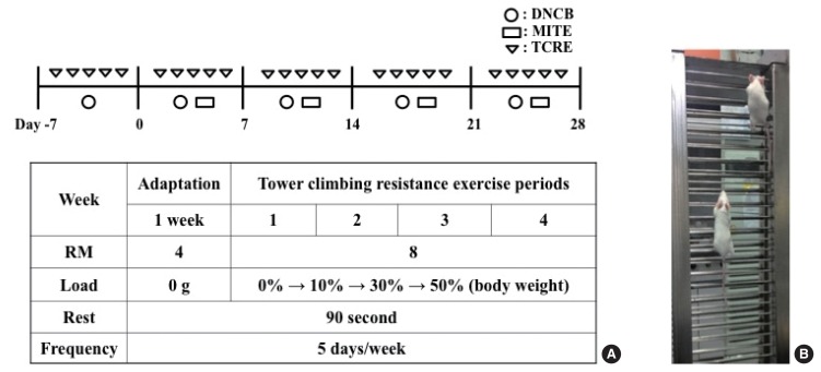
AD was induced in the mice by repeated local exposure of the ears to Dermatophagoides farinae extract (DFE; house dust mite extract) and 2,4-dinitrochlorobenzene (DNCB), as previously described (Kim et al., 2014). For the induction of AD, the mice were divided into four groups (control, TCRE, AD-only, AD+TCRE). The surfaces of both earlobes were stripped 5 times with surgical tape (Nichiban, Tokyo, Japan). After stripping, 20 μL of 1% DNCB was painted onto each ear, followed 4 days later by 20 μL of DFE (10 mg/mL). DFE or DNCB treatment was administered alternately once per week for 4 weeks.
The mice climbed the vertical ladder for 4 weeks. The exercise was accomplished utilizing a 1-m ladder with a 1.5-cm grid and inclined at 85°. Initially, the mice climbed with free weights for a week, to become accustomed. For the first training session, a free weight equivalent to 10% of their body weight was attached to the base of their tail, and the resistance was progressively increased to 30%, 50% for 4 weeks. When the mice reached the top of the ladder, they were allowed to rest for 90 sec. The training session was stopped when the mice succeeded to climb the ladder for eight repetitions.
Ear thickness measurement and blood sample preparation
Ear thickness was measured 24 hr after DNCB or DFE application with a dial thickness gauge (Kori Seiki MFG, Co., Tokyo, Japan). At days 14 and 28, blood samples were collected by the orbital puncture. Plasma samples were prepared from the blood samples and stored at −70°C for further analysis. After blood collection, the ears were removed and used for histopathological analysis. Serum IgE and IgG2a levels were measured at days 14 and 28 after the first induction, using an IgE enzyme-linked immunoassay kit (Bethyl Laboratories Inc., Montgomery, TX, USA) according to the manufacturer’s instructions.
Histological observations
Excised ears were fixed in 4% paraformaldehyde for 16 hr and embedded in paraffin. Thin (6 μm) sections were stained with hematoxylin and eosin (H&E). The thickness of the epidermis and dermis was measured under a microscope. For measurement of mast cell infiltration, skin sections were stained with toluidine blue, after which the number of mast cells was counted in five randomly chosen fields of view.
Real-time polymerase chain reaction
Quantitative real-time polymerase chain reaction (PCR) was carried out using a Thermal Cycler Dice TP850 (Takara Bio Inc., Shiga, Japan) according to the manufacturer’s protocol. Total RNA was isolated from the ear tissue and lymph nodes of each group. The PCR conditions were similar to those previously described (Kim et al., 2014). Briefly, 2 μL of cDNA (100 ng), 1 μL of sense and antisense primer solution (0.4 μM), 12.5 μL of SYBR Premix Ex Taq (Takara Bio Inc.), and 9.5 μL of dH2O were mixed to obtain a final 25-μL reaction mixture in each reaction tube. The amplification conditions were as follows: 10 sec at 95°C, 40 cycles of 5 sec at 95°C and 30 sec at 60°C, 15 sec at 95°C, 30 sec at 60°C, and 15 sec at 95°C. In each sample, the expression level of the analyzed gene was normalized to that of GAPDH and presented as a relative mRNA level.
Statistical analysis
Statistical analysis was carried out using SAS ver. 9.2 (SAS Institute, Cary, NC, USA). Multiple-group data were analyzed by one-way analysis of variance followed by Dunnett multiple range test. All results are expressed as the mean±standard deviation of comparative fold differences. Data are representative of three independent experiments. The threshold for significance was set at P<0.05.
RESULTS
Effect of TCRE on the ear thickness and histopathological observation and on serum Ig levels
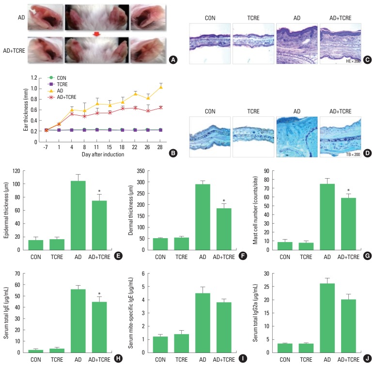
Experimental AD was induced in both earlobes of BALB/c mice by the alternative painting of DFE or DNCB for 4 weeks (Fig. 1A). To evaluate the immunomodulatory effects of TCRE, the mice climbed a vertical ladder for 4 weeks. For the first training session, a free weight equivalent of 10% of their body weight was attached to the base of the tail, and the resistance was progressively increased to 30%, 50% over 4 weeks (Fig. 1B). We found that DFE/DNCB application significantly increased ear thickness and AD lesions. On the contrary, following TCRE for 4 weeks, we observed that TCRE significantly reduced ear thickness (Fig. 2A, B). The TCRE group mice showed significantly reduced epidermal and dermal thickness (Fig. 2C, E, F). In addition, TCRE reduced numbers of infiltrating immune cells, such as mast cells, compared with the AD group (Fig. 2D, G).
Fig. 1.
(A) Experimental schedule for the induction of atopic dermatitis lesions. Mice climbed a vertical ladder for 4 weeks. The exercise was performed utilizing a 1-m ladder with a 1.5-cm grid inclined at 85°. Initially, the mice climbed without free weights for 1 week, to become accustomed to climbing. For the first training session, a free weight equivalent to 10% of their body weight was attached to the base of their tail, and the resistance was progressively increased to 30%, 50% over 4 weeks. (B) Tower climbing resistance exercise. DNCB, 2,4-dinitrochlorobenzene; TCRE, tower climbing resistance exercise; RM, Repetition maximum.
Fig. 2.
(A) Photographs of the ears of mice from each group on day 28. (B) Ear thickness was measured with a dial thickness gauge every 3 days after 2,4-dinitrochlorobenzene (DNCB) or Dermatophagoides farinae extract (DFE) application. Representative photomicrographs of ear sections stained with hematoxylin and eosin (C) or toluidine blue (D). Epidermal (E) and dermal (F) thickness was measured using the microphotographs of hematoxylin and eosin-stained tissue. (G) The number of infiltrating mast cells was determined on the basis of toluidine blue staining. Blood samples were collected by an orbital puncture on day 28. Serum total IgE (H), mite-specific IgE (I), and IgG2a (J) levels were quantified by enzyme-linked immunosorbent assay. Data are presented as the mean±standard deviation of triplicate determinations. *P<0.05, a significant difference from the value of the AD mice. AD induced by DFE and DNCB treatment. The pictures shown are representative of each group (n=3–6). The original magnification was ×100. CON, control; TCRE, tower climbing resistance exercise; AD, atopic dermatitis.
Mice in the TCRE group showed significantly reduced total and specific IgE (Fig. 2H, I) and IgG2a levels (Fig. 2J) in comparison with those of DFE/DNCB-treated mice. These data suggest that the effect of TCRE in AD progression is related to downregulation of serum Ig levels.
Effect of TCRE on the expression of various pro-inflammatory cytokines in vivo
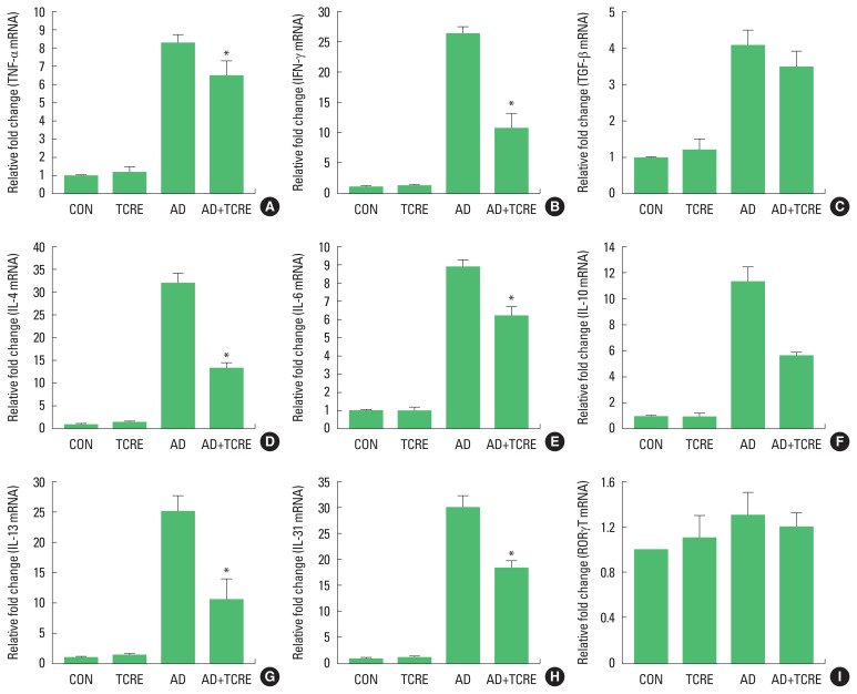
We further examined mRNA expression levels of AD-related pro-inflammatory cytokines from ear tissues by real-time PCR. As shown in Fig. 3, mRNA levels of all tested cytokines were upregulated in the ear tissue of AD mice. On the contrary, the expression of TNF-α, INF-γ, TGF-β1, and Th2-related cytokines such as IL-4, IL-6, IL-10, IL-13, and IL-31 were significantly suppressed in the AD+TCRE in the ear tissue. In addition, TCRE significantly reduced the expression of RORγT mRNA in the ear tissue (Fig. 3).
Fig. 3.
Effect of tower climbing resistance exercise (TCRE) on the expression of various pro-inflammatory cytokines in the ear. The ears were excised on day 28 and total RNA was isolated. (A–I) The quantitative real-time polymerase chain reaction was performed as described in the Materials and Methods section. Data are presented as the mean±standard deviation of triplicate determinations. *P<0.05, a significant difference from the value for atopic dermatitis (AD) mice respectively. CON, control; TNF, tumor necrosis factor; IFN, interferon; IL, interleukin; RORγT, retinoic acid-related orphan receptor gamma T.
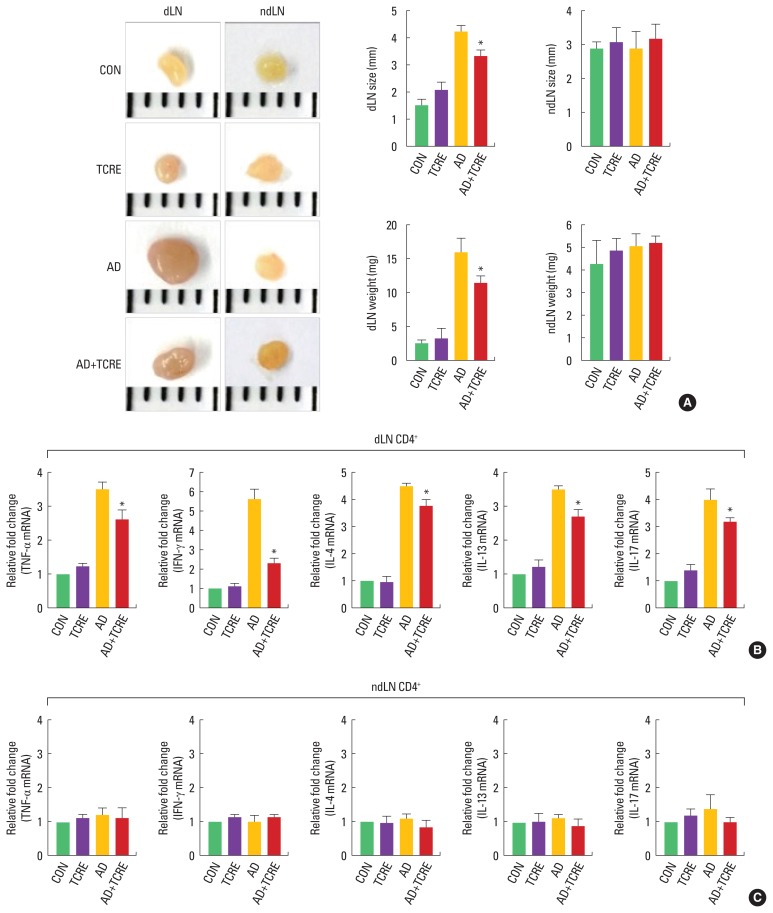
Accordingly, we examined the size and weight of dLNs and ndLNs as well as cytokine-related mRNA expression in CD4+ T cells from dLNs and ndLNs. We observed that AD mice had larger and heavier dLNs than healthy untreated control mice; after 4 weeks of TCRE, the size and weight of dLNs were reduced (Fig. 4A), and no significant differences were observed in the weight of ndLNs between mice in the TCRE and AD groups (Fig. 4A). Analysis of mRNA expression of pro-inflammatory cytokines by CD4+ T cells purified from dLNs showed that mice in the AD+TCRE group showed significantly lower dLN expression of TNF-α, IFN-γ, IL-4, IL-13, and IL-17 (Fig. 4B), whereas mRNA expression of pro-inflammatory cytokines was not changed in CD4+ T cells purified from ndLNs from the AD+TCRE group (Fig. 4C).
Fig. 4.
Effect of tower climbing resistance exercise (TCRE) on the size and weight of draining lymph nodes (dLNs) and nondraining lymph nodes (ndLNs) (A), as well as cytokine-related mRNA expression in CD4+ T cells from dLNs (B) and ndLNs (C). *P<0.05, a significant difference from the value for atopic dermatitis (AD) mice. CON, control; TNF, tumor necrosis factor; IFN, interferon; IL, interleukin.
DISCUSSION
The benefits of exercise have been well studied. Regular physical exercise reduces the risk of several diseases and improves antioxidant activity and immune function (Booth and Lees, 2007; Sen, 1999). However, patients with AD may show a reduced desire for physical exercise due to itching following intense exercise. Therefore, in the present study, we assessed the influence of low-intensity exercise in an AD model.
The allergic response is linked to mast cells, which originate from myeloid stem cells. Therefore, we performed histological analysis of atopic ears to confirm the visual evaluation of AD signs. Ears were removed from each group of mice, then stained with H&E or toluidine blue and infiltration of mast cells, and thickening of the epidermis and dermis were observed under a microscope. After TCRE for 4 weeks, TCRE significantly decreased ear thickness epidermal and dermal thickness. In addition, TCRE also reduced numbers of infiltrating mast cells.
Meanwhile, most patients with AD have increased total serum immunoglobulin IgE levels and specific IgE antibodies to environmental allergens (Dokmeci and Herrick, 2008; Schäfer et al., 1999). Mast cells are stimulated by cross-linking of IgE and an allergen, which stimulates release of chemical mediators including cytokines at the sites of allergic reactions (Borish and Joseph, 1992). Further, high levels of IgG antibodies are associated with chronic AD, and IgG is related to the Th1 response (Mathlouthi and Koenig, 1986). Several studies have also reported that AD is linked to both Th2 and Th1 cell-mediated lesions and causes increased total IgE levels and Th2/Th1-type cytokine expression (Chen et al., 2004; Grewe et al., 1998; Hamid et al., 1994; Neis et al., 2006). To determine whether these effects are mainly exerted via the Th1 or Th2 response, serum levels of IgE (total and DFE-specific) and IgG2a were measured from each treatment group. Total and specific IgE, and IgG2a levels of AD+TCRE group were significantly reduced compared with AD group. In addition, TCRE significantly reduced not only Th2 cytokines but also the Th1-related cytokine TNF-α, IFN-γ and the pro-inflammatory cytokine TGF-β1 which was found to be altered in several skin disorders (Khaheshi et al., 2011). Moreover, RORγT is essential for the induction of IL-17 transcription and is an indicator of Th17-dependent autoimmune disease in mice. Here, TCRE decreased the RORγT level.
Naive CD4+ T cells cause the generation of Th1 and Th2 cells in the presence of pathogens. Th1 cells produce TNF-α and IFN-γ, whereas Th2 cells secrete IL-4, IL-13, and IL-17 which promote humoral immunity and IgE production, as well as regulate Th1 response (Abbas et al., 1996). In particular, IL-4 plays a key role in transforming naive CD4+ T cells into Th2 cells (Rincón et al., 1997). Since AD often develops as a systemic disease (Darlenski et al., 2014), we next examined whether TCRE affected systemic immune responses. Larger dLNs were found in mice in the AD+TCRE group, compared with mice in the AD group, and no significant differences were observed in the weight of ndLNs between mice in the TCRE and AD groups. Further, mice in the AD+TCRE group showed reduced mRNA expression of TNF-α, IFN-γ, IL-4, IL-13, and IL-17.
Meanwhile, previous studies have reported that exercise and sweating are noteworthy exacerbating factors for the schoolchildren with AD (Stern et al., 1998; Williams et al., 2004). Tay et al. (2002) have also studied AD in Singaporean schoolchildren and reported that exercise, heat, and sweating are the most irritating factors for schoolchildren. Accordingly, Kim et al. (2014) showed that high-intensity swimming exercise increases AD symptoms in BALB/c mice by increasing Ig and cytokine expression. Contrary to these data, the results of our study show that low-intensity exercise decreases AD symptoms.
In this study, we showed that low-intensity exercise reduces the intensity of DFE/DNCB-induced AD-like skin lesions in BALB/c mice. TCRE downregulated the severity of histopathological symptoms, production of Ig, and mRNA expression of pro-inflammatory cytokines in ear tissue, and reduced the size and weight of dLNs, along with expression of pro-inflammatory cytokines in CD4+ T cells from dLNs. Taken together, our data suggest that TCRE may be beneficial for patients with AD.
Footnotes
CONFLICT OF INTEREST
No potential conflict of interest relevant to this article was reported.
REFERENCES
- Abbas AK, Murphy KM, Sher A. Functional diversity of helper T lymphocytes. Nature. 1996;383:787–793. doi: 10.1038/383787a0. [DOI] [PubMed] [Google Scholar]
- Albanesi C, Cavani A, Girolomoni G. IL-17 is produced by nickel-specific T lymphocytes and regulates ICAM-1 expression and chemokine production in human keratinocytes: synergistic or antagonist effects with IFN-γ and TNF-α. J Immunol. 1999;162:494–502. [PubMed] [Google Scholar]
- Bilsborough J, Leung DY, Maurer M, Howell M, Boguniewicz M, Yao L, Storey H, LeCiel C, Harder B, Gross JA. IL-31 is associated with cutaneous lymphocyte antigen-positive skin homing T cells in patients with atopic dermatitis. J Allergy Clin Immunol. 2006;117:418–425. doi: 10.1016/j.jaci.2005.10.046. [DOI] [PubMed] [Google Scholar]
- Boguniewicz M, Leung DY. Recent insights into atopic dermatitis and implications for management of infectious complications. J Allergy Clin Immunol. 2010;125:4–13. doi: 10.1016/j.jaci.2009.11.027. [DOI] [PMC free article] [PubMed] [Google Scholar]
- Booth FW, Lees SJ. Fundamental questions about genes, inactivity, and chronic diseases. Physiol Genomics. 2007;28:146–157. doi: 10.1152/physiolgenomics.00174.2006. [DOI] [PubMed] [Google Scholar]
- Borish L, Joseph BZ. Inflammation and the allergic response. Med Clin North Am. 1992;76:765–787. doi: 10.1016/s0025-7125(16)30325-x. [DOI] [PubMed] [Google Scholar]
- Chen L, Martinez O, Overbergh L, Mathieu C, Prabhakar BS, Chan LS. Early up-regulation of Th2 cytokines and late surge of Th1 cytokines in an atopic dermatitis model. Clin Exp Immunol. 2004;138:375–387. doi: 10.1111/j.1365-2249.2004.02649.x. [DOI] [PMC free article] [PubMed] [Google Scholar]
- Darlenski R, Kazandjieva J, Hristakieva E, Fluhr JW. Atopic dermatitis as a systemic disease. Clin Dermatol. 2014;32:409–413. doi: 10.1016/j.clindermatol.2013.11.007. [DOI] [PubMed] [Google Scholar]
- Dokmeci E, Herrick CA. The immune system and atopic dermatitis. Semin Cutan Med Surg. 2008;27:138–143. doi: 10.1016/j.sder.2008.04.006. [DOI] [PubMed] [Google Scholar]
- Grewe M, Bruijnzeel-Koomen CA, Schöpf E, Thepen T, Langeveld-Wildschut AG, Ruzicka T, Krutmann J. A role for Th1 and Th2 cells in the immunopathogenesis of atopic dermatitis. Immunol Today. 1998;19:359–361. doi: 10.1016/s0167-5699(98)01285-7. [DOI] [PubMed] [Google Scholar]
- Hamid Q, Boguniewicz M, Leung DY. Differential in situ cytokine gene expression in acute versus chronic atopic dermatitis. J Clin Invest. 1994;94:870–876. doi: 10.1172/JCI117408. [DOI] [PMC free article] [PubMed] [Google Scholar]
- Haskell WL, Lee IM, Pate RR, Powell KE, Blair SN, Franklin BA, Macera CA, Heath GW, Thompson PD, Bauman A, American College of Sports Medicine; American Heart Association Physical activity and public health: updated recommendation for adults from the American College of Sports Medicine and the American Heart Association. Circulation. 2007;116:1081–1093. doi: 10.1161/CIRCULATIONAHA.107.185649. [DOI] [PubMed] [Google Scholar]
- Khaheshi I, Keshavarz S, Imani Fooladi AA, Ebrahimi M, Yazdani S, Panahi Y, Shohrati M, Nourani MR. Loss of expression of TGF-βs and their receptors in chronic skin lesions induced by sulfur mustard as compared with chronic contact dermatitis patients. BMC Dermatol. 2011;11:2. doi: 10.1186/1471-5945-11-2. [DOI] [PMC free article] [PubMed] [Google Scholar]
- Kim SH, Kim EK, Choi EJ. High-intensity swimming exercise increases dust mite extract and 1-chloro-2,4-dinitrobenzene-derived atopic dermatitis in BALB/c mice. Inflammation. 2014;37:1179–1185. doi: 10.1007/s10753-014-9843-z. [DOI] [PubMed] [Google Scholar]
- Letterio JJ, Roberts AB. Regulation of immune responses by TGF-β. Annu Rev Immunol. 1998;16:137–161. doi: 10.1146/annurev.immunol.16.1.137. [DOI] [PubMed] [Google Scholar]
- Leung DY, Boguniewicz M, Howell MD, Nomura I, Hamid QA. New insights into atopic dermatitis. J Clin Invest. 2004;113:651–657. doi: 10.1172/JCI21060. [DOI] [PMC free article] [PubMed] [Google Scholar]
- Mathlouthi M, Koenig JL. Vibrational spectra of carbohydrates. Adv Carbohydr Chem Biochem. 1986;44:7–89. doi: 10.1016/s0065-2318(08)60077-3. [DOI] [PubMed] [Google Scholar]
- McKenzie BS, Kastelein RA, Cua DJ. Understanding the IL-23-IL-17 immune pathway. Trends Immunol. 2006;27:17–23. doi: 10.1016/j.it.2005.10.003. [DOI] [PubMed] [Google Scholar]
- Neis MM, Peters B, Dreuw A, Wenzel J, Bieber T, Mauch C, Krieg T, Stanzel S, Heinrich PC, Merk HF, Bosio A, Baron JM, Hermanns HM. Enhanced expression levels of IL-31 correlate with IL-4 and IL-13 in atopic and allergic contact dermatitis. J Allergy Clin Immunol. 2006;118:930–937. doi: 10.1016/j.jaci.2006.07.015. [DOI] [PubMed] [Google Scholar]
- Niebuhr M, Werfel T. Innate immunity, allergy and atopic dermatitis. Curr Opin Allergy Clin Immunol. 2010;10:463–468. doi: 10.1097/ACI.0b013e32833e3163. [DOI] [PubMed] [Google Scholar]
- Nilsson OB, Adedoyin J, Rhyner C, Neimert-Andersson T, Grundström J, Berndt KD, Crameri R, Grönlund H. In vitro evolution of allergy vaccine candidates, with maintained structure, but reduced B cell and T cell activation capacity. PLoS One. 2011;6:e24558. doi: 10.1371/journal.pone.0024558. [DOI] [PMC free article] [PubMed] [Google Scholar]
- Odhiambo JA, Williams HC, Clayton TO, Robertson CF, Asher MI, ISAAC Phase Three Study Group Global variations in prevalence of eczema symptoms in children from ISAAC Phase Three. J Allergy Clin Immunol. 2009;124:1251–1258. doi: 10.1016/j.jaci.2009.10.009. [DOI] [PubMed] [Google Scholar]
- Parisi CA, Smaldini PL, Gervasoni ME, Maspero JF, Docena GH. Hypersensitivity reactions to the Sabin vaccine in children with cow’s milk allergy. Clin Exp Allergy. 2013;43:249–254. doi: 10.1111/cea.12059. [DOI] [PubMed] [Google Scholar]
- Rincón M, Anguita J, Nakamura T, Fikrig E, Flavell RA. Interleukin (IL)-6 directs the differentiation of IL-4-producing CD4+ T cells. J Exp Med. 1997;185:461–469. doi: 10.1084/jem.185.3.461. [DOI] [PMC free article] [PubMed] [Google Scholar]
- Salzer B, Schuch S, Rupprecht M, Hornstein OP. Group sports as adjuvant therapy for patients with atopic eczema. Hautarzt. 1994;45:751–755. doi: 10.1007/s001050050165. [DOI] [PubMed] [Google Scholar]
- Savinko T, Matikainen S, Saarialho-Kere U, Lehto M, Wang G, Lehtimäki S, Karisola P, Reunala T, Wolff H, Lauerma A, Alenius H. IL-33 and ST2 in atopic dermatitis: expression profiles and modulation by triggering factors. J Invest Dermatol. 2012;132:1392–1400. doi: 10.1038/jid.2011.446. [DOI] [PubMed] [Google Scholar]
- Schäfer T, Heinrich J, Wjst M, Adam H, Ring J, Wichmann HE. Association between severity of atopic eczema and degree of sensitization to aeroallergens in schoolchildren. J Allergy Clin Immunol. 1999;104:1280–1284. doi: 10.1016/s0091-6749(99)70025-4. [DOI] [PubMed] [Google Scholar]
- Sen CK. Glutathione homeostasis in response to exercise training and nutritional supplements. Mol Cell Biochem. 1999;196:31–42. [PubMed] [Google Scholar]
- Stern UM, Salzer B, Schuch S, Hornstein OP. Sex-dependent differences in sweating of normal probands and atopic patients in cardiovascular stress. Hautarzt. 1998;49:209–215. doi: 10.1007/s001050050729. [DOI] [PubMed] [Google Scholar]
- Tay YK, Kong KH, Khoo L, Goh CL, Giam YC. The prevalence and descriptive epidemiology of atopic dermatitis in Singapore school children. Br J Dermatol. 2002;146:101–106. doi: 10.1046/j.1365-2133.2002.04566.x. [DOI] [PubMed] [Google Scholar]
- Vestergaard C, Yoneyama H, Murai M, Nakamura K, Tamaki K, Terashima Y, Imai T, Yoshie O, Irimura T, Mizutani H, Matsushima K. Overproduction of Th2-specific chemokines in NC/Nga mice exhibiting atopic dermatitis-like lesions. J Clin Invest. 1999;104:1097–1105. doi: 10.1172/JCI7613. [DOI] [PMC free article] [PubMed] [Google Scholar]
- Williams H, Stewart A, von Mutius E, Cookson W, Anderson HR, International Study of Asthma and Allergies in Childhood (ISAAC) Phase One and Three Study Groups Is eczema really on the increase worldwide? J Allergy Clin Immunol. 2008;121:947–954. doi: 10.1016/j.jaci.2007.11.004. [DOI] [PubMed] [Google Scholar]
- Williams JR, Burr ML, Williams HC. Factors influencing atopic dermatitis-a questionnaire survey of schoolchildren’s perceptions. Br J Dermatol. 2004;150:1154–1161. doi: 10.1111/j.1365-2133.2004.05869.x. [DOI] [PubMed] [Google Scholar]
- Yamada N, Wakugawa M, Kuwata S, Yoshida T, Nakagawa H. Chronologic analysis of in situ cytokine expression in mite allergen-induced dermatitis in atopic subjects. J Allergy Clin Immunol. 1995;96(6 Pt 2):1069–1075. doi: 10.1016/s0091-6749(95)70192-3. [DOI] [PubMed] [Google Scholar]
- Zheng Y, Danilenko DM, Valdez P, Kasman I, Eastham-Anderson J, Wu J, Ouyang W. Interleukin-22, a TH17 cytokine, mediates IL-23-induced dermal inflammation and acanthosis. Nature. 2007;445:648–651. doi: 10.1038/nature05505. [DOI] [PubMed] [Google Scholar]


