Abstract
Background
MicroRNA (miR)-21 is overexpressed in numerous types of malignancy and participates in the development of cancer. However, the basic mechanism of the influence of miR-21 on the malignant phenotype of pancreatic cancer remains unclear.
Purpose
The present study aimed to investigate the role of miR-21 in pancreatic cancer development and explore its molecular mechanism.
Patients and methods
The tissue samples were collected at the Second Hospital of Tianjin Medical University (Tianjin, China) between January 2013 and December 2015. The expression of VHL in tissue samples was evaluated by IHC staining. The expression of miR-21 was measured by quantitative real-time polymerase chain reaction (qRT-PCR). MiR-21 target gene was detected by real-time PCR, Western blot and the luciferase reporter assay. Cell viability, cell proliferation, cell migration and invasion were evaluated by the MTT assays, the colony formation assays and the transwell assays. The nude mouse tumor xenograft model was performed to detect the effect of miR-21 on tumor growth in vivo.
Results
Von Hippel-Lindau tumor suppressor (VHL) was downregulated in pancreatic cancer tissues compared with pancreatic non-tumor tissues. VHL was identified as a novel direct target of miR-21, by which it is negatively regulated. In PANC-1 cells, inhibition of miR-21 and upregulation of VHL significantly suppressed cell proliferation, migration and invasion. Knockdown of miR-21 inhibited the hypoxia-inducible factor (HIF)-1α/vascular endothelial growth factor (VEGF) pathway, while inhibiting the expression of matrix metallopeptidase (MMP)-2 and MMP-9. Silencing of miR-21 inhibited tumor growth in vivo.
Conclusion
Knockdown miR-21 increased the expression of VHL, and thus modulated the HIF-1α/VEGF pathway and the expression of MMP-2 and MMP-9, which led to the inhibition of the proliferation, migration and invasion of pancreatic cancer cells. All of these results suggest that the miR-21/VHL interaction may be a novel potential target for pancreatic cancer prevention and therapy.
Keywords: miR-21, pancreatic cancer, VHL, proliferation, migration, invasion
Introduction
Pancreatic cancer is one of the most lethal malignancies in the world, with mortality rates being close to the incidence rates. The incidence rates of pancreatic cancer is 3%.1,2 Most patients with pancreatic cancer are diagnosed at the advanced stage due to the deficiency of a standard program for screening patients at a high risk of pancreatic cancer,leading to a poor prognosis with a 5-year survival rate of <7%.1,2 Therefore, it is very important to clarify the mechanisms of pancreatic cancer progression and develop novel therapeutic strategies to improve the overall survival of affected patients.
Previous studies have demonstrated that mircoRNAs (miRNAs/miRs) are implicated in the development of pancreatic cancer as both oncogenes or tumor suppressors.3,4 miRNAs regulate gene expression at the post-transcriptional level by binding to the complementary 3′-untranslated regions (3′-UTR) of target genes.3 Studies have shown that miRNAs are involved in many biological processes, such as proliferation, migration and invasion, by regulating the expression of their target genes.3,4 Increasing evidence shows that miR-21 is markedly overexpressed in numerous types of cancer, including pancreatic cancer.5–7 It has been reported that miR-21 acts as an oncogene participating in the development of pancreatic cancers and may be utilized as a diagnostic or prognostic miRNA for pancreatic cancer.6–8 In pancreatic cancer, miR-21 decreased the expression of Slug and Fas ligand, and influenced the growth, apoptosis and invasion of pancreatic cancer cells.9,10 Another study indicated that miR-21 regulated the epithelial growth factor receptor/AKT signaling pathway through targeting Von Hippel-Lindau tumor suppressor (VHL) in glioblastomas;11 however, the function of the interaction of miR-21 and VHL in pancreatic cancer has remained elusive.
VHL is a component of the protein complex that includes elongin B, elongin C and cullin-2, and possesses ubiquitin ligase E3 activity. When oxygen supply is adequate, hypoxia-inducible factor (HIF)-1α is hydroxylated by prolyl hydroxylase proteins and is then recognized by VHL, leading to the ubiquitination and degradation of HIF-1α.12 However, numerous types of solid tumor are anoxic; HIF-1α may be upregulated due to inactivation of VHL, thus promoting the progression of tumors. VHL is a tumor suppressor inactivated in various types of tumor through diverse mechanisms, including the regulation by miRNAs. Numerous miRNAs have been reported to regulate the expression of VHL; for instance, miR-101 and13 miR-155.14 However, whether miR-21 and VHL contributed together to the development of the pancreatic cancer remained to be clarify. The present study demonstrated that VHL is a direct and functional target of miR-21 and is downregulated in pancreatic cancer cells. Knockdown of miR-21 increased the expression of VHL and modulated the HIF-1α/vascular endothelial growth factor (VEGF) pathway, leading to inhibition of the malignant phenotypes of pancreatic cancer. The present study may provide novel clues to improve the poor prognosis of pancreatic cancer.
Materials and methods
Tissue samples and ethics statement
A total of 16 pancreatic ductal adenocarcinoma tissues and 9 pancreatic non-tumorous samples were collected at the Second Hospital of Tianjin Medical University (Tianjin, China) between January 2013 and December 2015, and all tissues were paraffin-embedded. Written informed consent was obtained from each patient.and the consent procedure was in accordance with the Declaration of Helsinki. All of the experiments were approved by the ethics committee of the Tianjin Medical University (Tianjin, China).
Cell lines and culture
The PANC-1 and MiaPaca-2 human pancreatic cancer cell lines were purchased from the Cell Bank of the Chinese Academy of Sciences (Shanghai, China). The cells were cultured at 37°C in a humidified atmosphere of 5% CO2 and 95% air in Dulbecco’s modified Eagle’s medium (DMEM; Hyclone; GE Healthcare, Little Chalfont, UK) supplemented with 10% fetal bovine serum (FBS; Hyclone; GE Healthcare).
Public databases and prediction algorithms
Oncomine (Compendia Bioscience, Ann Arbor, MI, USA) and the Human Protein Atlas (http://www.proteinatlas.org/ENSG00000134086-VHL/tissue) were used for analysis and visualization the expression of VHL in pancreatic cancer and normal pancreas samples. The prediction algorithms of TargetScan (http://genes.mit.edu/targetscan.test/ucsc.html), PicTar (https://pictar.mdc-berlin.de/) and miRanda (http://www.microrna.org/microrna/home.do) were used to predict the potential targets of miR-21.
Immunohistochemical (IHC) analysis
The expression of VHL in tissue samples was evaluated by IHC staining. The paraffin-embedded tissue sections were used for examination of VHL expression using antibody against VHL. The sections were deparaffinized by heating at 55°C for 30 min and by 2 washes, 10 mins each, with xylene at room temperature, then rehydrated by a series of incubations in absolute alcohol for 5 min at room temperature, 95% alcohol for 2 min and 70% alcohol for 2 min at room temperature. Antigen retrieval was undertaken by boiling the sections in citrate antigen retrieval solution (Guangzhou Leader Bio-Technology) for 8 min. Endogenous peroxidase activity was blocked with 3% H2O2 for 10 min at room temperature. The sections were next blocked in 10% normal goat serum at room temperature for 30 min. Sections were incubated with primary antibodies against VHL (1:1,000 dilution; cat. no. ab140989; Abcam, Cambridge, UK) overnight at 4°C, followed by incubation with biotin-labeled secondary antibody (1:100 dilution; cat. no. ZB-2010; Zhongshan Golden Bridge Biological Technology Co., Beijing, China) for 1 h at room temperature. Sections were then incubated with avidin biotin complex-peroxidase (Solarbio, Beijing, China) and diaminobenzidine (Solarbio) for 30 min at room temperature, counterstained with hematoxylin (Solarbio) and visualized using a light microscope (Olympus, Tokyo, Japan). The VHL expression in tissue samples was semi-quantitated by measuring the integrated optical density (IOD) using the image analysis software Image-Pro Plus 6.0 (Media Cybernetics, Rockville, MD, USA). The mean density of each sample was used as an index of VHL expression, and was calculated as follows: Mean density=IOD/Area. We selected three samples per tissue specimen and evaluated five fields of view per slide. The clinical information and the mean density values of each sample are presented in Tables 1 and 2.
Table 1.
The mean density of immunohistochemistry staining for VHL in 16 pancreatic cancer tissues
| Sample ID | Age | Gender | Tumor size (cm) | Pathology | Mean density | Sample ID | Age | Gender | Tumor size (cm) | Pathology | Mean density |
|---|---|---|---|---|---|---|---|---|---|---|---|
| 1 | 59 | Female | 2 | PDAC | 218.7±38.82 | 9 | 64 | Male | 6 | PDAC | 110.5±29.45 |
| 2 | 47 | Male | 4 | PDAC | 50.9±11.34 | 10 | 72 | Female | 5 | PDAC | 235.6±28.53 |
| 3 | 72 | Female | 5 | PDAC | 163.5.±23.67 | 11 | 71 | Male | 5 | PDAC | 109.5±12.54 |
| 4 | 66 | Female | 2 | PDAC | 106.2±27.43 | 12 | 62 | Male | 7 | PDAC | 72.9±16.43 |
| 5 | 61 | Female | 4 | PDAC | 155.4±30.65 | 13 | 68 | Male | 2.5 | PDAC | 169.3±21.32 |
| 6 | 57 | Female | 2.5 | PDAC | 267.8±26.96 | 14 | 75 | Female | 6 | PDAC | 151.1±16.53 |
| 7 | 44 | Male | 2.5 | PDAC | 200.7±31.34 | 15 | 61 | Male | 4.5 | PDAC | 119.0±21.23 |
| 8 | 65 | Female | 4 | PDAC | 120.2±15.65 | 16 | 44 | Male | 7 | PDAC | 140.2±25.87 |
Abbreviation: PDAC, Pancreatic ductal adenocarcinoma.
Table 2.
The mean density of immunohistochemistry staining for VHL in 9 pancreatic non-tumor tissues
| Sample ID | Age | Gender | Mean density | Sample ID | Age | Gender | Mean density |
|---|---|---|---|---|---|---|---|
| 1 | 69 | Male | 360.8±51.32 | 6 | 64 | Female | 591.2±62.54 |
| 2 | 58 | Male | 510.7±71.43 | 7 | 68 | Male | 625.1±53.97 |
| 3 | 68 | Male | 391.4±42.34 | 8 | 63 | Male | 607.3±52.65 |
| 4 | 53 | Male | 298.4±24.54 | 9 | 60 | Male | 751.0±66.76 |
| 5 | 58 | Female | 364.1±34.87 |
Western blot analysis
Pancreatic cancer cells were washed with PBS and lysed with ice-cold radioimmunoprecipitation assay buffer (Pierce; Thermo Fisher Scientific, Inc., Waltham, MA, USA) containing the protease inhibitor phenylmethane sulfonylfluoride (Sigma-Aldrich; Merck KGaA, Darmstadt, Germany). The protein concentration was measured with a NanoDrop ND-1000 spectrophotometer (NanoDrop Technologies; Thermo Fisher Scientific, Inc.). Equal amounts of total protein were separated by 10%SDS-PAGE and transferred onto polyvinylidene difluoride membranes (Roche, Basel, Switzerland). The membranes were incubated with primary antibodies against VHL (1:1,000 dilution; cat. no. ab140989; Abcam, Cambridge, UK), MMP-2 (1:1,000 dilution; cat. no. M00286; Boster Biotechnology, Wuhan, China), MMP-9 (1:1,000 dilution; cat. no. BA0573; Boster Biotechnology, Wuhan, China), VEGF (1:1,000 dilution; cat. no. BM4256; Boster Biotechnology, Wuhan, China), HIF-1α (1:1,000 dilution; cat. no. ab16066; Abcam Cambridge, UK;) and GAPDH (1:1,000 dilution; cat. no. TA-08; Zhongshan Golden Bridge Biological Technology Co., Beijing, China) overnight at 4°C. Blots were washed with PBS containing 0.1% Tween-20 and labeled with horseradish peroxidase-conjugated secondary anti-mouse or anti-rabbit antibody (1:1,000 dilution; cat. no. ZB-2301; cat. no. ZB-2305; Zhongshan Golden Bridge Biological Technology Co; Beijing, China.). Antibody-labeled protein bands on the membranes were detected using a G:BOX F3 (Syngene, Cambridge, UK). The protein expression levels were quantified by optical densitometry using ImageJ software version6.0 (National Institutes of Health, Bethesda, MD, USA).
Lentiviral infection and gene transfection
Lentiviruses containing a miR-21 inhibitor segment (lenti-AS-21) or negative control (lenti-NC) segment were obtained from Genepharma (Shanghai, China). The human pancreatic cancer cell lines PANC-1 and MiaPaca-2 were seed in 6-well tissue plates and grown to 80–90% confluence prior to infection and transfection. Cells were infected with the viral suspension at a multiplicity of infection of 10. pcDNA3 and pcDNA3-VHL plasmids provided by Dr. ChunSheng Kang (Department of Neurosurgery, Tianjin Medical University General Hospital) were transfected using Lipofectamine 2000 (Invitrogen; Thermo Fisher Scientific, Inc.) following the manufacturer’s protocols. After 6 hrs, the transfection solution was replaced by normal medium. The cells were further cultured for another 48 h before RNA and protein extractions were performed.
Reverse transcription-quantitative polymerase chain reaction (RT-qPCR)
Total RNA was extracted from pancreatic cancer cells using TRIzol reagent (Invitrogen; Thermo Fisher Scientific, Inc.) according to the manufacturer’s protocols. To detect miR-21, stem-loop RT-PCR was performed with a one-step RNA PCR kit (cat. no. RR066A; Takara Bio Inc., Otsu, Japan) according to the manufacturer’s protocols. Real-time PCR was performed with SYBR green detection with a forward primer for the mature miRNA sequence and a universal adaptor reverse primer (miR-21 forward, 5′-GCCGCTAGCTTATCAGACTGATG-3′. and reverse, 5′-GTCGTATCCAGTGCAGGGTCCGAGGTATTCGCACTGGATACGACTCAACA-3′; U6 forward, 5′-CTCGCTTCGGCAGCACA-3′ and reverse, 5′-AACGCTTCACGAATTTGCGT-3′; miR-21-RT-primer:5′-GTCGTATCCAGTGCAGGGTCCGAGGTATTCGCACTGGATACGACTCAACA-3′;U6-RT-primer:5′-GTCGTATCCAGTGCAGGGTCCGAGGTGCACTGGATACACAAAATATGG-3′). The thermocycling conditions for PCR were as follows: 95°C for 30 sec, 40 cycles of 95°C for 5 sec, 60°C for 20 sec and 72°C for 20 sec, followed by 40°C for 20 min. The primer sets of U6 and miR-21 used for RT-PCR were purchased from GenePharma (Shanghai, China). Quantitative miR-21 expression data were calculated using the 2-ΔΔCq method.15 All miRNA expression data were normalized to U6 small nuclear RNA from the same sample. All reactions were performed in triplicate.
MTT assay
Cells were seeded into 96-well plates at 5×10³ cells per well after infection and transfection as described above. For each group, five wells with the same experimental conditions were tested at 24, 48 and 72 h after seeding using the MTT assay. Subsequently, 50 µl MTT solution (5 mg/mL; Sigma-Aldrich; Merck KGaA) was added to each well and the cells were incubated at 37°C for an additional 4 h. The supernatant was then discarded, and 200 µl dimethyl sulfoxide (Solarbio) was added to each well to dissolve the precipitate at room temperature for 10 min. The OD of each well was measured at the wavelength of 570 nm.
Colony formation assay
Cells were infected and transfected as described above and a single-cell suspension was prepared by detachment with trypsin (Hyclone; GE Healthcare). The treated cells were seeded into 6-well cell culture plates (200 cells per well) and incubated for 14 days at 37°C. Methanol was used to fix the cell colonies for 15 min at 4°C, followed by staining with 0.1% crystal violet (Solarbio) for 20 min at room temperature. The plates were washed and air-dried at room temperature, followed by capturing of images and counting of colonies. The efficiency of colony formation was calculated as the percentage of plated cells that formed colonies, and each colony with a minimum of 50 cells was counted.
Cell migration and invasion assay
A cell suspension contain PANC-1 cells (5×10⁴ for migration and 7×10⁴ for invasion) in 200 µl DMEM without FBS was seeded into each well of the upper compartment of a Transwell chamber (Corning, Inc., Corning, NY, USA), which was pre-coated with or without Matrigel® (BD Biosciences, San Jose, CA, USA). In the lower chamber, 600 µl DMEM with 10% FBS was added. After 48 h of incubation at 37°C with 5% CO2, cells that had migrated or invaded through the membrane were fixed with 20% methanol for 30 min at room temperature, and stained with 0.1% crystal violet for 30 min at room temperature. The cells that had transgressed through the membrane were counted under a microscope in 10 randomly selected visual fields.
Luciferase reporter assay
In order to validate that VHL mRNA is a target of miR-21, a reporter plasmid containing luciferase with the 3′-UTR sequence of VHL mRNA was generated. The 3′-UTR segment of VHL mRNA containing miR-21 binding sites was inserted into the XbaI site of pGL3-control vector (Promega Corp., Madison, WI, USA) and named pGL3-WT-VHL-3′UTR. To generate the pGL3-VHL-mutated reporter plasmid, 3′-UTR segment of VHL mRNA without miR-21 binding sites was inserted into the XbaI site of pGL3-control vector and named pGL3-MUT-VHL-3′UTR. In a 96-well plate, PANC-1 or MiaPaca-2 cells (5x10⁴ cells per well) were infected with lenti-AS-21 or lenti-NC, and co-transfected with 0.2 μg pGL3-WT-VHL-3′UTR plasmid using Lipofectamine 3000 (Thermo Fisher Scientific, Inc.). For another group, PANC-1 or MiaPaca-2 cells (5x10⁴ cells per well) infected with lenti-AS-21 or lenti-NC co-transfected with pGL3-MUT-VHL-3′UTR plasmid using Lipofectamine 3000. After 48 h of transfection, luciferase activity was measured using the Promega Bright-Glo™ Luciferase Assay Systerm (Promega Corp.).
Tumor xenograft model in nude mice
To investigate the effect of miR-21 on the proliferation of pancreatic cancer in vivo, a tumor xenograft model in nude mice was established. A total of 12 female BALB/c nude mice (age, 6 weeks) were purchased from the Guangdong Medical Laboratory Animal Center (Guangdong, China). PANC-1 cells that were infected with miR-21 inhibitor or scrambled oligonucleotide for 24 h were suspended by 100μl serum-free DMEM culture medium and were subcutaneously injected into the flanks of the nude mice at a concentration of 5×10⁷ cells/mL. After 4 days of injection, animal weight and tumor size were measured, and every three days after that. The tumor volume was calculated as follows: Tumor volume = (length x width2)/2. All mice were sacrificed by cervical dislocation at 21 days after implantation and the tumors were isolated from the mice for subsequent IHC analysis. Cryosections (4 mm) were stained with hematoxylin and eosin and used for IHC analysis of VHL expression. The study was performed according to the NIH guidelines for the humane treatment of animals and the protocol adhered to national and international standards. The animal experiments were approved by the ethics committee of the Tianjin Medical University.
Statistical analysis
All data included in the present study were obtained from at least three independent experiments for each condition. SPSS 19 statistical analysis software (IBM Corp., Armonk, NY, USA) and GraphPad Prism 7.0 were used for statistical analysis. The Student’s t-test was used to determine the significance of differences between the two groups. Differences were considered significant if P<0.05. Values are expressed as the mean ± standard deviation.
Results
VHL is downregulated in human pancreatic cancer tissues
The expression of VHL in 16 pancreatic cancer tissues and 9 pancreatic non-tumor tissues was assessed by IHC analysis. The basic information of the tissues is presented in Tables 1 and 2, and informed consent was obtained from each patient. IHC staining revealed that VHL is markedly downregulated in pancreatic cancer tissues compared to non-tumor tissues (Figure 1A), which was supported by a semi-quantitative analysis using an IOD/area index for the IHC staining (Figure 1B). This observation was confirmed by mining of public databases. The Oncomine expression data indicated downregulation of VHL transcript in pancreatic adenocarcinoma compared with that in normal pancreas tissues. Data from the Human Protein Atlas indicated positive staining for VHL in the cytosol and membrane of normal pancreas tissue and weak or negative staining in pancreatic cancer tissues (http://www.proteinatlas.org/ENSG00000134086-VHL/tissue; http://www.proteinatlas.org/ENSG00000134086-VHL/pathology). These results suggested that VHL is downregulated in pancreatic cancer tissues and may have an important role in the genesis and progression of pancreatic cancer.
Figure 1.
Expression levels of VHL in human pancreatic cancer tissues and pancreatic non-tumor tissues. (A) Representative images of VHL protein expression determined by immunohistochemical staining of pancreatic non-tumor tissues (left) and human pancreatic cancer tissues (right). (B) Semi-quantitative analysis of VHL protein expression using the mean density in human pancreatic cancer tissues and pancreatic non-tumor tissues. Values are expressed as the average values from several fields of view from several slices of the same sample and from several samples per group. Values are expressed as the mean ± standard deviation.
Notes: **P<0.01. The image color is RGB.
Abbreviations: VHL, Von Hippel-Lindau tumor suppressor.
VHL is a direct target of miR-21
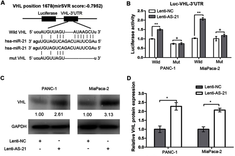
To investigate whether miR-21 contributed to the downregulation of VHL, a bioinformatics analysis was performed to predict the potential targets of miR-21. It was revealed found that the 3′-UTR of the VHL transcript has a putative miR-21 binding site (Figure 2A). To confirm whether miR-21 regulates the expression of VHL, luciferase reporter plasmids containing the wild-type (pGL3-WT-VHL-3′UTR) or mutant type (pGL3-MUT-VHL-3′UTR) binding sequence from the 3′-UTR of VHL mRNA were constructed (Figure 2A) and employed in subsequent luciferase reporter assays. Pancreatic cancer cells were co-transfected pGL3-WT-VHL-3′UTR or pGL3-MUT-VHL-3′UTR with lenti-AS-21 or lenti-NC and the luciferase activity was measured after 48 h of treatment. The results revealed that knockdown of miR-21 triggered a marked increase of pGL3-WT-VHL-3′UTR luciferase activity compared to the control group, while no significant change in luciferase activity was observed in the cells transfected with pGL3-MUT-VHL-3′UTR along with lenti-AS-21 or lenti-NC (Figure 2B). To determine whether VHL expression is indeed regulated by miR-21, miR-21 was silenced in PANC-1 and MiaPaca-2 cells and VHL expression levels were examined by Western blot analysis. The results indicated that VHL protein was significantly increased in lenti-AS-21-treated cells compared with that in lenti-NC-treated cells (Figure 2C and D). All of these results indicate that VHL is a direct target of miR-21 and downregulated by miR-21.
Figure 2.
VHL is a direct target of miR-21. (A) The 3′-UTR fragments containing the predicted miR-21 targeting sites of VHL (wild type or mutant type) were cloned into the reporter plasmid downstream of the firefly luciferase gene. (B) PANC-1 and MiaPaca-2 cells were co-transfected with human VHL 3′-UTR (wild type or mutant type) firefly luciferase reporter plasmid and lenti-NC or lenti-AS-21 as indicated. After 48 h, firefly luciferase activity was measured. (C) The expression levels of VHL protein in PANC-1 and MiaPaca-2 cells treated with lenti-NC or lenti-AS-21 were detected by Western blot analysis. (D) Quantification of VHL protein expression detected by Western blot and normalized to GAPDH. Values are expressed as the mean ± standard deviation from three independent experiments.
Notes: *P<0.05; **P<0.01; +No significance.
Abbreviations: VHL, Von Hippel-Lindau tumor suppressor; UTR, untranslated region; lenti-NC, negative control lentivirus; lenti-AS-21, microRNA-21 inhibitor vector; PANC-1, human pancreatic cancer cell; Mia Paca-2, human pancreatic cancer cell.
VHL inhibits the proliferation, migration and invasion of PANC-1 cells
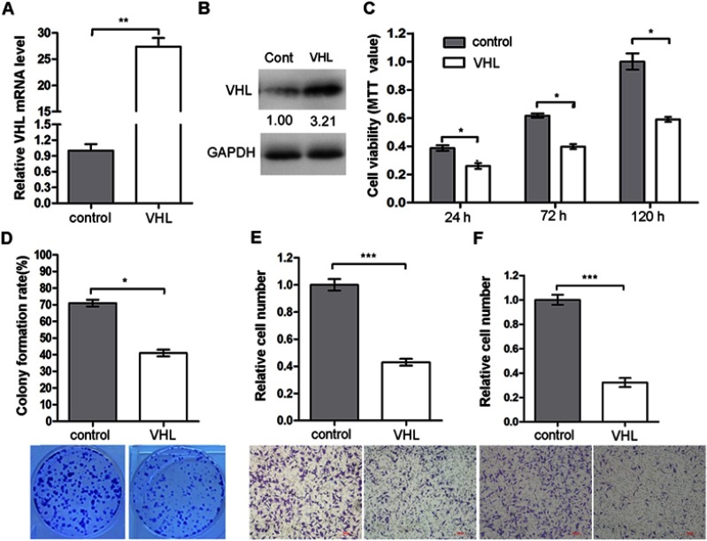
To confirm whether VHL is involved in the development of pancreatic cancer, the biological function of VHL in PANC-1 cells was explored. First, the effectiveness of the VHL expression plasmid pcDNA3-VHL was verified by RT-qPCT and Western blot assays (Figure 3A and B). Next, MTT, colony formation and Transwell migration/invasion assays were performed to examine the influence of VHL on cell viability, proliferation, migration and invasion. The results of the MTT assay demonstrated that ectopic expression of VHL reduced the cell viability (Figure 3C). The colony formation assays demonstrated the proliferation rate of PANC-1 cells transfected with pcDNA3-VHL was decreased to 40% compared with that of the controls (Figure 3D). Finally, transwell assays were performed to determine the effect of VHL on the migration and invasion abilities. The results revealed that the migration and invasion capacities of PANC-1 cells transfected with pcDNA3-VHL were decreased to 42% and 32%, respectively (Figure 3E and F). All of these results demonstrate that VHL acts as a tumor suppressor gene and inhibits the cell viability, proliferation, migration and invasion of PANC-1 cells.
Figure 3.
VHL inhibits the proliferation, migration and invasion of pancreatic cancer cells. (A) PANC-1 cells were transfected with either control plasmid or pcDNA3-VHL The mRNA expression levels of VHL at 24 h post-transfection were detected by reverse transcription-quantitative polymerase chain reaction analysis with normalization to β-actin. (B) The protein expression levels of VHL at 48 h post-transfection were detected by Western blot analysis. (C) Cell viability was measured using an MTT assay at 24, 72 and 120 h post-transfection. (D) Colony formation assay was performed at 24 h post-transfection. (E) Transwell migration and (F) invasion assays were carried out at 24 h post-transfection. Values are expressed as the mean ± standard deviation from three independent experiments.
Notes: *P<0.05; **P<0.01; ***P<0.001. The image color is RGB.
Abbreviations: VHL, Von Hippel-Lindau tumor suppressor; pcDNA3-VHL, overexpression plasmid for Von Hippel-Lindau tumor suppressor; PANC-1, human pancreatic cancer cell.
Silencing of miR-21 inhibits the proliferation, migration and invasion of PANC-1 cells
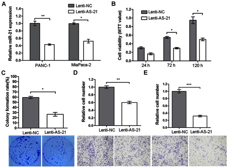
The present study demonstrated that VHL is a potential target of miR-21 and VHL acts as a tumor suppressor gene regulating the malignant phenotypes of PANC-1 cells. Thus, it was assumed that miR-21 is an oncogene that promotes the development of pancreatic cancer. Therefore, the impact of miR-21 on the cell viability, proliferation, migration and invasion of pancreatic cancer cells was evaluated. The endogenous expression of miR-21 was knocked down by infecting pancreatic cancer cells with lenti-AS-21, a lentiviral vector expressing a sponge inhibitor of miR-21, which was confirmed by RT-qPCR. The expression of miR-21 was decreased by 57 and 48%, respectively, in PANC-1 and MiaPaca-2 cells infected with lenti-AS-21 compared with that in the lenti-NC treated controls (Figure 4A). Furthermore, MTT and colony formation assays were used to evaluate the roles of miR-21 in cell viability and proliferation. The cells infected with lenti-AS-21 had a significantly lower viability and colony-forming ability than the controls (Figure 4B and C). The Transwell assays indicated that the migration and invasion capability of PANC-1 cells infected with lenti-AS-21 was significantly decreased to 60% and 31% respectively compared with that in the controls (Figure 4D and E). These results reveal that knockdown of miR-21 inhibited the cell viability, proliferation, migration and invasion of pancreatic cancer cells.
Figure 4.
Silencing of miR-21 suppresses the proliferation, migration and invasion of pancreatic cancer cells. (A) PANC-1 and MiaPaca-2 cells were infected with lenti-AS-21 or lenti-NC. Reverse transcription-quantitative polymerase chain reaction analysis was used to confirm the knockdown of miR-21 in PANC-1 and MiaPaca-2 cells after 24 h infection. (B) An MTT assay was used to detect the viability of PANC-1 cells infected with lenti-AS-21 or lenti-NC for 24, 72 or 120 h. (C) A colony formation assay was used to detect the colony formation rate of PANC-1 cells infected with lenti-AS-21 or lenti-NC. The colony formation rate was calculated using the following equation: Colony formation rate = (number of colonies/number of plated cells) x100%. (D) Transwell migration and (E) invasion assays were performed with PANC-1 cells infected with lenti-AS-21 or lenti-NC. Values are expressed as the mean ± standard deviation from three independent experiments.
Notes: *P<0.05; **P<0.01; ***P<0.001. The image color is RGB.
Abbreviations: miR, microRNA; lenti-NC, negative control lentivirus; lenti-AS-21, microRNA-21 inhibitor vector; PANC-1, human pancreatic cancer cell; Mia Paca-2, human pancreatic cancer cell.
Silencing of miR-21 inhibits the HIF-1α/VEGF pathway and the expression of MMP-2 and MMP-9
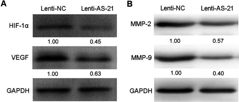
The above mentioned results confirmed that miR-21 regulates the malignant phenotypes of PANC-1 cells and negatively regulates the expression of VHL. To further explore the potential mechanism of miR-21 to promote the malignant phenotypes of pancreatic cancer, it was investigated whether miR-21 affects the downstream signaling pathways of VHL. The expression of HIF-1α, VEGF, MMP-2 and MMP-9 in PANC-1 cells with knockdown of miR-21 was detected by Western blot analysis. The results indicated that silencing of miR-21 inhibited the expression of HIF-1α, VEGF, MMP-2 and MMP-9 (Figure 5A and B). It was therefore suggested that miR-21 promotes the malignant phenotype of pancreatic cancer, at least partially by regulating the VHL/HIF-1α/VEGF pathway and the expression of MMP-2 and MMP-9.
Figure 5.
Silencing of miR-21 inhibits the HIF-1α/VEGF pathway, and the expression of MMP-2 and MMP-9. The expression levels of HIF-1α and VEGF (A), MMP-2 and MMP-9 (B) in PANC-1 cells treated with lenti-NC or lenti-AS-21 were determined by Western blot analysis with normalization to GAPDH.
Abbreviations: Lenti-NC, negative control lentivirus; lenti-AS-21, microRNA-21 inhibitor vector. HIF-1α, hypoxia-inducible factor-1α; VEGF, vascular endothelial growth factor; MMP, matrix metallopeptidase; PANC-1, human pancreatic cancer cell; GAPDH, internal control.
Knockdown of miR-21 inhibits tumor growth in vivo
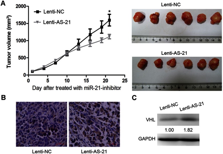
To further assess the effect of miR-21 on tumor growth in vivo, an animal experiment using the nude mouse tumor xenograft model was performed. PANC-1 cells were pre-treated with lenti-AS-21 and lenti-NC, and the pre-treated cells were subcutaneously injected into the flank of 6-week-old female nude mice. Compared with that in the lenti-NC groups, the average tumor volume in the lenti-AS-21 groups was lower (Figure 6A). Furthermore, IHC analysis and western bolt assays suggested that tumor tissues derived from lenti-AS-21-treated PANC-1 cells displayed an increased expression of VHL compared with those in the lenti-NC-treated group (Figure 6B and C).
Figure 6.
Knockdown miR-21 inhibits tumor growth in vivo. (A) The nude mouse tumor xenograft model was utilized to study the effect of miR-21 on tumor growth in vivo. The tumor growth curve is presented on the left. The tumor volume was calculated as follows: Tumor volume = (length x width2)/2. *P<0.05, n=6 (Student’s t-test). The mice were euthanized and the tumors were isolated 21 days after implantation (right panel). (B) The VHL expression levels in the tumors of the mice were detected by immunohistochemistry, and representative photomicrographs of the tumor sections are presented. (C) The VHL expression levels in the tumors of the mice were detected by Western blot with normalization to GAPDH. Values are expressed as the mean ± standard deviation from six nude mouse tumor samples.
Notes: GAPDH: internal control; nude mice: female BALB/c nude mice (age, 6 weeks). The image color is RGB.
Abbreviations: VHL, Von Hippel-Lindau tumor suppressor; miR, microRNA Lenti-NC, negative control lentivirus; lenti-AS-21, microRNA-21 inhibitor vector.
Discussion
Pancreatic cancer is a highly lethal malignancy with a poor prognosis owing to the lack of early diagnostic and effective therapeutic strategies.1 Extensive studies have demonstrated that certain miRNAs are involved in regulating the development of pancreatic cancer by controlling the expression of different oncogenes or tumor suppressors, which provides a novel basis for diagnostic and effective therapeutic strategies.16,17 miR-21 is known to be an oncogene and to have a key role in the genesis, recurrence and metastasis of various types of cancer.18 miR-21 has been reported to regulate the expression of PDCD4 to control the development of medullary thyroid carcinoma and colorectal cancer.19,20 miR-21, through targeting PTEN, RECK and B-cell lymphoma-2, regulates the cell proliferation, migration and invasion in lung squamous cell carcinoma.21 In addition, the oncogenic role of miR-21 has been well described in other cancer types, including glioblastoma22 and gastric cancer.23 A previous study reported that miR-21 is highly expressed in the serum of pancreatic cancer patients and may be a novel diagnostic marker and therapeutic target for pancreatic cancer.24 It has been indicated that miR-21 contributes to chemoresistance in pancreatic cancer,25 In the present study, we demonstrated that miR-21 acts as an oncogene in pancreatic cancer. The results indicate that silencing of miR-21 led to an inhibition of proliferation, migration and invasion of PANC-1 cells. Furthermore, VHL was confirmed as a direct functional target of miR-21 and it was indicated that the effect of miR-21 on the malignant phenotype of pancreatic cancer cells is, at least in part, mediated by downregulation of the expression of VHL.
The VHL tumor suppressor gene is located on the short arm of chromosome 3 and widely expressed in human tissue.26 The mutation or loss of VHL leading to VHL disease has been well studied.27 While recent research has indicated that the loss of VHL is not exclusive to VHL disease, certain cancer types are highly associated with a VHL mutation. For instance, VHL - elongin B and C protein complex components are important for the genesis of pheochromocytomas.28 VHL inactivation was observed in 80% of renal clear cell carcinomas.29 However, whether VHL is associated with the development of pancreatic cancer has yet to be explored. The present study demonstrated that VHL is downregulated in human pancreatic cancer tissues, and the results were further confirmed by mining of public databases. Thus, it is likely that VHL may regulate the development of pancreatic cancer. As expected, the present results suggested that ectopic overexpression of VHL inhibited the proliferation, migration and invasion of PANC-1 cells.
The tumor suppressor effect of VHL has been described to be due to its ability to induce the ubiquitination and proteasomal destruction of HIF-1α.30,31 HIF-1α has an important role in tumor development through inducing tumor cells to become more aggressive to better adapt to the hypoxic environment of solid tumors.32 It has been reported that under hypoxic stress, HIF-1α regulates tumor angiogenesis by regulating the expression of several pro-angiogenic factors, including VEGF.33 VEGF contributes to tumor growth and metastasis in pancreatic cancer.34 In the present study, it was demonstrated that knockdown of miR-21 resulted in reduced expression of HIF-1α, which in turn inhibited the expression of VEGF. Furthermore, additional downstream molecules of VHL, including MMP-2 and MMP-9, were also downregulated by miR-21 inhibition. These results demonstrate that miR-21 regulates the malignant phenotypes of pancreatic cancer, at least partially by regulating the expression of VHL, thus modulating the HIF-1α/VEGF pathway or other downstream molecules.
Conclusion
Taken together, the present study demonstrated that knockdown of miR-21 and overexpression of VHL inhibited the proliferation, migration and invasion of pancreatic cancer cells. VHL is downregulated by miR-21 as its direct and functional target. miR-21 exerts it oncogenic effect to promote the malignant phenotypes of pancreatic cancer by repressing the expression of VHL and modulating the HIF-1α/VEGF pathway.
Acknowledgment
This work was supported by the Natural Science Foundation of Tianjin (No.16JCYBJC27100).
Abbreviations
miR-21, microRNA-21; miRNAs, microRNAs; 3′-UTR, 3′-untranslated regions; VHL, Von Hippel-Lindau tumor suppressor; MMP-2, matrix metallopeptidase 2; MMP-9, matrix metallopeptidase 9; VEGF, vascular endothelial growth factor; IHC, immunohistochemical staining.
Ethics approval and informed consent
Ethics approval was obtained from ethics committee of the Tianjin Medical University. Written informed consent was obtained from all individual participants included in the study. The consent procedure was in accordance with the Declaration of Helsinki. The animal experiments were performed according to the NIH guidelines for the humane treatment of animals and the protocol adhered to national and international standards. All procedures performed in studies involving animals were in accordance with the ethical standards of the institution or practice at which the studies were conducted.
Data availability
All data generated and analysed during this study are included in this published article.
Disclosure
The authors declare that they have no conflicts of interest in this work.
References
- 1.Kamisawa T, Wood LD, Itoi T, Takaori K. Pancreatic cancer. Lancet. 2016;388(10039):73–85. doi: 10.1016/S0140-6736(16)00141-0 [DOI] [PubMed] [Google Scholar]
- 2.Siegel RL, Miller KD, Jemal A. Cancer statistics, 2017. CA Cancer J Clin. 2017;67(1):7–30. doi: 10.3322/caac.21387 [DOI] [PubMed] [Google Scholar]
- 3.Farazi TA, Hoell JI, Morozov P, Tuschl T. MicroRNAs in human cancer. Adv Exp Med Biol. 2013;774:1–20. doi: 10.1007/978-94-007-5590-1_1 [DOI] [PMC free article] [PubMed] [Google Scholar]
- 4.Bartel DP. MicroRNAs: genomics, biogenesis, mechanism, and function. Cell. 2004;116(2):281–297. doi: 10.1016/s0092-8674(04)00045-5 [DOI] [PubMed] [Google Scholar]
- 5.Moriyama T, Ohuchida K, Mizumoto K, et al. MicroRNA-21 modulates biological functions of pancreatic cancer cells including their proliferation, invasion, and chemoresistance. Mol Cancer Ther. 2009;8(5):1067–1074. doi: 10.1158/1535-7163.MCT-08-0592 [DOI] [PubMed] [Google Scholar]
- 6.Zhang H, Feng X, Zhang M, et al. Long non-coding RNA CASC2 upregulates PTEN to suppress pancreatic carcinoma cell metastasis by downregulating miR-21. Cancer Cell Int. 2019;19:18.. [DOI] [PMC free article] [PubMed] [Google Scholar]
- 7.Lubezky N, Loewenstein S, Ben-Haim M, et al. MicroRNA expression signatures in intraductal papillary mucinous neoplasm of the pancreas. Surgery. 2013;153(5):663–672. doi: 10.1016/j.surg.2012.11.016 [DOI] [PubMed] [Google Scholar]
- 8.Calatayud D, Dehlendorff C, Boisen MK, et al. Tissue MicroRNA profiles as diagnostic and prognostic biomarkers in patients with resectable pancreatic ductal adenocarcinoma and periampullary cancers. Biomark Res. 2017;5:8. doi: 10.1186/s40364-017-0087-6 [DOI] [PMC free article] [PubMed] [Google Scholar]
- 9.Liu Z, Zhang J, Hong G, Quan J, Zhang L, Yu M. Propofol inhibits growth and invasion of pancreatic cancer cells through regulation of the miR-21/Slug signaling pathway. Am J Transl Res. 2016;8(10):4120–4133. [PMC free article] [PubMed] [Google Scholar]
- 10.Wang P, Zhuang L, Zhang J, et al. The serum miR-21 level serves as a predictor for the chemosensitivity of advanced pancreatic cancer, and miR-21 expression confers chemoresistance by targeting FasL. Mol Oncol. 2013;7(3):334–345. doi: 10.1016/j.molonc.2012.10.011 [DOI] [PMC free article] [PubMed] [Google Scholar]
- 11.Zhang KL, Han L, Chen LY, et al. Blockage of a miR-21/EGFR regulatory feedback loop augments anti-EGFR therapy in glioblastomas. Cancer Lett. 2014;342(1):139–149. doi: 10.1016/j.canlet.2013.08.043 [DOI] [PubMed] [Google Scholar]
- 12.de Paulsen N, Brychzy A, Fournier MC, et al. Role of transforming growth factor-alpha in von Hippel–Lindau (VHL)(-/-) clear cell renal carcinoma cell proliferation: a possible mechanism coupling VHL tumor suppressor inactivation and tumorigenesis. Proc Natl Acad Sci U S A. 2001;98(4):1387–1392. doi: 10.1073/pnas.031587498 [DOI] [PMC free article] [PubMed] [Google Scholar]
- 13.Liu N, Xia WY, Liu SS, et al. MicroRNA-101 targets von Hippel-Lindau tumor suppressor (VHL) to induce HIF1alpha mediated apoptosis and cell cycle arrest in normoxia condition. Sci Rep. 2016;6:20489. doi: 10.1038/srep20489 [DOI] [PMC free article] [PubMed] [Google Scholar]
- 14.Kong W, He L, Richards EJ, et al. Upregulation of miRNA-155 promotes tumour angiogenesis by targeting VHL and is associated with poor prognosis and triple-negative breast cancer. Oncogene. 2014;33(6):679–689. doi: 10.1038/onc.2012.636 [DOI] [PMC free article] [PubMed] [Google Scholar]
- 15.Bustin SA, Benes V, Garson JA, et al. The MIQE guidelines: minimum information for publication of quantitative real-time PCR experiments. Clin Chem. 2009;55(4):611–622. doi: 10.1373/clinchem.2008.112797 [DOI] [PubMed] [Google Scholar]
- 16.Hu H, Zhang Q, Chen W, et al. MicoRNA-301a promotes pancreatic cancer invasion and metastasis through the JAK/STAT3 signaling pathway by targeting SOCS5. Carcinogenesis. Epub 2019 Jun 22. doi: 10.1093/carcin/bgz121 [DOI] [PubMed] [Google Scholar]
- 17.Feng SD, Mao Z, Liu C, et al. Simultaneous overexpression of miR-126 and miR-34a induces a superior antitumor efficacy in pancreatic adenocarcinoma. Onco Targets Ther. 2017;10:5591–5604. doi: 10.2147/OTT.S149632 [DOI] [PMC free article] [PubMed] [Google Scholar] [Retracted]
- 18.Liu J, Zhu H, Yang X, et al. MicroRNA-21 is a novel promising target in cancer radiation therapy. Tumour Biol. 2014;35(5):3975–3979. doi: 10.1007/s13277-014-1623-8 [DOI] [PubMed] [Google Scholar]
- 19.Pennelli G, Galuppini F, Barollo S, et al. The PDCD4/miR-21 pathway in medullary thyroid carcinoma. Hum Pathol. 2015;46(1):50–57. doi: 10.1016/j.humpath.2014.09.006 [DOI] [PubMed] [Google Scholar]
- 20.Peacock O, Lee AC, Cameron F, et al. Inflammation and MiR-21 pathways functionally interact to downregulate PDCD4 in colorectal cancer. PLoS One. 2014;9(10):e110267. doi: 10.1371/journal.pone.0110267 [DOI] [PMC free article] [PubMed] [Google Scholar]
- 21.Xu LF, Wu ZP, Chen Y, Zhu QS, Hamidi S, Navab R. MicroRNA-21 (miR-21) regulates cellular proliferation, invasion, migration, and apoptosis by targeting PTEN, RECK and Bcl-2 in lung squamous carcinoma, Gejiu City, China. PLoS One. 2014;9(8):e103698. doi: 10.1371/journal.pone.0103698 [DOI] [PMC free article] [PubMed] [Google Scholar]
- 22.Yang CH, Yue J, Pfeffer SR, et al. MicroRNA-21 promotes glioblastoma tumorigenesis by down-regulating insulin-like growth factor-binding protein-3 (IGFBP3). J Biol Chem. 2014;289(36):25079–25087. doi: 10.1074/jbc.M114.593863 [DOI] [PMC free article] [PubMed] [Google Scholar]
- 23.Uozaki H, Morita S, Kumagai A, et al. Stromal miR-21 is more important than miR-21 of tumour cells for the progression of gastric cancer. Histopathology. 2014;65(6):775–783. doi: 10.1111/his.12491 [DOI] [PubMed] [Google Scholar]
- 24.Passadouro M, Pedroso de Lima MC, Faneca H. MicroRNA modulation combined with sunitinib as a novel therapeutic strategy for pancreatic cancer. Int J Nanomedicine. 2014;9:3203–3217. doi: 10.2147/IJN.S64456 [DOI] [PMC free article] [PubMed] [Google Scholar]
- 25.Garajova I, Le Large TY, Frampton AE, Rolfo C, Voortman J, Giovannetti E. Molecular mechanisms underlying the role of microRNAs in the chemoresistance of pancreatic cancer. Biomed Res Int. 2014;2014:678401. doi: 10.1155/2014/678401 [DOI] [PMC free article] [PubMed] [Google Scholar]
- 26.Latif F, Tory K, Gnarra J, et al. Identification of the von Hippel-Lindau disease tumor suppressor gene. Science. 1993;260(5112):1317–1320. doi: 10.1126/science.8493574 [DOI] [PubMed] [Google Scholar]
- 27.Haddad NM, Cavallerano JD, Silva PS. Von hippel-lindau disease: a genetic and clinical review. Semin Ophthalmol. 2013;28(5–6):377–386. doi: 10.3109/08820538.2013.825281 [DOI] [PubMed] [Google Scholar]
- 28.Rowbotham DA, Enfield KS, Martinez VD, et al. Multiple components of the VHL tumor suppressor complex are frequently affected by DNA copy number loss in pheochromocytoma. Int J Endocrinol. 2014;2014:546347. doi: 10.1155/2014/546347 [DOI] [PMC free article] [PubMed] [Google Scholar]
- 29.Moore LE, Nickerson ML, Brennan P, et al. Von Hippel-Lindau (VHL) inactivation in sporadic clear cell renal cancer: associations with germline VHL polymorphisms and etiologic risk factors. PLoS Genet. 2011;7(10):e1002312. doi: 10.1371/journal.pgen.1002138 [DOI] [PMC free article] [PubMed] [Google Scholar]
- 30.Semenza GL. Hypoxia-inducible factor 1 (HIF-1) pathway. Sci STKE 2007;2007(407):cm8. [DOI] [PubMed] [Google Scholar]
- 31.Paltoglou S, Roberts BJ. HIF-1α and EPAS ubiquitination mediated by the VHL tumour suppressor involves flexibility in the ubiquitination mechanism, similar to other RING E3 ligases. Oncogene. 2007;26(4):604–609. doi: 10.1038/sj.onc.1209818 [DOI] [PubMed] [Google Scholar]
- 32.Semenza GL. Defining the role of hypoxia-inducible factor 1 in cancer biology and therapeutics. Oncogene. 2010;29(5):625–634. doi: 10.1038/onc.2009.441 [DOI] [PMC free article] [PubMed] [Google Scholar]
- 33.Manalo DJ, Rowan A, Lavoie T, et al. Transcriptional regulation of vascular endothelial cell responses to hypoxia by HIF-1. Blood. 2005;105(2):659–669. doi: 10.1182/blood-2004-07-2958 [DOI] [PubMed] [Google Scholar]
- 34.You WK, Sennino B, Williamson CW, et al. VEGF and c-Met blockade amplify angiogenesis inhibition in pancreatic islet cancer. Cancer Res. 2011;71(14):4758–4768. doi: 10.1158/0008-5472.CAN-10-2527 [DOI] [PMC free article] [PubMed] [Google Scholar]
Associated Data
This section collects any data citations, data availability statements, or supplementary materials included in this article.
Data Availability Statement
All data generated and analysed during this study are included in this published article.


