Abstract
Discoidin Domain Receptor 2 (DDR2) is a receptor tyrosine kinase which has been shown to regulate cell migration upon binding its ligand, collagen. Expression studies determined that DDR2 mRNA and protein are present in the atrioventricular canal during epithelial-mesenchymal transformation (EMT) and the receptor is expressed in both activated endothelial and migrating mesenchymal cells in vivo.
Keywords: discoidin domain receptor, atrioventricular canal, epithelial-mesenchymal cell transformation
Early in cardiac development, the extracellular matrix (ECM) inside the atrioventricular (AV) canal region expands and swells within the two concentric heart tube layers. During this expansion a series of molecules are secreted from the myocardium and move through the matrix to signal the overlying endothelium. A subset of cells in the endothelium then undergoes a process known as epithelial-mesenchymal transformation (EMT). This transformation is restricted to the AV canal and produces an invasive subpopulation of mesenchymal cells that fills the ECM space. These mesenchymal cells differentiate into fibroblasts that eventually contribute to the formation of the membranous septa and cardiac valves. EMT consists of defined steps such as migration of endothelium, the subsequent “activation” of the endothelium and cellular transformation, and invasion of mesenchymal cells into the underlying ECM which are all recapitulated in an in vitro collagen gel assay (Runyan et al., 1990; Zuk and Hay, 1994; Newgreen and Minichiello, 1995; Boyer et al., 1999). Endothelial cell activation is accompanied by cellular hypertrophy, polarization, and a change in morphology, from a cuboidal shaped cell to an elongated spindle-like cell that appears fusiform (Zuk et al., 1989; Hay, 1995). Although a number of genes are expressed specifically in the endothelium overlying the AV cushions (Nakajima et al., 2000; Gitler et al., 2003), markers which delineate activated endothelial cell phenotype remain limited.
The Discoidin Domain Receptor 2 (DDR2) is a receptor tyrosine kinase whose ligand is the extracellular matrix protein collagen (Shrivastava et al., 1997; Vogel et al., 1997). DDR2 expression has been detected in both rat and mouse heart (Lai and Lemke, 1991, 1994; Morales et al., 2005), where it is primarily expressed on cardiac fibroblasts (Goldsmith et al., 2004). Specific activation of the tyrosine kinase domain in DDR2 by collagen binding was demonstrated by stimulating the receptor with various types of collagen (Shrivastava et al., 1997; Vogel et al., 1997) and results in the up-regulation of multiple matrix metalloproteinases (MMPs; Vogel et al., 1997; Olaso et al., 2001; Ferri et al., 2004). Activation of hepatic stellate cells induced DDR2 expression and resulted in increased expression of MMP-2 creating more invasive cells with increased levels of proliferation (Olaso et al., 2001). DDR2 also regulates dermal fibroblast proliferation and MMP-2 mediated migration in a reconstituted basement membrane (Olaso et al., 2002).
Since mesenchymal cells in the AV canal synthesize Type I procollagen (Sinning et al., 1988) and undergo an up-regulation of MMP activity (Hoffman et al., 1999), we sought to determine whether DDR2 was present in the AV canal at the time of EMT. DDR2 protein distribution was localized in Stage 17–23 chicken embryos (Hamburger and Hamilton, 1956) (ISE Newberry, Inc., Newberry, SC). Embryos were isolated and staged, fixed in 4% paraformaldehyde at 4°C overnight and subsequently embedded in acrylamide, vibratome-sectioned (100 μm), stained, and examined by confocal microscopy. Reverse transcriptase polymerase chain reaction (RT-PCR) was used to confirm the presence of the DDR2 transcript. Total RNA was isolated from 30 stage 17 AV canals and mesenchymal cells derived from 30 stage 22 AV canals as previously described (Ramsdell et al., 1998) using the RNeasy Mini prep kit (Qiagen). Reverse transcription reactions were performed using Superscript II reverse transcriptase (Gibco BRL) and 1.5 μg of total RNA. Two different 5′ primers (5.1 and 5.2) and a single 3′primer (3.1) were used to ensure specific amplification of the DDR2 mRNA. The primer sequences were 5.1: 5′ TCCCCATCCTGCTCCG-GACT 3′ and 5.2: 5′ GCAGCCAGAGCCCAGGTCAA 3′ and primer 3.1: 5′ TCAATCTGCAGGAAACTCCTT 3′. The PCR reaction had a 5 min hot start at 95C followed by 80°C for 2 min. Next the reaction cycled for 30 cycles with a 1-min denaturing step at 95°C, a 1 min annealing step at 56°C, and a 1 min 30 sec elongation step at 72°C. PCR products were visualized by agarose gel electrophoresis on a 1.8% TAE agarose gel.
The amount of DDR2 transcript present during AV canal development was determined by real time PCR. Total RNA (1 μg) was obtained from AV canals from 30 stage 13 through stage 23 hearts and relative quantitation of gene expression was performed using a Bio-Rad I cyler system (Bio-Rad, Hercules, CA) and SYBR Green reagent method according to the manufacturer’s instructions (PE-Applied Biosystems). The comparative CT method was used to assess fold-change differences in expression following previous published methods (Parapuram et al., 2003, Valarmathi et al., 2008). The primers used for real time are as follows. DDR2: forward primer: TCCCCATCCTGCTCCGGACT; reverse primer: TCAATC TGCAGGAACTCCTT; and ARBP forward primer: TAAA CCCCGCGTGGCAATC; reverse primer: CCACGTTCCC CCGGATGTGA.
DDR2 protein was detected by immunolocalization using confocal scanning laser microscopy on stage 17–20 AV canal explants cultured on collagen gels for 24 hr as previously described (Potts et al., 1991; Romano and Runyan, 2000). Western blotting was used to confirm the presence of DDR2 in AV canals and cultured mesenchymal cells. Stage 17 AV canals were placed in lysis buffer (0.1% SDS, 1 mM EDTA, 200 μM sodium vanadate, 5 mg/mL leupeptin, 10 mg/mL aprotinin, and 1 mM levamisol in PBS) immediately upon dissection. A pure population of mesenchymal cells from stage 22 AV canals was prepared as previously described (Ramsdell et al., 1998). Stage 22 AV canals were isolated from chick embryos and needle dissection (25 Gauge) used to separate cushion tissue from myocardium followed by digestion with 0.05% trypsin in M199 media. After centrifugation, the resultant cell pellet was resuspended in M199 supplemented with 10% fetal bovine serum, placed in a single well of a 96-well plate and incubated at 37°C in a tissue culture incubator. After 48 hr, mesenchymal cells were lysed in lysis buffer and a BCA protein assay (Pierce) performed to determine concentration. Totally, 10 μg of protein was loaded per lane and subject to electrophoresis in 4–15% gradient Tris-HCL gels (Bio-Rad). The gels were transferred to a nitrocellulose and probed for DDR2 using two polyclonal antibodies (1:250 for Santa Cruz DDR2; SC7555; and 100 μg/mL of EC-DDR2 antisera). The blots were stripped and reprobed with antibodies to ß-actin to assure normalized protein loading. Protein was detected using an HRP-conjugated secondary antibody and chemiluminescent detection.
RESULTS
DDR2 Transcript Is Present in the Developing AV Canal
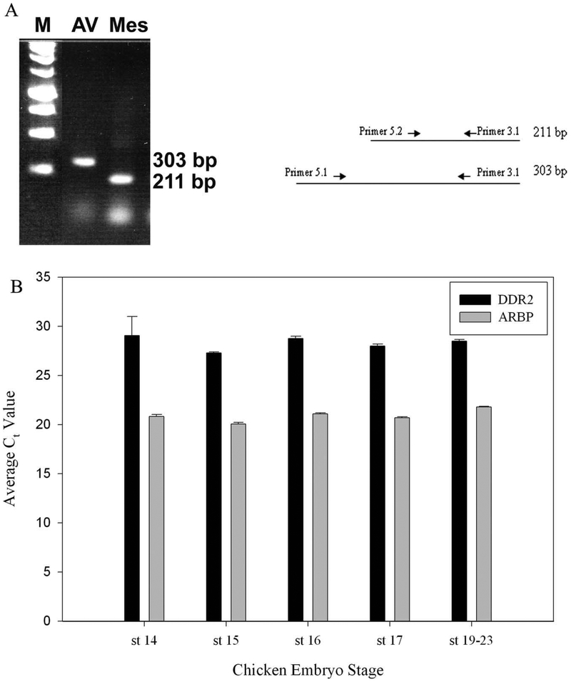
Recent studies have shown that the temporal expression of DDR2 within the heart overlaps the window during which EMT occurs (Goldsmith et al., 2004). To determine if the transcript coding for DDR2 was present in the early AV canal at the time of EMT, RT-PCR was performed on RNA from stage 17 AV canals and cultured AV cushion mesenchyme. In both cases, a transcript coding for chicken DDR2 was detected (Fig. 1A). To ensure that the chicken homolog of DDR2 was amplified, PCR reactions were performed using two separate 5° primers paired with a single 3° primer that generated two distinct sized PCR products (5.1 and 3.1−303 bp; 5.2 and 3.1−211 bp). Both products were sequenced to confirm that the amplicon was DDR2. (Genebank accession number BM 489098 and BM 427091). The chicken DDR2 shows a 97% identity to the rodent DDR2 sequences at the amino acid level in the extracellular domain. Using RT-PCR, the change in DDR2 transcript level in stage 14–23 AV canal RNA was determined. DDR2 transcripts were detected in every stage examined and the level of DDR2 transcript remained virtually unchanged in all stages studied (Fig. 1B). These data clearly indicated that the mRNA encoding DDR2 is present in the AV canal and throughout the EMT process.
Fig. 1.
Expression of DDR2 in the Developing AV Canal. (A) Reverse transcriptase–polymerase chain reaction analyses of chicken DDR2 using specific primers designed to amplify either a 211 bp or 303 bp sequence from the early heart is shown in the schematic. Using either of the 5′primers with the 3.1 primer will result in amplification of the same region of mRNA. Two 5′ primers were used to ensure specific amplification of the chicken DDR2. Expression pattern from Stage 17 chicken AV canals (AV) and isolated mesenchymal cells (Mes). Both size products were observed in both the AV canal and mesenchymal cells. A 1 KB DNA marker (M) was used for sizing purposes (Promega, Corp.). RNA was isolated from 30 AV canals at each stage. The avian DDR2 EST used for primer production has been given the Genbank accession number BM 426366. (B) Real time PCR analysis was done to determine if the level of DDR2 transcript changed during the EMT process. AV canals were isolated from Stages 14–22 and reactions run for DDR2 mRNA and ARBP mRNA (control). The stages examined by real time PCR represent various time point during the EMT process: Stage 14 signal for EMT is being secreted; Stage 15 early endothelial cell activation; Stage 16 endothelial cell activation, Stage 17 endocardial cells begin transformation, and Stages 19–23 abundant migration and proliferation of mesenchyme within the AV canal. No significant changes in DDR2 or ARBP transcript levels were detected as previously described (Valaramathi et al., 2008). For each stage a minimum of 30 AV canals were used to generate RNA. Three individual isolations were performed for each stage.
DDR2 Protein Is Present in the Developing AV Canal
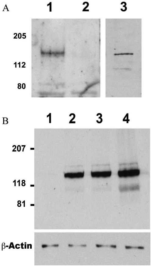
To confirm the presence of DDR2 protein in the AV canal at the time of EMT, proteins were extracted from isolated Stage 17–19 AV canals. These times correspond to the period when mesenchymal cells are first being formed and are actively being produced, respectively. Western blotting analysis using both a commercial antibody to DDR2 (Santa Cruz Biotechnology) and a polyclonal antibody generated to the extracellular domain of DDR2, (EC-DDR2), was performed. Protein isolated from the AV canal showed a band of the correct size (~130 kDa) when probed using the commercial anti-DDR2 antibody (Fig. 2A Lane 1). In contrast, when protein from isolated competent endothelium was also probed with the same antibody no band was visible (Fig. 2A Lane 2). When the same protein from the AV canal was probed with the polyclonal anti EC-DDR2 antisera, again a band of the correct size was obtained (Fig. 2A Lane 3), indicating that both antibodies recognize the same protein. Western blot analysis of isolated AV canals from Stages 14–22 revealed that DDR2 protein levels increased slightly as the AV canal continued to develop (Fig. 2B). These data demonstrate the presence of DDR2 protein on mesenchymal cells in the AV canal during EMT.
Fig. 2.
DDR2 Protein Levels during AV Canal Development. (A) Western blotting of protein (10 μg per lane) from cultured AV canal mesenchyme (Lane 1) and competent endothelium (Lane 2) using a commercial anti-DDR2 antibody demonstrated that DDR2 protein was only present in the mesenchymal cells (Lane 1) with no band detected in protein extracted from competent endothelium. Lane 3 indicates that a similar sized band was also detected from an identical blot containing the same AV canal mesenchymal cell protein and incubated with an antibody generated against the extracellular domain of DDR2 (EC-DDR2). (B) Protein from AV canals from Stages 14–15 (Lane 2), Stages 18–19 (Lane 3), and Stages 21–22 (Lane 4) were probed using DDR2 antibodies. A slight increase in the amount of DDR2 protein was observed with increasing AV canal age. No expression was observed in the protein lysate that was preabsorbed with the antibody (Lane 1). The blot was then stripped and reprobed with antibodies to ß-actin to make sure equal amounts to protein were loaded in each lane.
Expression of DDR2 the Embryonic Heart
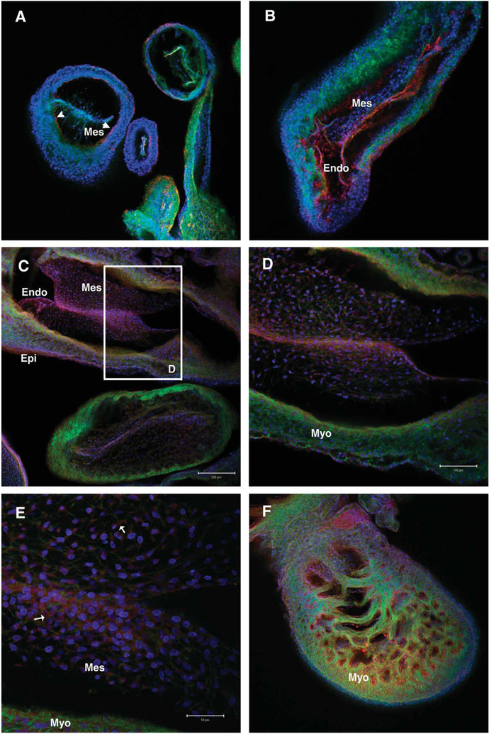
To determine the in vivo expression pattern of DDR2 during cardiac development, sections of Stages 17, 19, and 23 chicken hearts were stained using a commercial DDR2 antibody. Expression of DDR2 in the embryonic heart was observed in the migrating mesenchyme of the AV canal (Fig. 3A–E). In Stage 17 AVCs, DDR2 expression was seen on migrating mesenchymal cells (Fig. 3A, Mes) with limited expression also detected near the myocardium. A similar expression pattern was observed in Stage 19 AVCs (Fig. 3B), with more pronounced DDR2 staining noted in the endocardium (Endo Fig. 3B) and near the myocardium. Figure 3C demonstrates that DDR2 expression still persisted in the endocardium and migrating mesenchyme of Stage 23 AV canals and also showed that DDR2 was now expressed on epicardial cells (Epi in Fig. 3C). Higher magnification (Fig. 3D,E) of the cushions showed that DDR2 was localized on the membrane of migrating mesenchymal cells and also within the cell cytoplasm, largely around the cell nucleus. This peri-nuclear staining is likely the accumulation of newly synthesized DDR2 within the golgi apparatus in preparation for transport to the cell membrane. Similar staining was observed when identical sections were stained with the EC-DDR2 antibody (data not shown). Examination of Stage 23 chicken heart illustrated that DDR2 was also expressed on endocardial cells overlying trabeculae within the myocardium (Fig. 3F).
Fig. 3.
Localization of DDR2 in the Developing AV Canal. Laser scanning confocal micrographs of Stages 17, 19, 23 chicken heart vibratome sections (100 μm) stained with DDR2 antibodies. Expression of DDR2 was observed in the early AV canal (A-E). (A) A stage 17 heart cut through the AV canal shows migrating mesenchymal cells (Mes). DDR2 (red) is observed with faint expression seen near the myocardium. (B) A later staged AV canal (st. 19) showing much stronger DDR2 staining in the endocardium (Endo) and cushion mesenchyme. (C). Similarly, another older staged AV canal (st. 23) shows expression of DDR2 in the endothelium (Endo) and mesenchyme (Mes) as well as the epicardium (Epi). (D). A higher magnification of panel C inset showing the strong DDR2 localization to the endocardium and migrating mesenchyme. (E). Even higher magnification of AV cushion mesenchyme, similar to that shown in D, demonstrating cellular expression of DDR2 in the mesenchyme was both membrane bound and cytoplasmic with a principally perinuclear location. (F). A section through the ventricle of the st. 23 heart illustrates the staining within the trabeculated myocardium (Myo). Negative controls using no primary antibody showed an absence of staining in all stages (Not shown). The sections were also stained with phalloidin (green) to visualize the actin cytoskeleton and DAPI (blue) to visualize the nuclei. Scale bar = 50 μm in E, 200 μm in C and 100 μm in D. Panels A, B, F are at the same magnification as in C. All images were collected using a Zeiss 510 Meta confocal system (Zeiss, Germany).
DDR2 Is Expressed During AV Canal Endothelium Activation
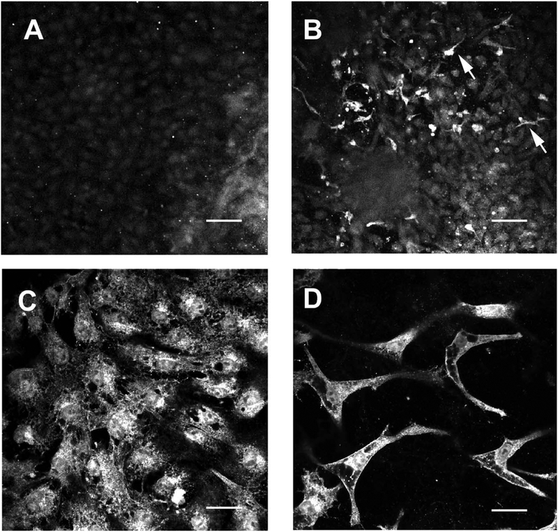
The AV canal explant model system that accurately mimics the EMT process in vitro, including the timing of endothelial cell activation, was examined using confocal scanning laser microscopy to determine the expression pattern of DDR2 during the EMT. As the endothelial cells began to migrate away from the AV canal explant there was virtually no expression of DDR2 (Fig. 4A). However, when the endothelial cells were examined 12 hr later, expression of DDR2 was observed in those endothelial cells that had begun the activation process (Fig. 4B). When competent AV endothelium was treated with myocardium-conditioned media (MCM) to induce the EMT process, DDR2 was observed in the newly formed mesenchymal cells and activated endothelial cells (Fig. 4C). In addition, when migrating mesenchymal cells in the collagen gel were examined there was intense DDR2 staining which appeared as punctate cytoplasmic staining, likely due to increased synthesis of DDR2 protein, with strong membrane localization (Fig. 4D). The stellate mesenchymal cells within the collagen gel show staining that outline individual cells supporting the notion that membrane staining is present in these cells.
Fig. 4.
Expression of DDR2 during the EMT process. AV canal explants were placed on collagen gels and either removed 16 hr later or allowed to go through the EMT process. Antibodies directed against DDR2 were used to stain isolated endothelial monolayers and examined by confocal microscopy. (A). Stage 14 AV competent endothelial cells fail to show any DDR2 expression. (B). Removing the AV explant after mesenchyme has formed shows DDR2 staining in the mesenchyme and the activated endothelial cells. The arrowheads highlight stained mesenchyme within the collagen gel (C). When the explant is allowed to remain on the gel, endothelial cells migrate away and become activated. As these endothelial cells become activated they begin to express DDR2. (D). Migrating mesenchyme within the collagen gel demonstrate a punctate cytoplasmic staining pattern as well as membrane localization. Scale bar = 50 μm for each panel. All images were collected using a Zeiss 510 Meta confocal system (Zeiss, Germany).
DISCUSSION
During the EMT process, a variety of changes in both cell-cell and cell-ECM interactions occur (Reviewed in Mjaatvedt et al., 1999; Person et al., 2005). A subpopulation of endocardial cells within the AV canal become activated and transform into migrating mesenchymal cells. These mesenchymal cells migrate into the underlying ECM and begin the process of ultimately remodeling cushions into valve leaflets. By a variety of methods the presence of DDR2 has been demonstrated at specific steps during EMT in AV canals. The presence of both the DDR2 transcript and protein in the AV canal is suggestive of a role in mesenchymal cell regulation. Previous studies have shown that DDR2 is expressed on mesenchymal cells within rodent cushions and is associated with cells expressing Type I collagen (Morales et al., 2005). However, it is interesting to note that the DDR2 transcript was detected in the AV canal as early as Stage 13. This is prior to any mesenchymal cells being produced. It is also before any potential fibroblasts would be present as the result of epicardial cells migrating into the myocardium. DDR2 is found in early epicardium and in migrating cells entering the heart at the AV sulcus, as well as covering the trabeculae in the late embryonic rodent heart (Morales et al., 2005). It is possible that DDR2 is being produced by the early myocardium as these cells begin to create the matrix milieu present in the AV canal or that early endocardial cells are making DDR2 transcripts. Protein isolated from competent endothelium failed to show any DDR2 reactivity by western blotting analysis while increased DDR2 protein in later stage AV canals suggests that DDR2 expression may be regulated post-transcriptionally. It is also possible that the increased size of the AV canal during development contributes to the increase in DDR2 staining observed in Figure 3. Expression data presented suggests that most endothelial cells begin to express DDR2 in preparation for losing their cell–cell contacts and maintain expression as they begin to transform into mesenchyme and invade the underlying ECM. Thus, it is likely that DDR2 is a marker for an early phase of endothelial cell activation. Identification of DDR2 as a marker for the activated endocardial cell phenotype begins to unify previous data regarding cell-ECM interactions during the EMT process. For instance, DDR2 is a receptor for Type I collagen, which is synthesized in AV canal migrating mesenchymal cells in response to activation (Sinning et al., 1988), and has been shown in be involved in mediating collagen fibrillogenesis (Mihai et al., 2006).
The EMT process is dependent upon secreted molecules originating in the myocardium (Mjaatvedt and Markwald, 1989; Potts and Runyan, 1989). Data presented indicates that DDR2 expression is also dependent upon myocardial stimulation and supports the hypothesis that activated endothelial cells begin to express DDR2 prior to the time when they begin to express Type I collagen. In contrast to Type I procollagen, where no expression was observed in the any cardiac cells until Stage 24, DDR2 expression was observed in both activated endothelial cells which did not undergo transformation into mesenchyme as well as mesenchymal cells present at Stage 22. Conversely, only the AV canal mesenchyme showed any Type I procollagen expression and it was not until Stage 28 that the AV canal myocardium began to express Type I procollagen (Sinning et al., 1988). It is possible that these endocardial cells, by expressing DDR2, are priming themselves either for the stimulation that they receive from binding to Type I collagen or that they need DDR2 expressed before they can produce collagen. Why DDR2 is expressed so early in the myocardium is uncertain, but it may be in response to the production of collagen from the newly formed migrating mesenchyme as a mechanism to regulate collagen fibril size (Mihai et al., 2006). Thus by binding to newly synthesized collagen an autocrine feedback loop may be established that maintains mesenchyme expression of DDR2, production of collagen I and continued cell proliferation and migration as has been shown for skin fibroblasts (Olaso et al., 2002). Future experiments to establish if such an autocrine pathway exists for DDR2 in the migrating mesenchymal cells are planned. In addition, studies will examine whether DDR2 is important in other aspects of AV canal formation such cellular proliferation and regulating collagen production.
ACKNOWLEDGMENTS
The authors thank Mary Morales for technical support and Jack Goldsmith for proofreading this manuscript.
Grant sponsor: American Heart Association; Grant numbers: 0060217U, 0160361U; Grant sponsor: NIH; Grant numbers: HL73937, HL072958.
LITERATURE CITED
- Boyer AS, Erickson CP, Runyan RB. 1999. Epithelial-mesenchymal transformation in the embryonic heart is mediated through distinct pertussis toxin-sensitive and TGFbeta signal transduction mechanisms. Dev Dyn 214:81–91. [DOI] [PubMed] [Google Scholar]
- Ferri N, Carragher NO, Raines EW. 2004. Role of discoidin domain receptors 1 and 2 in human smooth muscle cell-mediated collagen remodeling. Am J Pathol 164:1575–1585. [DOI] [PMC free article] [PubMed] [Google Scholar]
- Gitler AD, Lu MM, Jiang YQ, Epstein JA, Gruber PJ. 2003. Molecular markers of cardiac endocardial cushion development. Dev Dyn 228:643–650. [DOI] [PubMed] [Google Scholar]
- Goldsmith EC, Hoffman A, Morales MO, Potts JD, Price RL, McFadden A, Rice M, Borg TK. 2004. Organization of fibroblasts in the heart. Dev Dyn 230:787–794. [DOI] [PubMed] [Google Scholar]
- Hamburger V, Hamilton HL. 1956. A series of normal stages in the development of the chick embryo. J Morphol 88:49–92. [PubMed] [Google Scholar]
- Hay ED. 1995. An overview of epithelio-mesenchymal transformation. Acta Anat (Basel) 154:8–20. [DOI] [PubMed] [Google Scholar]
- Hoffman LM, Kulyk WM. 1999. Alcohol promotes in vitro chondrogenesis in embryonic facial mesenchyme. Int J Dev Biol 43:167–174. [PubMed] [Google Scholar]
- Lai C, Lemke G. 1991. An extended family of protein-tyrosine kinase genes differentially expressed in the vertebrate nervous system. Neuron 6:691–704. [DOI] [PubMed] [Google Scholar]
- Lai C, Lemke G. 1994. Structure and expression of the Tyro 10 receptor tyrosine kinase. Oncogene 9:877–883. [PubMed] [Google Scholar]
- Mihai C, Iscru DF, Druhan LJ, Elton TS, Agarwal G. 2006. Discoidin domain receptor 2 inhibits fibrillogenesis of collagen type I. J Mol Biol 361:864–876. [DOI] [PubMed] [Google Scholar]
- Mjaatvedt C, Markwald RR. 1989. Induction of an epithelial-mesenchymal transition by an in vivo adheron- like complex. Dev Biol 136:118–128. [DOI] [PubMed] [Google Scholar]
- Mjaatvedt CJ, Yamamura H, Wessels A, Ramsdell A, Turner D, Markwald RR. 1999. Mechanisms of segmentation, septation, and remodeling of the tubular heart: endocardial cushion fate and cardiac looping In: Harvey R, Rosenthal N, editors. Heart development. San Diego: Academic Press; p 159–177. [Google Scholar]
- Morales MO, Price RL, Goldsmith EC. 2005. Expression of discoidin domain receptor 2 (DDR2) in the developing heart. Microsc Microanal 11:260–267. [DOI] [PubMed] [Google Scholar]
- Nakajima Y, Yamagishi T, Hokari S, Nakamura H. 2000. Mechanisms involved in valvuloseptal endocardial cushion formation in early cardiogenesis: roles of transforming growth factor (TGF)-b and bone morphogenetic protein (BMP). Anat Rec 258:119–127. [DOI] [PubMed] [Google Scholar]
- Newgreen DF, Minichiello J. 1995. Control of epitheliomesenchymal transformation. I. Events in the onset of neural crest cell migration are separable and inducible by protein kinase inhibitors. Dev Biol 170:91–101. [DOI] [PubMed] [Google Scholar]
- Olaso E, Ikeda K, Eng FJ, Xu L, Wang LH, Lin HC, Friedman SL. 2001. DDR2 receptor promotes MMP-2-mediated proliferation and invasion by hepatic stellate cells. J Clin Invest 108:1369–1378. [DOI] [PMC free article] [PubMed] [Google Scholar]
- Olaso E, Labrador JP, Wang L, Ikeda K, Eng FJ, Klein R, Lovett DH, Lin HC, Friedman SL. 2002. Discoidin domain receptor 2 regulates fibroblast proliferation and migration through the extracellular matrix in association with transcriptional activation of matrix metalloproteinase-2. J Biol Chem 277:3606–3613. [DOI] [PubMed] [Google Scholar]
- Parapuram SK, Ganti R, Hunt RC, Hunt DM. 2003. Vitreous induces components of the prostaglandin E2 pathway in human retinal pigment epithelial cells. Invest Ophthalmol Vis Sci 44:1767–1774. [DOI] [PubMed] [Google Scholar]
- Person AD, Klewer SE, Runyan RB. 2005. Cell biology of cardiac cushion development. Int Rev Cytol 243:287–335. [DOI] [PubMed] [Google Scholar]
- Potts JD, Runyan RB. 1989. Epithelial-mesenchymal cell transformation in the embryonic heart can be mediated, in part, by transforming growth factor beta. Dev Biol 134:392–401. [DOI] [PubMed] [Google Scholar]
- Potts JD, Dagle JM, Walder JA, Weeks DL, Runyan RB. 1991. Epithelial-mesenchymal transformation of embryonic cardiac endothelial cells is inhibited by a modified antisense oligodeoxynucleotide to transforming growth factor beta 3. Proc Natl Acad Sci USA 88:1516–1520. [DOI] [PMC free article] [PubMed] [Google Scholar]
- Ramsdell AF, Moreno-Rodriguez RA, Wienecke MM, Sugi Y, Turner DK, Mjaatvedt CH, Markwald RR. 1998. Identification of an autocrine signaling pathway that amplifies induction of endocardial cushion tissue in the avian heart. Acta Anat (Basel) 162:1–15. [DOI] [PubMed] [Google Scholar]
- Romano LA, Runyan RB. 2000. Slug is an essential target of TGFbeta2 signaling in the developing chicken heart. Dev Biol 223:91–102. [DOI] [PubMed] [Google Scholar]
- Runyan RB, Potts JD, Sharma RV, Loeber CP, Chiang JJ, Bhalla RC. 1990. Signal transduction of a tissue interaction during embryonic heart development. Cell Regul 1:301–313. [DOI] [PMC free article] [PubMed] [Google Scholar]
- Shrivastava A, Radziejewski C, Campbell E, Kovac L, McGlynn M, Ryan TE, Davis S, Goldfarb MP, Glass DJ, Lemke G, Yancopoulos GD. 1997. An orphan receptor tyrosine kinase family whose members serve as nonintegrin collagen receptors. Mol Cell 1:25–34. [DOI] [PubMed] [Google Scholar]
- Sinning AR, Lepera RC, Markwald RR. 1988. Initial expression of type I procollagen in chick cardiac mesenchyme is dependent upon myocardial stimulation. Dev Biol 130:167–174. [DOI] [PubMed] [Google Scholar]
- Valarmathi MT, Yost MJ, Goodwin RL, Potts JD. 2008. A three-dimensional tubular scaffold that modulates the osteogenic and vasculogenic differentiation of rat bone marrow stromal cells. Tissue Eng Part A 14:491–504. [DOI] [PubMed] [Google Scholar]
- Vogel W, Gish GD, Alves F, Pawson T. 1997. The discoidin domain receptor tyrosine kinases are activated by collagen. Mol Cell 1:13–23. [DOI] [PubMed] [Google Scholar]
- Zuk A, Kleinman HK, Hay ED. 1989. Culture on basement membrane does not reverse the phenotype of lens derived mesenchyme-like cells. Int J Dev Biol 33:487–490. [PubMed] [Google Scholar]
- Zuk A, Hay ED. 1994. Expression of beta 1 integrins changes during transformation of avian lens epithelium to mesenchyme in collagen gels. Dev Dyn 201:378–393. [DOI] [PubMed] [Google Scholar]


