ABSTRACT
A 56 year old African-American female with history of well-controlled hypertension and hyperlipidemia presented to the office for an annual physical examination. She did not have any complaints. She reported being compliant with her medications, exercised daily at her local gym, and maintained a low salt diet. She visits her dentist every 6 months and has had a few fillings in her premolars in the past.
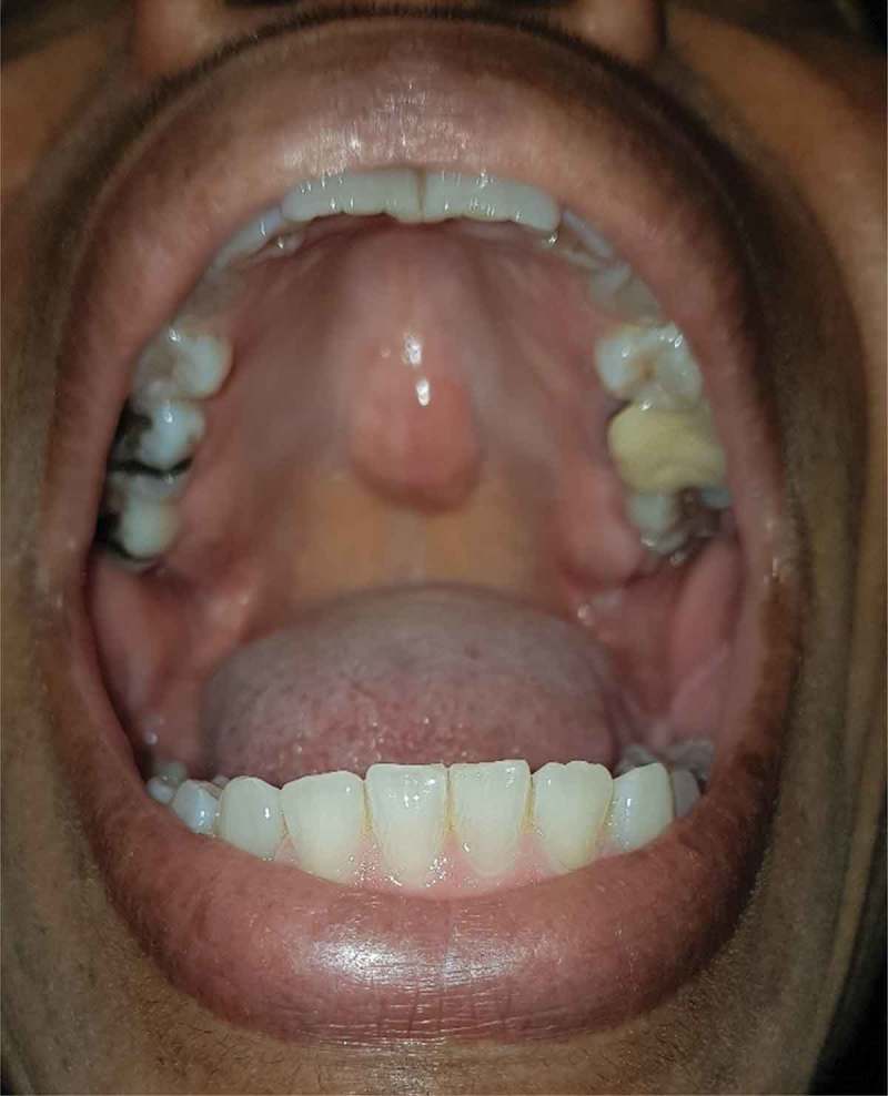
On physical examination, her vital signs were normal and the entirety of her physical examination was normal with the exception of her oropharynx. Throat examination revealed a 2 × 1 cm midline hard palate swelling that was bony hard in consistency and covered by normally appearing oral mucosa. It was diagnosed as a torus palatinus. The patient was unaware of its presence and denied being informed about it by her dentist on any visit. She was also educated about the diagnosis and safety-netted by being informed about red-flags that would prompt investigation such as change in size or shape.
KEYWORDS: Torus, palatinus, oral, tori, maxillofacial, palate, dental, mass, primary, benign
Oral tori (Figure 1) are common exostoses with a prevalence of about 20–30% in the general USA population. [1] Torus palatinus is more common in women and in people of Asian and Inuit ancestry. [2,3] The pathogenesis is not well-understood, and appears to be a complex interplay of occlusive (biting) forces, genetics, and environmental factors. [3–5] Medical conditions associated with bone disruption, e.g., hyperparathyroidism (of any type), have also been found to be associated with torus development[6]. This slowly growing benign lesion can take decades to grow into a noticeable size and patients generally do not report them as they are asymptomatic. [4] In the cases in which a torus interferes with dental work, such as fitting dentures or prosthetic devices, or in the process of mastication, surgical removal might be warranted. [3–5]
Figure 1.

Midline swelling seen protruding for the hard palate.
Beyond the interference with dental work, complications of oral tori are rare, and generally at the case report level. Oral tori have also been linked to low bone mineral density, but are felt to be secondary to the underlying process. [7] This may explain why torus palatinus is found more commonly in women than men. Tori have been reported to be subject to bisphosphonate-induced osteonecrosis, so this should be included in the differential of oral pain in the appropriate population[8].
1. Conclusion
A Torus Palatinus is a common, benign exophytic growth in the midline of the bony palate. Patients should be reassured about it when incidentally found to avoid any anxiety or unnecessary investigation.
Disclosure statement
No potential conflict of interest was reported by the authors.
References
- [1].Ladizinski B, Lee KC.. A Nodular Protuberance on the Hard Palate. JAMA. 2014;311(15):1558–1559. [DOI] [PubMed] [Google Scholar]
- [2].Hiremath VK, Husein A, Mishra N. Prevalence of torus palatinus and torus mandibularis among Malay population. J Int Soc Prev Community Dent. 2011. Jul-Dec;1(2):60–64. [DOI] [PMC free article] [PubMed] [Google Scholar]
- [3].Loukas M, Hulsberg P, Tubbs RS, et al. The tori of the mouth and ear: a review. Clin Anat. 2013. November;26(8):953–960. [DOI] [PubMed] [Google Scholar]
- [4].Komori T, Takato T. Time-related changes in a case of torus palatinus. JOMOS. 1998;56(4):492–494. [DOI] [PubMed] [Google Scholar]
- [5].Morrison MD, Tamimi F. Oral tori are associated with local mechanical and systemic factors: A case-control study. JOMOS. 2013;71(1):14–22. [DOI] [PubMed] [Google Scholar]
- [6].Padbury AD, Tözüm TF, Taba M, et al. The Impact of primary hyperparathyroidism on the oral cavity. J Clin Endocrinol Metab. 2006;91(9):3439–3445. [DOI] [PubMed] [Google Scholar]
- [7].Lo JC, O’Ryan F, Yang J, et al. Oral Health Considerations in Older Women Receiving Oral Bisphosphonate Therapy. J Am Geriatr Soc. 2011;59(5):916–922. [DOI] [PubMed] [Google Scholar]
- [8].Goldman ML, Denduluri N, Berman AW, et al. A Novel Case of Bisphosphonate-Related Osteonecrosis of the Torus Palatinus in a Patient with Metastatic Breast Cancer. Oncology. 2006;71:306–308. [DOI] [PubMed] [Google Scholar]


