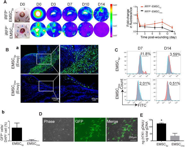
Figure 2.
Viability and engraftment of EMSC following their transplantation to wounds. (A) Representative bright-field images of wounds with transplanted iRFP-expressing EMSCSp (upper row) or EMSCDiss (bottom row) were taken on day 0, followed by fluorescent images acquired using the In-Vivo Xtreme System on various days following wound formation. The colored bar indicates the fluorescence intensity. The fold change in the fluorescent intensity is shown in the graph on the right. (B) (a) Immunostaining for GFP+ cells in wounds with transplanted Envy hESC-derived EMSCSp and EMSCDiss 14 days after wound formation. Boxed areas are amplified and shown on the right. (b) The percentage of GFP+ cells (determined via DAPI staining of the nucleus) per wound section. (C) Flow cytometry to detect GFP+ cells in wounds with transplanted Envy hESC-derived EMSCSp and EMSCDiss 7 and 14 days after wound formation. Values represent the percentage of GFP+ cells from 3 biological repeats. (D) Cultured cells were isolated following the digestion of wounds with transplanted EMSCSp (Envy) 7 days after wound formation. Bright-field (left) and fluorescent (middle) images of the cultured cells were taken and merged (right). Scale bar = 200 μm. (E) Detection of human TK1 gDNA in mice 14 days after EMSC transplantation. One x 106 EMSCSp and EMSCDiss cells were transplanted to the skin wound. After 14 days, wounded skin was dissected, and total gDNA was extracted for PCR to detect human TK1. n = 3 biological repeats. * P < 0.05 for EMSCSp versus EMSCDiss per student t-test.

