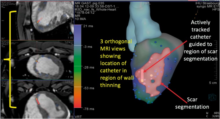
Figure 2.
Representative depiction of image-guidance platform showing three orthogonal MRI views demonstrating location of catheter in relation to LV endocardium, three-dimensional segmentation of the left ventricle derived from MRI and scar segmentation from LGE images to guide EAM. EAM, electroanatomic mapping; LGE, late gadolinium enhancement; LV, left ventricular; MRI, magnetic resonance imaging.

