Abstract
Calculus is a mineralized bacterial plaque that is formed on natural teeth surfaces where there is constant supply of saliva. Dental calculus is commonly seen over the buccal surfaces of maxillary molars and lingual surfaces of mandibular anterior teeth where the salivary duct opens into the oral cavity. This case report presents an unusual presentation of a large hard calcified mass in the left side of retromolar region associated with partially erupted tooth; hard mass was excised and examined histochemically which suggested the presence of calculus. Elimination of such nidus shall prevent formation of such calculus in such unusual position. This can also be achieved with proper oral hygiene measures.
Keywords: Dental calculus, nidus/foci, retromolar region
INTRODUCTION
Calculus is a calcified mass, most commonly seen in areas where the salivary duct opens into the oral cavity. It consists of mineralized dental plaque that forms on the natural teeth and dental prosthesis. It can be seen either supragingival or subgingival, and it is mainly composed of 80%–85% of inorganic content.[1] Supragingival calculus is whitish yellow and is usually clay-like in consistency, whereas subgingival calculus is not visible clinically but can be evaluated by tactile sensation. It is usually typically dark-brown or green or black and dense in consistency. Usually, the formation of dental calculus is mainly by mineral precipitation from a local rise in the degree of saturation of calcium and phosphate ions and also due to inducing of seeding agents to form small foci for calcification of dental plaque.
It is usually seen in young age and continues to be deposited till 25–30 years where they exhibit maximal deposition. The areas to exhibit calculus deposits were the facial aspect of maxillary molars and lingual surface of mandibular teeth. In spite of these regular places, calculus can be presented in an unusual location of the oral cavity where maintenance is very difficult.[2] This case report reveals an unusual presentation of dental calculus in the left side of retromolar region associated with a partially erupted left mandibular third molar tooth.
CASE REPORT
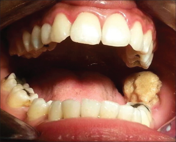
A 25-year-old male patient reported with a history of discomfort and pain in the left lower tooth region for the past 3 months along with difficulty in closing his mouth. On examination, there was a large yellowish hard mass extending from the distal of first molar to the retromolar region with an approximate size of 3 cm × 2 cm in diameter, which was associated with inflammation and purulent discharge [Figure 1].
Figure 1.

Hard mass of calculus in the retromolar area
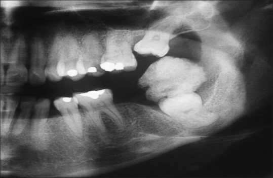
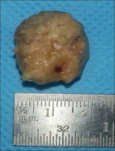
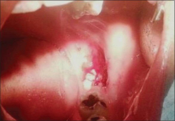
Orthopantomograph [Figure 2] revealed a radiopaque mass involving the embedded tooth (tooth 38) along with incomplete eruption of tooth 28. After routine blood investigations, the calcified mass was surgically removed [Figure 3] under local anesthesia and sent for histochemical and biochemical study. Histochemical and biochemical examination of the specimen and salivary examination revealed the presence of calcium (42%), phosphates (18%), and calcium phosphate (70%), which was suggestive of dental calculus. The patient was recalled and educated for proper maintenance of oral hygiene and advised to report for regular follow-up [Figure 4].
Figure 2.

Orthopantomogram showing the radiopaque mass of calculus impinging on the unerupted molar
Figure 3.

Hard mass of 3 cm × 2 cm of calculus
Figure 4.

Postoperative view
DISCUSSION
Dental calculus is a calcified dental plaque, which usually occurs between 1 and 14 days of plaque formation, usually reaching 60%–90% of calcification by 12 days.[3] The main source of mineralization is from saliva. Several reasons have been proposed such as increase in pH of saliva and precipitation of colloidal proteins in saliva and seeding agent inducing the foci of calcification. Dental calculus does not contribute directly to gingival inflammation, but it provides nidus for the continued accumulation of plaque.
In the early 17th century, Pierre Fauchard[4] in his classic treatise “Le Chirugren Dentiste” reported a completely submerged molar tooth in a large piece of calculus, of 20 times the size of the molar itself. Only in ancient times, such types of massive and unusual deposition of calculus have been reported in dentistry. This is a rare phenomenon to be seen today with our ever-changing lifestyles, with the availability of top-notch dental care systems, highly effective oral hygiene products besides dignity in oral cleanliness, and personal pride.
In this case report, the patient had a myth that the deposition was related to some other lesion and also was hesitant to meet the dentist until he felt difficulty in closing his mouth. The unusual amount of formation of calculus seen in this patient goes well beyond the classification of heavy calculus formation as reported by Mandel.[5] The deposition near the floor of the mouth supports the fact that saliva from submandibular gland shall have provided the inorganic content for calculus formation. Hence, the presence of calculus in this site correlates with McGaughey et al., who proposed that calcium and potassium formation in calculus is due to the protein bond in saliva.[6] However, in this case report, the partially erupted tooth, which was embedded in the retromolar area, provided a nidus for the formation of dental plaque, because of difficulty in tooth brushing, leading to continuous deposition of plaque and mineralization leading to the formation of calculus as there is a constant supply of saliva.[7]
The mass formation can be because of partially erupted 38 and also due to the absence of opposing teeth (27, 28) and also the adjoining tooth (37). Further, there are missing teeth 37 and 27 which was not prosthetically replaced may have led the patient not to utilize the left side ultimately leading to the formation of such a large mass of calculus without any hindrances. Presence of large chunk of calculus made the patient, further difficulty in cleansing the area and thereby leading to formation of calculus to the present size. Moreover, tooth eruption is hampered by various reasons such as cyst, thickened bone, thickened gingiva, and supernumerary teeth, but calculus has not been mentioned in the literature.[8] The presence of large hard mass of calculus could have been a confounding factor along with mesial drift of 28, due to missing 27 and also leading to incomplete eruption, which has not been reported so far in literature.
Schroeder in his classic in vitro studies showed biochemical evidence of the presence of calcium and phosphates in the calculus specimen he examined. Saliva has been the source of the inorganic content and also upon biochemical analysis of saliva revealed more concentration of inorganic content such as calcium, phosphates, and oxylates which are more suggestive of calculus (Moskow). Saliva is the source for mineralization of supragingival calculus. It has been shown that the level of calcium in calculus is 20% more than in the saliva In this case report, when the hard mass which is present supragingivally, when analyzed biochemically it was found out that there was the presence of higher concentrations of calcium and phosphates which correlates well with the studies of Schroeder[9] and Hidaka et al.[10]
With the availability of state-of-the-art dental care and more effective oral hygiene practices along with personal pride, the report of calculus deposition in such grandeur is rarely seen nowadays. This case report is one of the rarest and unusual presentations especially in the retromolar area along with an embedded molar tooth which led to the deposition of calculus, which is normally seen in natural teeth positions. Moreover, the size of the calculus is the largest to best of our knowledge as reported in the literature so far.
CONCLUSION
This case report gives us a glimpse about the deposition of calculus not only in regular tooth positions but also in such unusual areas too. Elimination of predisposing factors such as embedded deciduous teeth or broken fragments and regular oral prophylaxis and maintenance shall prevent such formation of calculus in the oral cavity. Regular follow-up and maintenance should be the utmost aim of any periodontal treatment for treating such patients with poor oral hygiene.
Declaration of patient consent
The authors certify that they have obtained all appropriate patient consent forms. In the form the patient(s) has/have given his/her/their consent for his/her/their images and other clinical information to be reported in the journal. The patients understand that their names and initials will not be published and due efforts will be made to conceal their identity, but anonymity cannot be guaranteed.
Financial support and sponsorship
Nil.
Conflicts of interest
There are no conflicts of interest.
REFERENCES
- 1.White DJ. Dental calculus: Recent insights into occurrence, formation, prevention, removal and oral health effects of supragingival and subgingival deposits. Eur J Oral Sci. 1997;105:508–22. doi: 10.1111/j.1600-0722.1997.tb00238.x. [DOI] [PubMed] [Google Scholar]
- 2.Whittaker DK, Molleson T, Nuttall T. Calculus deposits and bone loss on the teeth of Romano-British and eighteenth-century Londoners. Arch Oral Biol. 1998;43:941–8. doi: 10.1016/s0003-9969(98)00086-7. [DOI] [PubMed] [Google Scholar]
- 3.Mandel ID. Biochemical aspects of calculus formation. I. Comparative studies of plaque in heavy and light calculus formers. J Periodontal Res. 1974;9:10–7. doi: 10.1111/j.1600-0765.1974.tb00647.x. [DOI] [PubMed] [Google Scholar]
- 4.Spielman AI. The birth of the most important 18th century dental text: Pierre Fauchard's le Chirurgien Dentiste. J Dent Res. 2007;86:922–6. doi: 10.1177/154405910708601004. [DOI] [PubMed] [Google Scholar]
- 5.Moskow BS. A case report of unusual dental calculus formation. J Periodontol. 1978;49:326–31. doi: 10.1902/jop.1978.49.6.326. [DOI] [PubMed] [Google Scholar]
- 6.Schroeder HE. Formation and inhibition of dental calculus. J Periodontol. 1969;40:643–6. doi: 10.1902/jop.1969.40.11.643. [DOI] [PubMed] [Google Scholar]
- 7.Gaare D, Rølla G, Aryadi FJ, van der Ouderaa F. Improvement of gingival health by toothbrushing in individuals with large amounts of calculus. J Clin Periodontol. 1990;17:38–41. doi: 10.1111/j.1600-051x.1990.tb01045.x. [DOI] [PubMed] [Google Scholar]
- 8.Hicks MJ, Greer RO, Jr, Flaitz CM. Delayed eruption of maxillary permanent first and second molars due to an ectopically positioned maxillary third molar. Pediatr Dent. 1985;7:53–6. [PubMed] [Google Scholar]
- 9.Sidaway DA. A microbiological study of dental calculus. III. A comparison of the in vitro calcification of viable and non-viable microorganisms. J Periodontal Res. 1979;14:167–72. doi: 10.1111/j.1600-0765.1979.tb00787.x. [DOI] [PubMed] [Google Scholar]
- 10.Hidaka S, Okamoto Y, Abe K. Possible regulatory roles of silicic acid, silica and clay minerals in the formation of calcium phosphate precipitates. Arch Oral Biol. 1993;38:405–13. doi: 10.1016/0003-9969(93)90212-5. [DOI] [PubMed] [Google Scholar]


