Abstract
Granulocyte colony-stimulating factor (G-CSF)-producing bladder cancer is rare, with only 75 cases reported in Japan. A 67-year-old woman was referred to our institution for the further examination of gross hematuria. Cystoscopy revealed a 7-cm bladder tumor. The initial white blood cell count was 17,100/μL, and a transurethral resected specimen showed G-CSF expression. CT revealed that the tumor had invaded the colon. As the patient had uncontrollable schizophrenia, radical cystectomy was abandoned. We herein report a case of G-CSF-producing bladder tumor.
Keywords: G-CSF, Bladder tumor, Granulocyte colony-stimulating Factor
Introduction
Granulocyte colony-stimulating factor (G-CSF) is a hematopoietic growth factor required for the proliferation and differentiation of hematopoietic precursors of neutrophil granulocytes [1].
G-CSF-producing bladder tumor is still a rare entity, and only 75 cases have been reported in Japan, all showing a poor prognosis [2, 3, 4, 5]. Due to the rarity of this disease, there are no established treatments.
We herein report a rare case of G-CSF-producing bladder tumor.
Case Presentation
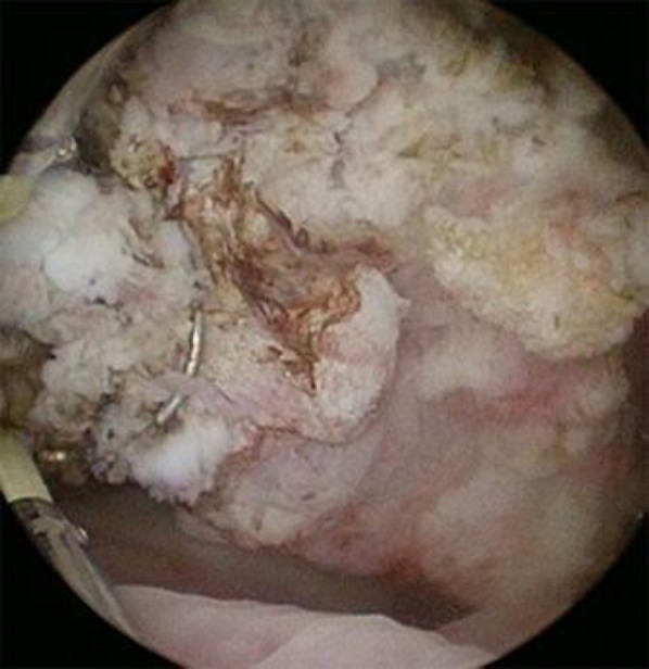
A 67-year-old woman was referred to our hospital for the further examination of gross hematuria. Cystoscopy revealed a 7-cm bladder tumor in her left bladder wall (Fig. 1). Urinary cytology showed class IIIb. She had a history of schizophrenia for 45 years. Laboratory data showed almost normal findings, except for an elevated white blood cell (WBC) count (17,100/μL; Neutro 79.8%, Baso 1.1%, Eos 1.1%, Mono 6.8%, Lymph 11.2%). CT and MRI revealed a 2.2-cm left iliac lymph node metastasis and suspected colon invasion (Fig. 2).
Fig. 1.

Cystoscopy findings.
Fig. 2.
MRI (a) diffusion and (b) axial and (c) coronal images of MRI T2 weighted image.
Two months after her initial visit, transurethral resection of bladder tumor (TUR-Bt) was performed. The tumor occupied from the left side wall to the anterior wall, with a 7- to 8-cm diameter. A pathological examination revealed highly invasive urothelial carcinoma with squamous cell differentiation (Grade 3, High Grade) with partial expression of G-CSF (Fig. 3). Due to her schizophrenia and lymph node metastasis, she and her family sought no further treatment. She ultimately died of bladder cancer 6 months after her initial visit (Fig. 4).
Fig. 3.
(a) HE staining and (b) immunohistochemistry of G-CSF of the tumor. (a) High grade urothelial carcinoma cells were seen in the left, and keratinizing squamous components in the right. (b) Immunohistochemically, G-CSF was focally observed in the tumor cells.
Fig. 4.
Clinical course.
Discussion
Colony-stimulating factors promote WBC differentiation and proliferation through stem cells in the bone marrow [6]. G-CSF is one such colony-stimulating factor produced by macrophages and fibroblasts.
G-CSF-producing tumors secrete cytokines and increase the WBC count. The lung is the most frequent site of G-CSF-producing tumor formation, followed by the stomach, thyroid, and liver. G-CSF-producing tumors are diagnosed by the following four criteria: an elevated WBC count in the peripheral blood, an elevated serum G-CSF level, G-CSF expression in the tumor tissue, and a reduction in the WBC count or G-CSF expression in the serum following tumor resection or treatment [6]. Recent technology has facilitated the confirmation of the expression of G-CSF using an enzyme immunoassay (EIA) to detect serum G-CSF levels [3]. The present case showed an elevated WBC count, and immunohistochemistry detected G-CSF expression in the tumor tissue.
Previous reports have shown that the WBC count and serum G-CSF level were correlated with G-CSF-producing cancer progression, so these two factors have been considered tumor markers. Most reported G-CSF-producing tumors have shown a poor outcome. G-CSF-producing tumors tend to display G-CSF receptors, so autocrine tumor progression and resulting in introducing tumor progression.
A low pre-therapeutic serum G-CSF level and control of the WBC count after tumor resection are thought to be favorable outcome factors [7]. The present case showed a fever and elevated serum CRP level. While some previously reported cases have shown systemic inflammation, G-CSF itself does not induce a fever or CRP elevation. The mechanism underlying the appearance of a fever and CRP elevation is thought to involve the production of systemic cytokines, including IL-1 and IL-6, by this aggressive tumor. Some inflammation was also observed [4].
Availability of Data and Material
Due to ethical restrictions, the raw data underlying this paper are available upon request to the corresponding author.
Statement of Ethics
Written informed consent to participate and for publication was obtained from the patient and all methods were followed by the ethical standards of the Declaration of Helsinki.
Disclosure Statement
The authors declare no conflicts of interest
Funding Sources
None.
Author Contributions
RM, TK. SK, YI are responsible for the concept and drafted the manuscript.
HU provided the intellectual content and critically reviewed the manuscript.
References
- 1.Xu S, Höglund M, Hâkansson L, Venge P. Granulocyte colony-stimulating factor (G-CSF) induces the production of cytokines in vivo. Br J Haematol. 2000 Mar;108((4)):848–53. doi: 10.1046/j.1365-2141.2000.01943.x. [DOI] [PubMed] [Google Scholar]
- 2.Osaka K, Kobayashi M, Takano T, Tsuchiya F, Iwasaki A, Ishizuka E, et al. [Two cases of granulocyte-colony stimulating factor-producing infiltrating urothelial carcinoma of the kidney] Hinyokika Kiyo. 2009 Apr;55((4)):223–7. [PubMed] [Google Scholar]
- 3.Shimamura K, Fujimoto J, Hata J, Akatsuka A, Ueyama Y, Watanabe T, et al. Establishment of specific monoclonal antibodies against recombinant human granulocyte colony-stimulating factor (hG-CSF) and their application for immunoperoxidase staining of paraffin-embedded sections. J Histochem Cytochem. 1990 Feb;38((2)):283–6. doi: 10.1177/38.2.1688901. [DOI] [PubMed] [Google Scholar]
- 4.Tachibana M, Murai M. G-CSF production in human bladder cancer and its ability to promote autocrine growth: a review. Cytokines Cell Mol Ther. 1998 Jun;4((2)):113–20. [PubMed] [Google Scholar]
- 5.Ohtaka M, Kawahara T, Ishiguro Y, Sharma M, Yao M, Miyamoto H, et al. Expression of receptor activator of nuclear factor kappa B ligand in bladder cancer. Int J Urol. 2018 Oct;25((10)):901–2. doi: 10.1111/iju.13756. [DOI] [PubMed] [Google Scholar]
- 6.Asano S, Urabe A, Okabe T, Sato N, Kondo Y. Demonstration of granulopoietic factor(s) in the plasma of nude mice transplanted with a human lung cancer and in the tumor tissue. Blood. 1977 May;49((5)):845–52. [PubMed] [Google Scholar]
- 7.Matsuzaki K, Okumi M, Kishimoto N, Yazawa K, Miyagawa Y, Uchida K, et al. [A case of bladder cancer producing granulocyte colony-stimulating factor and interleukin-6 causing respiratory failure treated with neoadjuvant systemic chemotherapy along with sivelestat] Hinyokika Kiyo. 2013 Jul;59((7)):443–7. [PubMed] [Google Scholar]
Associated Data
This section collects any data citations, data availability statements, or supplementary materials included in this article.
Data Availability Statement
Due to ethical restrictions, the raw data underlying this paper are available upon request to the corresponding author.


