Abstract
It has emerged that neutrophils can play important roles in the host response following infection with helminth parasites. Mice infected with the tapeworm, Hymenolepis diminuta, are protected from some inflammatory conditions, accompanied by reduced neutrophil tissue infiltration. Thus, the ability of a phosphate-buffered saline-soluble extract of the worm (H. diminuta extract [HdE]) was tested for (1) its ability to activate murine neutrophils (Ca2+ mobilization, reactive oxygen species (ROS) and cytokine production); and (2) affect neutrophil chemotaxis in vitro to the penta-peptide, WKYMVm, the chemokine, KC, and leukotriene B4. HdE was not cytotoxic to neutrophils, elicited a Ca2+ response and ROS, but not, cytokine (KC, interleukin-10, tumour necrosis factor-α) generation. HdE is not a chemotactic stimulus for murine neutrophils. However, a heat- and trypsin-sensitive, acid-insensitive proteoglycan (sensitive to sodium metaperiodate) in the HdE significantly reduced neutrophil chemotaxis towards WKYMVm or KC, but not LTB4. The latter suggested that the HdE interfered with p38 mitogen-activated protein kinase signalling, which is important in WKYMVm chemotaxis. Corroborating this, immunoblotting revealed reduced phosphorylated p38, and the downstream signal heat-shock protein-27, in protein extracts from HdE + WkYMVm treated cells compared to those exposed to the penta-peptide only. We speculate that HdE can be used to modify the outcome of neutrophilic disease and that purification of the bioactive proteoglycan(s) from the extract could be used as a template to design immunomodulatory drugs targeting neutrophils.
Keywords: Chemotaxis, Neutrophils, Parasite-host interactions
Introduction
The neutrophil has traditionally been considered a first-line responder to microbial infections, arriving pre-armed with proteases and rapid reactive oxygen species (ROS) generation to kill the invader and with the potential to do considerable collateral damage to host tissue. This is but one of the neutrophils roles, with studies demonstrating important functions in innate immunity and interaction with components of adaptive immunity [1]. Kubes recently detailed the importance of the often-maligned neutrophil in immunity, drawing attention to issues that require clarification such as commonalities and differences in responding to bacterial products versus damaged host tissue, tissue-specific neutrophil activity and the possibility of neutrophil subtypes, among others [1]. With respect to inflammation, recruitment and activation of the neutrophil is often a critical component of acute inflammation; however, in the setting of chronic inflammation, where their capacity to do damage goes unchecked, the neutrophil can contribute to disease. Thus, there can be benefits to promoting neutrophil responses as an immediate reaction to infection and benefits to inhibiting their activity in the setting of chronic inflammatory disease [1].
Based on its classification as an anti-microbial cell, the neutrophil is often overlooked, or dismissed, when considering effector mechanisms mobilized in response to infection with parasitic helminths. This position needs to be re-evaluated. Neutrophils have been implicated in an effective response to the larval stages of the nematodes Lithomosoides sigmodontis, Haemonchus contortus, and Strongyloides steroralis, which can involve myeloperoxidase (MPO) and the release of extracellular traps (i.e., proteases embedded in a chromatin net) [2, 3, 4, 5]. Mice infected with Ascaris have increased numbers of neutrophils in their lungs at the peak of helminth migration (and tissue damage) through the airways [6]. Similar findings were presented for Nippostronglyus brasiliensis-infected mice, and suppression of this neutrophil response resulted in reduced mobilization of macrophages capable of killing the parasite and alternatively activated macrophages (AAMs) [7]. In accordance with this, human and mouse neutrophils have been shown to collaborate with macrophages to kill S. steroralis larvae in vitro [8]. Finally, in the reciprocal direction of communication, the chitinase-like protein Ym1, that characterizes the murine AAM, has been shown to recruit neutrophils that contributed to the control of N. brasiliensis [9].
A substantial body of evidence illustrates the benefit of infection with helminth parasites in murine models of colitis, diabetes, multiple sclerosis and arthritis [10]. Suppression of inflammatory disease in these model systems can also be achieved by systemic delivery of extracts of the parasites or helminth-derived excretory-secretory products [11]. Using the non-permissive mouse host, we have shown that infection with the rat tapeworm, Hymenolepis diminuta, significantly reduced the severity of dinitrobenzene sulphonic acid-induced colitis (DNBS) [12] and complete Freund's adjuvant (CFA)-induced arthritis [13]. The amelioration of disease in both models was accompanied by lower tissue levels of the neutrophil marker, MPO, and in the case of CFA-induced arthritis, the H. diminuta-infected mice had fewer blood neutrophils. The reduced neutrophilia could reflect the impact of host-derived factors (e.g. interleukin [IL]-10), or be mediated by bioactive molecules from the helminth. Consequently, we designed the current study to test the hypothesis that an extract of H. diminuta would directly affect neutrophils and their chemotaxis.
Materials and Methods
Animals
Male BALB/c mice (6–8 weeks old) were purchased from Charles River Laboratories (Quebec, Canada) and housed under standard conditions with free access to food and water. Animal experiments were conducted with approval from the University of Calgary Health Science Animal Care Committee conforming to national guidelines, under protocol AC13-0015 issued to D.M. McKay.
Preparation of Hymenolepis diminuta Crude Extract
Adult H. diminuta from rats were rinsed in 0.9% NaCl, and ∼20 g wet weight homogenized in 20 mL of sterile phosphate-buffered saline (PBS; 5 min, maximum speed, Polytron PT1200 homogenizer [Kinematica, Inc., New York, NY, USA]). The homogenate was centrifuged (3,220 g, 30 min, 4°C), pelleted material discarded, the supernatant collected and subjected to 2 additional rounds of centrifugation. The PBS-soluble supernatant was collected, designated H. diminuta extract (HdE), the protein concentration determined by the Bradford assay, and aliquots stored at −80°C [14]. Bioactivity of the HdE was confirmed by its ability to suppress LPS-stimulated production of tumour necrosis factor-α (TNF-α) by human THP-1 macrophages (2 × 10⁵ PMA [10 nM, 24 h]-activated THP-1 cells were exposed to HdE [100 μg/mL] for 30 min and then activated with 10 ng/mL Escherichia coli-derived LPS. Cell-free conditioned medium was collected 24 h later and TNF-α measured by enzyme-linked immunosorbent assay [ELISA]) [14]. The HdE preparation contains < 100 pg LPS/1 mg of HdE protein. The in vivo (1 mg) and in vitro (100 μg) doses of HdE were based on previous studies [13, 14] and a dose-response experiment (see Results).
Assessing the nature of the bioactive molecule in the HdE, the following manipulations were performed: (1) boiled for 15 min; (2) trypsin-treated (1 unit/μg of HdE protein [Sigma-Aldrich] while rocking for 6 h at room temperature [RT], followed by neutralization with soybean trypsin inhibitor [1 unit inhibitor: 1 unit trypsin; 6 h rocking at RT]); (3) acidified with 2N HCL to pH 2 (30 min at RT followed by normalizing to pH 7 with 2N NaOH); or, (4) treated with sodium metaperiodate (SMP; Sigma-Aldrich) to disrupt glycans (HdE treated with 10 mM SMP for 30 min with rocking, following by repeated dialysis [Thermo Fisher] to remove excess SMP) [14]. In all instances, time-matched control HdE was treated identically with the absence of the active reagent.
LPS-Induced Neutrophil Recruitment to Mouse Knee
After hair removal around the knee, mice received either an intra-articular (ia.) injection of 50 ng E. coli lipopolysaccharide (LPS; 10 μL) (Sigma-Aldrich) or 0.9% NaCl (pyrogen free [Baxter Healthcare]) as a control. Some mice were co-treated with HdE (1 mg, intraperitoneal [ip.] in 1 mL saline). Six hours after LPS ± HdE, the knee and peritoneal cavity were washed with PBS and lavages collected, and blood and bone marrow (1 femur) were collected. The total number of leukocytes was determined by counting in a Neubauer chamber after staining with Turk's solution. Differential counts were obtained from cytospin (Shandon III, Thermo Shandon, Frankfurt, Germany) preparations by evaluating the percentage of each class of leukocyte based on nuclear morphology following staining with May-Grünwald-Giemsa [15].
Neutrophil Function in vitro
Cell Viability
Neutrophil necrosis was assessed via staining with trypan blue (0.4%), and measurement of released lactate dehydrogenase using the CytoTox 96® kit and following the manufactures instructions (Promega).
Calcium Mobilization
Mice were humanely euthanized, bone-marrow extracted from the femurs and tibias under sterile conditions and neutrophils isolated via a differential Percoll gradient following the protocol of Swamydas et al. [16] Neutrophils (2.5 × 10⁶ cells/mL) were incubated with the intracellular Ca2+ indicator dye Fluo-4 (2.5 μM, Sigma-Aldrich) for 45 min at RT while gently shaking, then centrifuged at 300 g for 10 min. The supernatant was discarded and the cells re-suspended in Hank's balanced salt solution ± Ca2+. Aliquots of 105 cells (100 μL) in 1 mL cuvettes were placed in a fluorescence spectrophotometer (Cole-Parmer, Montreal, Canada) and treated with HdE (100 μg/mL), the pentapeptide WKYMVm (1.0 μM, Tocris Bioscience), calcium ionophore, A23187 (2.5 μM, Sigma-Aldrich) or zymosan (1 μg/mL, Sigma Aldrich), as positive controls. Cells were excited at 480 nM and fluorescence measured at 530 nM, and recorded as the peak increase to occur within 3 min of treatment.
Mitochondrial Respiration
Neutrophil bioenergetics was determined using the Seahorse Bioscience extracellular flux analyzer (Agilent, Santa Clara, CA, USA), which measures O2 consumption and proton flux. Overnight hydrated XFe24 probes in calibrant solution were loaded with oligomycin (0.5 μg/mL; inhibits ATP synthase), carbonyl cyanide-4-(trifluoromethoxy)phenylhydrazone (FCCP, 0.6 μg/mL; H+ ionophore uncoupling oxidative phosphorylation) and antimycin (10 μM; binds to the Qi site of cytochrome c reductase (compel III in electron transport chain)) in ports B, C and D respectively. Port A was filled with HdE (100 μg/mL). Experimental media was used as control. Neutrophils were plated on XFe24 plates. The cartridge and cells were equilibrated in a 37°C, non-CO2 incubator for 1 h prior to assay. The oxygen consumption rate was monitored continuously and proton leak and ATP levels determined following the manufacturer's instructions and as described by Dranka et al. [17], with slight modification. We replaced the mixing step with longer incubation to avoid neutrophil aggregation. The results were plotted using Wave software from Agilent.
Reactive Oxygen Species Generation
Neutrophils (108 cells/mL) were re-suspended in 20 μM 2',7'-dichlorofluorescin diacetate (Abcam, Eugene, OR, USA), a fluorogenic dye that measures hydroxyl, peroxyl and other ROS, for 30 min at 37°C [18]. Then 105 cells/100 μL were added to each well of a dark, clear-bottom 96 well plate (Thermo Fisher) and treated with HdE (100 μg/mL), E. coli-derived LPS (1 μg/mL; Sigma-Aldrich) or both agents. In addition, some wells received polymyxin B (10 μg/mL; Sigma-Aldrich) to neutralize LPS. Plates were incubated at 37°C for 4 h and fluorescence read in a Victor-5 multi-label plate reader (excitation 535 nm, emission 485 nm) (Perkin Elmer).
Enzyme-Linked Immunosorbent Assay
Neutrophils (105/mL) were exposed to HdE (100 μg/mL), E. coli-derived LPS (1 μg/mL) or E. coli (strain HD5-α; 106 colony-forming units [America Type Culture Collection]). Four h later, supernatants were collected and levels of TNF-α, IL-10, and KC measured in duplicate samples by ELISA with DuoSet® kits from R&D Systems (Minneapolis, MN, USA), and following the manufacturer's instructions.
In other experiments, PBS or HdE (100 μg in 100 μL) was injected into the peritoneal cavity of mice and 6 h later the cavity washed with PBS, a 1 mL lavage retrieved and KC levels determined by ELISA.
Neutrophil Chemotaxis
Under Agarose Gel Assay
An ultrapure agarose gel was prepared as previously described [19] and 3 mL aliquots were added to plasma-treated 35 mm diameter Petri culture dishes (Waltham), which were left to harden for 45 min at RT, at which time a centre well and one to the right and left of centre were cut. The chemoattractant, WKYMVm (1 μM) or HdE (100–1,000 μg/mL) was added into the centre well and neutrophils (104) placed in the outer wells. In other experiments, neutrophils were treated with HdE (100 μg/mL) for 30 min, then rinsed prior to use in the chemotaxis assay. The number of neutrophils that moved into the space between neutrophil and WKYMVm chambers at the end of a 4 h incubation at 37°C was counted (duplicate neutrophil samples were used from each mouse and the average number of migrating neutrophils determined).
Transwell Assay
Chemoattractants (WKYMVm [2 μM], KC [keratinocyte-derived chemokine, or CXCL1 or GROα; 20 nM] and leukotriene B4 [LTB4, 100 nM]) were diluted in serum-free RPMI culture medium and added to the basal chamber of 24-well culture plates that contained 8-μm porous filter supports (Fisher, Scientific). Neutrophils (2 × 10⁵) were placed in the upper compartment of the transwell, the plate placed at 37oC for 4 h and subsequently neutrophils that had migrated into the basal chamber were retrieved and counted on a hemocytometer [20]. Neutrophils were either pre-treated with HdE (100 μg/mL) for 30 min prior to use or HdE was added to the apical chamber of the transwell coincident with the application of the neutrophils (duplicate neutrophil samples were assessed from each mouse).
In separate experiments, the pan-caspase inhibitor, Z-VAD (20 μM), LPS (1 μg/mL), A23187 (2.5 μM), the general antioxidant, n-acetylcystine (NAC, 1 μM) or the mitochondria-targeted antioxidant, MitoTEMPO (10 μM, Sigma-Aldrich) were used to assess roles for apoptosis, activation and ROS in the HdE-elicited suppression of neutrophil chemotaxis respectively.
Protein Detection on Immunoblotting
Protein samples from neutrophil whole-cell extracts were assessed using a standard immunoblotting protocol, with the following antibodies: rabbit polyclonal phospho-p38 primary antibody (1: 1,000, Cell Signalling, #9211); rabbit polyclonal β-actin antibody (1: 1,000, Abcam, #8227); rabbit polyclonal phospho-heat-shock protein (HSP)27 (1: 1,000, Cell Signaling, #2401).
Statistics and Data Presentation
Data comprised of mean ± SD. Data sets with n ≥5 were subjected to the Brown-Forsythe test for normal distribution and were analyzed by parametric one-way analysis of variance with Tukey's post-test for multiple group comparison or non-parametric Kruskal-Wallis test with Dunn's post-test or Student t test for two data sets using Graph Prism 6 statistical package. p < 0.05 was accepted as a level of statistically significant difference.
Results
HdE Attenuates LPS-Induced Neutrophil Recruitment to the Knee
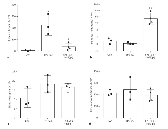
Lavage of knees showed that LPS induced robust recruitment of immune cells into the cavity by 6 h post-treatment, with differential cell counts revealing that the increase in leucocytes was almost entirely due to neutrophils: co-treating animals with HdE attenuated this neutrophil recruitment (Fig. 1a). Analysis revealed that HdE resulted in significant accumulation of neutrophils in the peritoneal cavity (Fig. 1b). As a percentage of total cells in the peritoneum, neutrophils were control = 1 ± 0.3%, LPS (ia.) = 3 ± 1.2%, and LPS (ia.) + HdE (ip.) = 76 ± 4%. The neutrophil recruitment in response to injection of HdE could be due, in part, to evoked production of KC by resident cells in the peritoneum (PBS treated mice = 168 ± 113 compared to HdE [100 μg, 6 h] treated mice = 1,891 ± 200* KC pg/mL [mean ± SEM, n = 3, * p < 0.05]). However, neither the numbers nor percentages of neutrophils in the blood and bone marrow were different between the experimental groups (Fig. 1c, d), suggesting that the HdE-evoked reduction in neutrophils into the knee following injection of LPS was not simply due to diversion of the cells to the peritoneal cavity.
Fig. 1.
H. diminuta extract (HdE) attenuates LPS-induced neutrophil recruitment to the knee. Lipopolysaccharide (LPS, 50 ng/10 μL) or sterile phosphate-buffered saline (PBS) were injected into the knee cavity (intra-articular [ia.]) ± co-treatment with HdE (1 mg/1 mL intraperitoneal [ip.]) or vehicle. Six hours later the knee cavity was washed to collect cells (a; representative of 3 experiments). Cells were also collected from the peritoneal cavity by washing (b), peripheral blood (c) and bone marrow from one femur (d). Neutrophils were identified by Wright-Giemsa staining of cytospin total cell preparations (data are mean ± SD; each datum point is an individual mouse; * and #, p < 0.05 compared to control [con] and LPS respectively, by one-way analysis of variance with Tukey's post-test).
HdE Is Not Cytotoxic on Murine Neutrophils
Giemsa staining and nuclear morphology analysis revealed that the population of cells isolated from bone marrow was 88–94% neutrophils (n = 4). Neutrophils tend not to survive well in vitro (hence the 4-h time-point in the following experiments); however, we observed no additional cytotoxic effects of HdE on murine bone marrow-derived neutrophils as gauged by exclusion of the vital dye, trypan blue, or lactate dehydrogenase release (Table 1).
Table 1.
HdE is not preferentially cytotoxic to murine bone marrow-derived neutrophils over a 16-h period
| Control | HdE, μg/mL |
Staurosporine 1 μM | Distilled H2O | ||||||||
|---|---|---|---|---|---|---|---|---|---|---|---|
| 100 |
500 |
1,000 |
|||||||||
| 4h | 16h | 4h | 16h | 4h | 16h | 4h | 16h | 4h | 16h | 4h | |
| % viable | 92±2 | 47±4 | 90±4 | 45±9 | 90±4 | 43±4 | 90±5 | 45±6 | 88±4 | 10±5 | nt |
| LDH (AU ×105) | 3.0±0.8 | nt | 3.4±0.9 | nt | 3.5±0.7 | nt | 2.7±0.7 | nt | nt | nt | 5.1±1.0 |
Data is mean ± SD, n = 4 mice for trypan blue % viability and n = 6 mice for lactate dehydrogenase (LDH); AU, arbitrary units; nt, not tested; h, hour; duplicate samples of neutrophils/mouse; HdE, Hymenolepis diminuta extract.
HdE Activates Murine Neutrophils
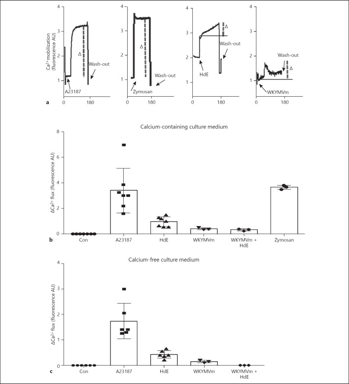
Calcium mobilization was chosen as a general indicator of cell responsiveness, with A23187- and zymosan-treated neutrophils displaying a robust response; WYKMVm evoked a response of lesser magnitude (Fig. 2a, b). Preliminary observations revealed that the addition of HdE alone to the cuvette (no neutrophils) gave an increase in signal in the spectrophotometer most likely due turbidity of the solution, and so the neutrophil response to HdE is the gradual increase in signal after the initial spike; a very different Ca2+ mobilization event as compared to A23187 or zymosan (Fig. 2a). HdE also evoked a Ca2+ response when experiments were conducted in Ca2+-free medium, suggesting the capacity to mobilize Ca2+ from intracellular stores (Fig. 2c).
Fig. 2.
H. diminuta extract (HdE) mobilizes calcium in murine bone marrow neutrophils. Neutrophils (105) were loaded with Furo-4 (2.5 μM) and then challenged with the calcium ionophore, A231877 (2.5 μM), zymosan (1 μg/mL), HdE (100 μg/mL) or the peptide WKYMVm (1 μM) and the maximum increase in fluorescence measured in arbitrary units (AU) that occurred within 3 min was recorded. a Representative tracings from the spectrophotometer. b and c show the peak responses (see Δ and dashed line on a) recorded in Ca2+-containing and Ca2+-free medium, respectively (data are mean ± SD; each datum is the average of 2 neutrophil replicates from an individual mouse).
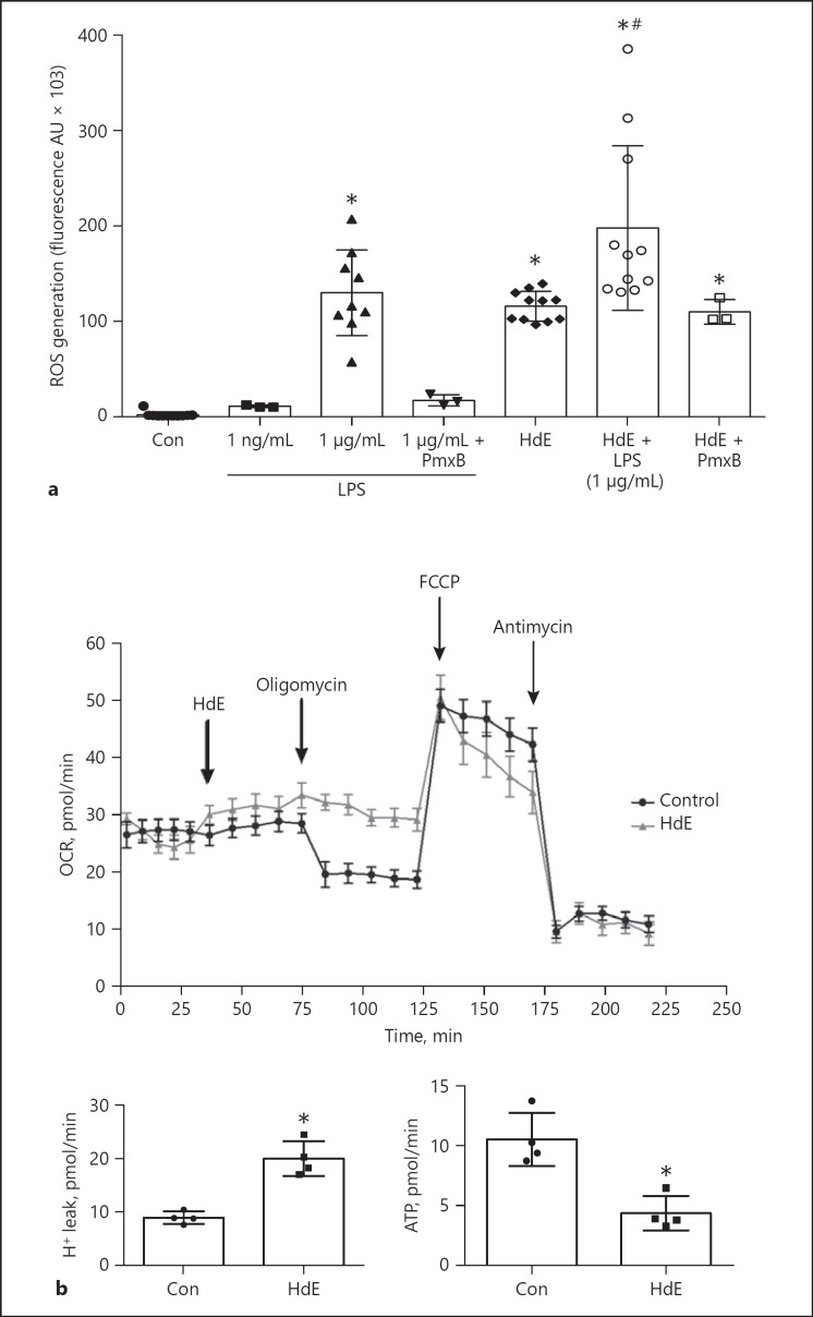
Calcium mobilization is important in ROS generation and HdE (100 μg/mL) was as effective as LPS (1 μg/mL) in evoking ROS in neutrophils: treatment with both agents induced a significantly greater respiratory burst than either agent alone (Fig. 3). Assessing the potential confounding factor of LPS contamination in HdE, the worm extract was treated with polymyxin B (neutralizes LPS8). Polymyxin B treatment did not inhibit HdE-induced ROS production but did inhibit the ability of LPS to induce ROS (Fig. 3).
Fig. 3.
H. diminuta extract (HdE) elicits a reactive oxygen species (ROS) response from murine bone-marrow neutrophils. a Neutrophils (105) were treated with the ROS detector 2',7'-dichlorofluorescin diacetate (DCFDA; 20 μM), then challenged with E. coli-derived lipopolysaccharide (LPS; 1 μg/mL) or HdE (100 μg/mL) ± polymyxin B (PmxB; 10 μg/mL) to neutralize LPS. Four hours later, ROS generation was measured as the increase in fluorescence and is presented as arbitrary units (AU). b Reduction in mitochondrial respiration induced by HdE as gauged by oxygen consumption rates (OCR), proton leak and ATP production analyzed by the Seahorse assay (data are mean ± SD; each datum point is the average of 2 neutrophil replicates from an individual mouse; * and # p < 0.05 compared to control [con] and LPS, respectively, by one-way analysis of variance with Tukey's post-test).
Analysis of mitochondrial respiration revealed that neutrophils treated with HdE where compromised in their response to oligomycin, and the perturbation of the ATP synthase resulted in decreased ATP production and a corresponding increase in proton leak across the mitochondrial inner membrane (Fig. 3b).
In contrast to ROS production, a 4 h exposure to HdE did not evoke any significant increases in TNF-α, KC or IL-10 from neutrophils (Table 2). The inability of HdE to affect the output of these cytokines does not dismiss the possibility that the worm extract could affect neutrophil synthesis and release of other cytokines, lipid-mediators or enzymes, such as elastase. This possibility should be considered in additional studies.
Table 2.
HdE does not evoke a murine neutrophil cytokine response
| Control | HdE | LPS | LPS and HdE | E. coli | E. coli and HdE | |
|---|---|---|---|---|---|---|
| KC | 44±5 | 3±4 | 338±119* | 450±188* | nt | nt |
| IL-10 | 33±9 | 20±12 | nt | Nt | 392±85* | 447±50* |
| TNF-α | 36±31 | 68±32 | 701±456* | 499±117* | nt | nt |
Data is pg/mL mean ± SD measured 4 h after treatment; n = 6; HdE at 100 μg/mL; LPS at 1 μg/mL; E. coli strain DH5-α at 106 colony forming units; nt, not tested;
p < 0.05 compared to control.
HdE, Hymenolepis diminuta extract; IL, interleukin; TNF-α, tumor necrosis factor-α.
HdE Is Not Directly Chemotactic for Neutrophils but Blocks Responses to WKYMVm and KC, but Not LTB4
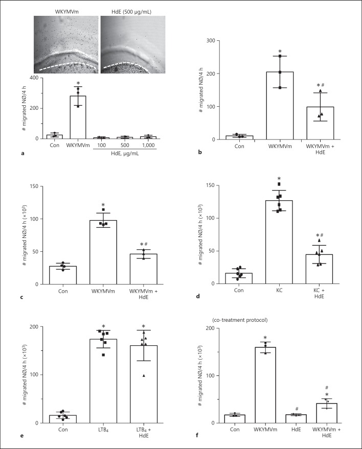
Employing the under-agarose assay, neutrophil migration in response to WKYMVm was obvious but did not occur in response to HdE (100–1,000 μg/mL; Fig. 4a). Moreover, when neutrophils were pre-treated with HdE (100 μg/mL, 37°C, 30 min) they showed significantly reduced chemotaxis to WKYMVm (Fig. 4b). Reduced chemotaxis to WKYMVm by HdE-treated neutrophils was also observed in the transwell migration assay (Fig. 4c), as was chemotaxis to KC (Fig. 4d). Neutrophil chemotaxis towards LTB4 was unaffected by HdE pre-treatment (Fig. 4e).
Fig. 4.
H. diminuta extract (HdE) suppresses WKYMVm and KC-induced chemotaxis in murine bone marrow neutrophil. Freshly isolated neutrophils (2 × 10⁵ NØs) were placed in the outside wells of an ultrapure agarose gel and chemotaxis towards HdE or WKYMVm (1 μM) assessed (a upper panels are representative images showing extensive migration of neutrophils out of the well (demarcated by the dashed line) in response to WKYMVm but not HdE). b shows that NØs pretreated with HdE (100 μg/mL, 30 min) had reduced chemotaxis to WKYMVm. Using a transwell (8 μm pore size) migration system revealed that HdE inhibited NØ chemotaxis to WKYMVm (c) and KC (20 ng/mL; d), but not leukotriene B4 (LTB4, 100 nM; e). Treatment of NØs with HdE at the time of application of WYKMVm to the culture well (co-treatment) also suppressed chemotaxis (f; data are mean ± SD; each datum point is the average of 2 NØ replicates from an individual mouse; * and # p < 0.05 compared to control [con] or the appropriate chemotactic stimulus only, respectively, by one-way analysis of variance with Tukey's post-test).
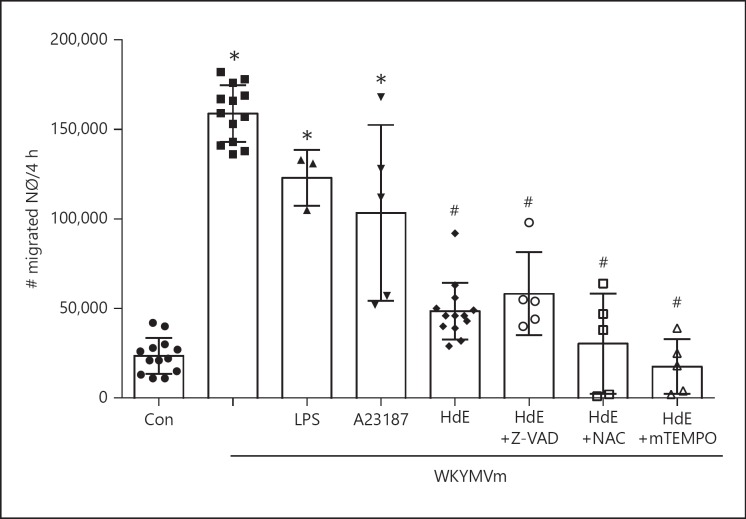
Neutrophils co-treated with HdE at the time of addition of WKYMVm to the basolateral chamber of the culture transwell displayed reduced chemotaxis (Fig. 4f). The reduced chemotaxis of HdE-treated neutrophils was not due to apoptosis, a generalized activation event causing stasis, or ROS, as determined by the use Z-VAD, LPS, A23187, NAC or MitoTEMPO, respectively (Fig. 5). With HdE being effective in a pre-treatment regimen (HdE rinsed from neutrophils prior to use) in the WKYMVm chemotaxis and reducing KC-induced neutrophil migration, it is unlikely that the HdE directly bound WKYMVm, although this possibility has not unequivocally been ruled out.
Fig. 5.
H. diminuta extract (HdE) suppression of neurophil chemotaxis is not dependent on reactive oxygen species (ROS). Murine bone marrow neutrophil (2 × 10⁵ NØs) migration towards WKYMVm (1 μM) in a transwell assay (8 μm pore size filter) is unaffected by a 30 min pre-treatment with LPS (1 μg/mL) or the calcium ionophore, A23187 (2.5 μM). Moreover, suppression of NØ chemotaxis by Hymenolepis diminuta extract (HdE, 100 μg/mL) was unaffected by co-treating the NØs with the anti-apoptotic pan-caspase inhibitor, Z-VAD (20 μM) or the antioxidants, n-acetylcystine (NAC, 1 μM) or mitoTEMPT (mTEMPO, 10 μM) (data are mean ± SD; each datum point is the average of 2 NØ replicates from an individual mouse, * and #, p < 0.05 compared to control (con) and WKYMVm only, respectively, by one-way analysis of variance with Tukey's post-test).
A Heat- and Trypsin-Sensitive Glycoprotein in the HdE Suppressed Neutrophil Migration
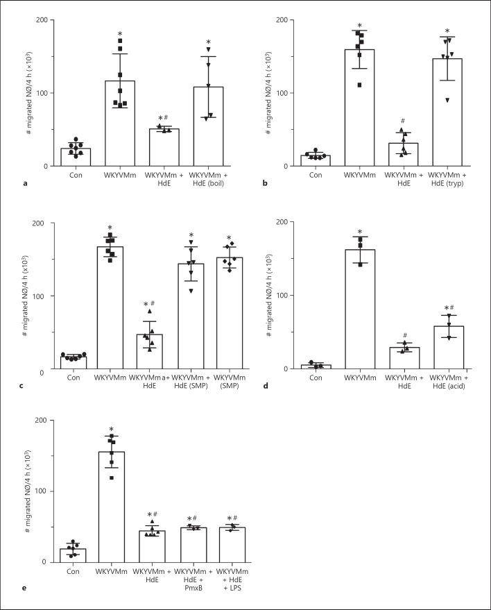
Considering the physico-chemical nature of the bioactive molecules(s) within the HdE, boiling, trysinization and sodium-metaperiodate (to disrupt glycans) treatment of the HdE ablated its ability to inhibit WKYMVm-induced chemotaxis, whereas acidification did not affect HdE's ability to reduce neutrophil chemotaxis (Fig. 6a–d).
Fig. 6.
A heat-, trypsin-sensitive glycoprotein mediates the effect of H. diminuta extract (HdE) on neutrophil chemotaxis. Murine bone marrow neutrophils (2 × 10⁵ NØs) were treated with (a) boiled (boil), (b) trypsinized (tryp), (c) sodium metaperiodate (SMP) or (d) acidified (acid) HdE (100 μg/mL) and placed in the upper chamber of the transwell (8 μm pore size filter) separated from WKYMVm (1 μM) and NØ migration assessed 4 h later. Panel D shows the lack of an effect of applying polymyxin B (PmxB; 10 μg/mL) or E. coli-derived LPS (1 ng/mL) on the HdE-suppression of chemotaxis (data are mean ± SD; each datum point is the average of 2 NØ replicates from an individual mouse, * and # p < 0.05 compared to control (con) and WKYMVm only, respectively, by one-way analysis of variance with Tukey's post-test).
Cognizant of low level of LPS contamination in HdE, we speculated that this may contribute to the inhibition of neutrophil chemotaxis. However, the addition of polymyxin B to the HdE did not affect its ability to inhibit murine neutrophil migration, and pre-treatment of neutrophils with HdE and LPS did not enhance further the suppression of chemotaxis in response to WKYMVm (Fig. 6e).
The Molecular Mechanism
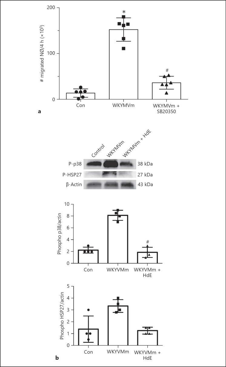
As reported [21], we also find that WKYMVm-evoked chemotaxis in neutrophils is completely blocked by the selective pharmacological inhibitor of p38 mitogen-activated protein kinase (MAPK), SB20350 (Fig. 7a). The inhibitor of the ERK1/2 MAPK pathway, U0126, blocked neutrophil by ∼40% in the transwell migration assay (data not shown). Focusing on the p38 MAPK pathway, we observed that the activation (i.e., phosphorylation) of p38 MAPK and the downstream signalling molecule, HSP27, by WKYMVm (10 min treatment) was substantially reduced by co-treatment with HdE (Fig. 7b).
Fig. 7.
H. diminuta extract (HdE) suppression of neutrophil chemotaxis occurs via p38 MAP kinase. a Murine bone marrow neutrophil (2 × 10⁵, NØs) migration in a transwell (8 μm pore size filter) in response to the peptide WKYMVm (1 μM) is completely blocked by pre-treatment (30 min, 37°C) with the selective pharmacologic inhibitor of the p38 mitogen-activated protein kinase (MAPK) pathway, SB20350 (2.5 μM) (data is mean ± SD; each datum point is the average of 2 NØ replicates from an individual mouse; * and # p < 0.05 compared to control (con) or WKYMVm only, respectively, by one-way analysis of variance with Tukey's post-test). b Immunoblots show that activation of p38 in NØs and the downstream signal heat-shock protein (HSP) 27 by WKYMVm is inhibited by the co-treatment with HdE (100 μg/mL; 10 min, 37°C; representative immunoblot and densitometric analysis where data are mean ± SD, # p < 0.05 compared to WKYMVm only, by Kruskal-Wallis test with Dunn's post-test).
Discussion
Often overlooked in the context of infection with parasitic helminths, the potential of neutrophils to kill larval nematodes in vitro was observed in 1979 [22]. Since then neutrophils have been shown to participate in the host response to helminths, but it is not always clear if they are mobilized because of concomitant tissue damage [7], the parasite (or its eggs/products) per se [6, 23], or bacteria from the helminth as described for Wolbachia endobacteria causing neutrophil accumulation around the filarial nematode, Onchocerca volvulus [24].
Infection with H. diminuta suppressed the severity of DNBS-induced colitis and CFA-induced arthritis in mice, one indicator of which was reduced MPO and hence neutrophil infiltration into the tissue [12, 13]. Whether this was due to reduced chemokine production by the host and/or related to bioactive molecules from the worm was not determined. Here, the injection of LPS into the mouse knee resulted in an accumulation of neutrophils that was significantly reduced by co-treatment with a PBS-soluble extract of H. diminuta (HdE). While consistent with the findings in the DNBS and CFA models of inflammation, reduced neutrophil recruitment to the knee of HdE-treated mice does not differentiate between direct and indirect effects of the HdE on the neutrophil. It was possible that the HdE was acting as a distraction, attracting neutrophils into the peritoneal cavity (noting that HdE evoked increases in KC in the peritoneal cavity), but this is unlikely to be the full explanation, since neither bone marrow nor blood levels of neutrophils were reduced in the HdE-treated mice.
Murine neutrophils were responsive to the HdE as gauged by Ca2+ mobilization, ROS generation (a component of which would arise from the noted H+ leak in mitochondria) and reduced mitochondrial capacity to make ATP, although, and perhaps surprisingly, the HdE did not elicit KC, IL-10 or TNF-α production directly from neutrophils. HdE-evoked ROS generation was not affected by use of polymyxin B to absorb LPS, suggesting that the effect was not because of this bacterial contaminant in the HdE. The scientific literature is replete with examples of helminth-derived excretory/secretory products, extracts or semi-purified molecules directly affecting a variety of functions of innate immune cells [25, 26]. Indeed, products from nematode and trematode parasites can be chemoattractants for neutrophils in vitro [27, 28, 29]: thus, it was unexpected when HdE failed to elicit neutrophil chemotaxis in under-agarose or transwell systems. Contrarily, when added to neutrophils as a pre- or co-treatment, HdE significantly suppressed migration towards the chemotactic penta-peptide, WKYMVm. Inhibition of neutrophil chemotaxis by HdE also occurred with the murine chemokine KC, but not the lipid mediator, LTB4. This reveals that the neutrophil retained the ability to rearrange its cytoskeleton and migrate (and that neutrophil viability was not an issue), suggesting that the HdE effect was restricted to peptide chemoattractants. Moreover, this was not because of neutrophil exhaustion due to the HdE-evoked oxidative burst or Ca2+ mobilization, since WKYMVm chemotaxis was unaffected by LPS exposure or treatment with the Ca2+ ionophore, A23187. Mechanistically, ROS neutralization did not affect the HdE suppression of WKYMVm-induced neutrophil migration.
The suppression of neutrophil chemotaxis is intriguing, particularly since other reports on helminth-derived molecules describe them as neutrophil chemoattractants [27, 28, 29, 30]. Schroeder et al. reported that live microfilariae of Brugia malayi blocked transendothelial migration of neutrophils but did not determine if this was via the release of a molecule from the nematode [31]. We add 2 speculative points when interpreting our data. One, identification of the bioactive molecule(s) within the HdE could yield a blueprint for the development of a new drug to direct neutrophil movements in the treatment of disease. Two, the HdE by acting as a “don't leave” signal could retain neutrophils in a tissue, potentially prolonging their activity and, where following apoptosis, they act as an important second signal to promote the reparative function of AAMs [32].
With few exceptions [33, 34], the identification of molecules from parasitic helminths has proved challenging. Characterization of the nature of the HdE revealed that the component that blocked neutrophil chemotaxis was a heat- and trypsin-sensitive glycan (SMP-sensitive), most likely a proteoglycan that was resistant to acidification. Similarly, a H. diminuta glycan was found to suppress LPS-evoked TNF-α production from macrophages [14], and numerous findings support the ubiquity of helminth-derived glycans in the modulation of host immunity [25, 35]. So while the challenge remains to identify the structure of the anti-chemotactic molecule(s) from H. diminuta, the search has been narrowed to a heat- and trypsin-sensitive proteoglycan.
Cell chemotaxis is a coordinated process [36], in which the p38 MAPK directs neutrophil migration in response to KC/IL-8 and formyl-peptide receptor ligands such as WKYMVm [37], but not LTB4 [38]: the leukotriene may be an important synergist stimulus of neutrophil chemotaxis [39] and has been shown to amplify eosinophil accumulation in response to infection with nematodes [40]. Following confirmation that pharmacological blockade of p38 MAPK signalling prevented WKYMVm-induced neutrophil chemotaxis, immunoblotting revealed that HdE-treated neutrophils displayed diminished activation of p38 MAPK and the downstream signal, HSP27 (important in chemotaxis [36]), in response to the challenge with WKYMVm. Subsequent research efforts need to be directed towards more fully elucidating HdE's mechanism of action that could involve binding with and blocking the FPR and KC receptors, or HdE mobilizing a signalling cascade that inhibits p38 MAPK phosphorylation directly or targets up-stream molecules such as phosphatidylinositol 3′-kinase [38].
What of the paradox of HdE injection resulting in accumulation of neutrophils in the peritoneum of mice compared to the suppression of chemotaxis in vitro. This most likely reflects the target cells of the HdE. With direct access to the neutrophil (in vitro analyses), the HdE will suppress neutrophil chemotaxis to WKYMVm or KC. However, in vivo HdE can evoke the production of neutrophil chemoattractants (KC was increased in the peritoneum) by other cells, possibly myeloid cells, which would attract neutrophils that have not been affected by direct contact with the HdE.
Often considered a pariah because of its capacity for collateral tissue damage upon activation, the neutrophil, alive or dead, is an important player in host immunity [1]: alive it performs critical anti-microbial functions, while apoptotic neutrophils may be important in promoting a tissue-healing phenotype in murine AAMs [32]. Here we show that a PBS-soluble extract of the cestode H. diminuta activates murine neutrophils and that a trypsin-sensitive proteoglycan in the extract blocks WKYMVm- and KC-induced chemotaxis via suppression of p38 MAPK signalling. While the exact nature of the bioactive molecule(s) of the helminth-derived molecules awaits determination, and the full significance of these changes in neutrophil function for host immunity needs to be defined, the current study reinforces the folly of overlooking the role that neutrophils play in the host response to infection with parasitic helminths.
Disclosure Statement
The authors declare that they have no disclosures to disclose.
Acknowledgements
Funding provided by a Natural Science and Engineering Research Council of Canada (NSERC) grant to Derek M. McKay. Nicholas Graves was supported by a Graduate Studentship from the NSERC CREATE Host-Parasite Interactions Program at University Calgary. Fernando Lopes was supported by Canadian Institutes of Health Research (CIHR)/Canadian Association of Gastroenterology (CAG)/Allergan Inc. and Alberta Innovates-Health solutions (AI-HS) Post-Doctoral Fellowships. Derek M. McKay holds a Canada Research Chair (CRC: Tier 1) in Intestinal Immunophysiology in Health and Disease. We thank Dr. M. Hollenberg and M. Saifeddine (University Calgary) for access to and assistance with the calcium measurement assay.
References
- 1.Kubes P. The enigmatic neutrophil: what we do not know. Cell Tiss Res. 2018;371:399–406. doi: 10.1007/s00441-018-2790-5. [DOI] [PubMed] [Google Scholar]
- 2.Pionnier N, Brotin E, Karadjian G, et al. Neutropenic mice provide insight into the role of skin-infiltrating neutrophils in the host protective immunity against filarial infective larvae. PLoS Negl Trop Dis. 2016;10:e0004605. doi: 10.1371/journal.pntd.0004605. [DOI] [PMC free article] [PubMed] [Google Scholar]
- 3.Munoz-Caro T, Rubio RM, Silva LM, et al. Leucocyte-derived extracellular trap formation significantly contributes to Haemonchus contortus larval entrapment. Parasit Vectors. 2015;8:607. doi: 10.1186/s13071-015-1219-1. [DOI] [PMC free article] [PubMed] [Google Scholar]
- 4.O'Connell AE, Hess JA, Santiago GA, et al. Major basic protein from eosinophils and myeloperoxidase from neutrophils are required for protective immunity to Strongyloides stercoralis in mice. Infect Immunity. 2011;79:2770–2778. doi: 10.1128/IAI.00931-10. [DOI] [PMC free article] [PubMed] [Google Scholar]
- 5.Vatta AF, Dzimianski M, Storey BE, et al. Ivermectin-dependent attachment of neutrophils and peripheral blood mononuclear cells to Dirofilaria immitis microfilariae in vitro. Vet Parasitol. 2014;206:38–42. doi: 10.1016/j.vetpar.2014.02.004. [DOI] [PubMed] [Google Scholar]
- 6.Gazzinelli-Guimaraes PH, Gazzinelli-Guimaraes AC, Silva FN, et al. Parasitological and immunological aspects of early Ascaris spp. infection in mice. Internat J Parasitol. 2013;43:697–706. doi: 10.1016/j.ijpara.2013.02.009. [DOI] [PubMed] [Google Scholar]
- 7.Chen F, Wu W, Millman A, et al. Neutrophils prime a long-lived effector macrophage phenotype that mediates accelerated helminth expulsion. Nat Immunol. 2014;15:938–946. doi: 10.1038/ni.2984. [DOI] [PMC free article] [PubMed] [Google Scholar]
- 8.Bonne-Annee S, Kerepesi LA, Hess JA, et al. Human and mouse macrophages collaborate with neutrophils to kill larval Strongyloides stercoralis. Infect Immunity. 2013;81:3346–3355. doi: 10.1128/IAI.00625-13. [DOI] [PMC free article] [PubMed] [Google Scholar]
- 9.Sutherland TE, Logan N, Ruckerl D, et al. Chitinase-like proteins promote IL-17-mediated neutrophilia in a tradeoff between nematode killing and host damage. Nat Immunol. 2014;15:1116–1125. doi: 10.1038/ni.3023. [DOI] [PMC free article] [PubMed] [Google Scholar]
- 10.Lopes F, Matisz C, Reyes JL, et al. Helminth regulation of immunity: a three-pronged approach to treat colitis. Inflamm Bowel Dis. 2016;22:2499–2512. doi: 10.1097/MIB.0000000000000889. [DOI] [PubMed] [Google Scholar]
- 11.Navarro S, Pickering DA, Ferreira IB, et al. Hookworm recombinant protein promotes regulatory T cell responses that suppress experimental asthma. Sci Translat Med. 2016;8:362ra143. doi: 10.1126/scitranslmed.aaf8807. [DOI] [PubMed] [Google Scholar]
- 12.Hunter MM, Wang A, Hirota CL, McKay DM. Neutralizing anti-IL-10 antibody blocks the protective effect of tapeworm infection in a murine model of chemically induced colitis. J Immunol. 2005;174:7368–7375. doi: 10.4049/jimmunol.174.11.7368. [DOI] [PubMed] [Google Scholar]
- 13.Shi M, Wang A, Prescott D, et al. Infection with an intestinal helminth parasite reduces Freund's complete adjuvant-induced monoarthritis in mice. Arthritis Rheum. 2011;63:434–444. doi: 10.1002/art.30098. [DOI] [PubMed] [Google Scholar]
- 14.Johnston MJ, Wang A, Catarino ME, et al. Extracts of the rat tapeworm, Hymenolepis diminuta, suppress macrophage activation in vitro and alleviate chemically-induced colitis in mice. Infect Immunity. 2010;78:1364–1375. doi: 10.1128/IAI.01349-08. [DOI] [PMC free article] [PubMed] [Google Scholar]
- 15.Lopes F, Coelho FM, Costa VV, et al. Resolution of neutrophilic inflammation by H2O2 in antigen-induced arthritis. Arthritis Rheum. 2011;63:2651–2660. doi: 10.1002/art.30448. [DOI] [PubMed] [Google Scholar]
- 16.Swamydas M, Luo Y, Dorf ME, Lionakis MS. Isolation of mouse neutrophils. Curr Protocol Immunol. 2015;110:3.20.1–3.20.15. doi: 10.1002/0471142735.im0320s110. [DOI] [PMC free article] [PubMed] [Google Scholar]
- 17.Dranka BP, Benavides GA, Diers AR, et al. Assessing bioenergetic function in response to oxidative stress by metabolic profiling. Free Rad Biol Med. 2011;51:1621–1635. doi: 10.1016/j.freeradbiomed.2011.08.005. [DOI] [PMC free article] [PubMed] [Google Scholar]
- 18.Yang L, Na CL, Luo S, et al. The phosphatidylcholine transfer protein stard7 is required for mitochondrial and epithelial cell homeostasis. Scient Rep. 2017;7:46416. doi: 10.1038/srep46416. [DOI] [PMC free article] [PubMed] [Google Scholar]
- 19.Heit B, Kubes P. Measuring chemotaxis and chemokinesis: the under-agarose cell migration assay. Sci STKE. 2003;2003:PL5. doi: 10.1126/stke.2003.170.pl5. [DOI] [PubMed] [Google Scholar]
- 20.Lopes F, Graepel R, Reyes JL, et al. Involvement of mast cells in α7 nicotinic receptor agonist exacerbation of Freund's complete adjuvant-induced monoarthritis in mice. Arthritis Rheum. 2016;68:542–552. doi: 10.1002/art.39411. [DOI] [PubMed] [Google Scholar]
- 21.de Oliveira S, Rosowski EE, Huttenlocher A. Neutrophil migration in infection and wound repair: going forward in reverse. Nat Rev Immunol. 2016;16:378–391. doi: 10.1038/nri.2016.49. [DOI] [PMC free article] [PubMed] [Google Scholar]
- 22.Bass DA, Szejda P. Mechanisms of killing of newborn larvae of Trichinella spiralis by neutrophils and eosinophils. Killing by generators of hydrogen peroxide in vitro. J Clin Invest. 1979;64:1558–1564. doi: 10.1172/JCI109616. [DOI] [PMC free article] [PubMed] [Google Scholar]
- 23.Chuah C, Jones MK, Burke ML, McManus DP, Owen HC, Gobert GN. Defining a pro-inflammatory neutrophil phenotype in response to schistosome eggs. Cell Microbiol. 2014;16:1666–1677. doi: 10.1111/cmi.12316. [DOI] [PubMed] [Google Scholar]
- 24.Tamarozzi F, Wright HL, Johnston KL, Edwards SW, Turner JD, Taylor MJ. Human filarial Wolbachia lipopeptide directly activates human neutrophils in vitro. Parasite Immunol. 2014;36:494–502. doi: 10.1111/pim.12122. [DOI] [PMC free article] [PubMed] [Google Scholar]
- 25.Johnston MJ, MacDonald JA, McKay DM. Parasitic helminths: a pharmacopeia of anti-inflammatory molecules. Parasitology. 2009;136:125–147. doi: 10.1017/S0031182008005210. [DOI] [PubMed] [Google Scholar]
- 26.Harnett W. Secretory products of helminth parasites as immunomodulators. Mol Biochem Parasitol. 2014;195:130–136. doi: 10.1016/j.molbiopara.2014.03.007. [DOI] [PubMed] [Google Scholar]
- 27.Jefferies JR, Corbett E, Barrett J, Turner RJ. Polarization and chemokinesis of ovine and human neutrophils in response to Fasciola hepatica excretory-secretory products. Internat J Parasitol. 1996;26:409–414. doi: 10.1016/0020-7519(95)00136-0. [DOI] [PubMed] [Google Scholar]
- 28.Reinhardt S, Scott I, Simpson HV. Neutrophil and eosinophil chemotactic factors in the excretory/secretory products of sheep abomasal nematode parasites: NCF and ECF in abomasal nematodes. Parasitol Res. 2011;109:627–635. doi: 10.1007/s00436-011-2305-8. [DOI] [PubMed] [Google Scholar]
- 29.Wu C, Chen Q, Fang Y, et al. Schistosoma japonicum egg specific protein SjE16.7 recruits neutrophils and induces inflammatory hepatic granuloma initiation. PLoS Negl Trop Dis. 2014;8:e2703. doi: 10.1371/journal.pntd.0002703. [DOI] [PMC free article] [PubMed] [Google Scholar]
- 30.O'Connell AE, Redding KM, Hess JA, Lok JB, Nolan TJ, Abraham D. Soluble extract from the nematode Strongyloides stercoralis induces CXCR2 dependent/IL-17 independent neutrophil recruitment. Microbes Infect. 2011;13:536–544. doi: 10.1016/j.micinf.2011.01.016. [DOI] [PMC free article] [PubMed] [Google Scholar]
- 31.Schroeder JH, Simbi BH, Ford L, et al. Live Brugia malayi microfilariae inhibit transendothelial migration of neutrophils and monocytes. PLoS Negl Trop Dis. 2012;6:e1914. doi: 10.1371/journal.pntd.0001914. [DOI] [PMC free article] [PubMed] [Google Scholar]
- 32.Bosurgi L, Cao YG, Cabeza-Cabrerizo M, et al. Macrophage function in tissue repair and remodeling requires IL-4 or IL-13 with apoptotic cells. Science. 2017;356:1072–1076. doi: 10.1126/science.aai8132. [DOI] [PMC free article] [PubMed] [Google Scholar]
- 33.Ahmed UK, Maller NC, Iqbal AJ, Al-Riyami L, Harnett W, Raynes JG. The carbohydrate-linked phosphorylcholine of the parasitic nematode product ES-62 modulates complement activation. J Biol Chem. 2016;291:11939–11953. doi: 10.1074/jbc.M115.702746. [DOI] [PMC free article] [PubMed] [Google Scholar]
- 34.Everts B, Hussaarts L, Driessen NN, et al. Schistosome-derived omega-1 drives Th2 polarization by suppressing protein synthesis following internalization by the mannose receptor. J Exp Med. 2012;209:1753–1767. doi: 10.1084/jem.20111381. [DOI] [PMC free article] [PubMed] [Google Scholar]
- 35.Velupillai P, Harn DA. Oligosaccharide-specific induction of interleukin 10 production by B220+ cells from schistosome-infected mice: a mechanism for regulation of CD4+ T-cell subsets. Proc Nat Acad Sci U S A. 1994;91:18–22. doi: 10.1073/pnas.91.1.18. [DOI] [PMC free article] [PubMed] [Google Scholar]
- 36.Cuenda A, Rousseau S. p38 MAP-kinases pathway regulation, function and role in human diseases. Biochim Biophys Acta. 2007;1773:1358–1375. doi: 10.1016/j.bbamcr.2007.03.010. [DOI] [PubMed] [Google Scholar]
- 37.Cara DC, Kaur J, Forster M, McCafferty DM, Kubes P. Role of p38 mitogen-activated protein kinase in chemokine-induced emigration and chemotaxis in vivo. J Immunol. 2001;167:6552–6558. doi: 10.4049/jimmunol.167.11.6552. [DOI] [PubMed] [Google Scholar]
- 38.Heit B, Tavener S, Raharjo E, Kubes P. An intracellular signaling hierarchy determines direction of migration in opposing chemotactic gradients. J Cell Biol. 2002;159:91–102. doi: 10.1083/jcb.200202114. [DOI] [PMC free article] [PubMed] [Google Scholar]
- 39.Lammermann T, Afonso PV, Angermann BR, et al. Neutrophil swarms require LTB4 and integrins at sites of cell death in vivo. Nature. 2013;498:371–375. doi: 10.1038/nature12175. [DOI] [PMC free article] [PubMed] [Google Scholar]
- 40.Patnode ML, Bando JK, Krummel MF, Locksley RM, Rosen SD. Leukotriene B4 amplifies eosinophil accumulation in response to nematodes. J Exp Med. 2014;211:1281–1288. doi: 10.1084/jem.20132336. [DOI] [PMC free article] [PubMed] [Google Scholar]


