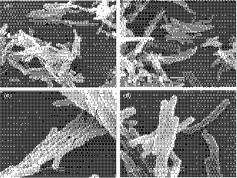
Figure 3.

Representative SEM micrographs of Mycobacterium smegmatis cells. Cells were grown with 20 ng/ml tetracycline. The Z rings are indicated by arrows. (a) M. smegmatis cells carrying pMind‐Sm‐ddlA‐AS (magnification 8,000×); (b) M. smegmatis cells carrying pMind (magnification of 8,000×); (c) M. smegmatis cells carrying pMind‐Sm‐ddlA‐AS (magnification 20,000×); (d) M. smegmatis cells carrying pMind (magnification 20,000×)
