Abstract
A 4-month-old Holstein Friesian calf was presented to the Ontario Veterinary College with progressive respiratory distress. The calf was diagnosed with tracheal collapse following perinatal rib fractures. Tracheal collapse has been infrequently reported in calves and is a possible sequela after delivery by forced extraction. Clinical signs can appear from days to months after birth, making the connection between clinical signs and dystocia more challenging. Multiple imaging modalities were used to diagnose and determine the severity of the tracheal collapse, and to establish the most likely cause and prognosis.
Résumé
Modalités multiples d’imagerie pour le diagnostic de collapse trachéal chez un veau : rapport de cas. Un veau de race Holstein Friesian âgé de 4 mois fut présenté au Ontario Veterinary College pour détresse respiratoire progressive. Un diagnostic de collapse trachéal à la suite de fractures de côtes périnatales fut posé. Le collapse de la trachée n’a été rapporté que très peu fréquemment chez les veaux et serait une séquelle possible d’une mise-bas par extraction forcée. Les signes cliniques peuvent apparaitre des jours jusqu’à des mois après la naissance, rendant l’association entre les signes cliniques et la dystocie encore plus difficile. Des modalités multiples d’imagerie furent utilisées pour diagnostiquer et déterminer la sévérité du collapse trachéal, et afin d’établir la cause la plus probable et le pronostic.
(Traduit par Dr Serge Messier)
Tracheal collapse occurs in several species, but is a particularly common disorder in small dog breeds (1). Tracheal collapse has been less frequently reported in calves. A congenital etiology has been proposed (2), but most reported cases are presented with a history of thoracic trauma from dystocia and forced extraction during delivery (3–7). Forced extraction causes dorsoventral compression of the trachea, and injury to the tracheal rings. In cases of rib fractures, the injury to the tracheal rings could be exacerbated further by bony callus formation during healing of the fractures (6). Most of the calves reported with tracheal collapse were born in breech position and were assisted mechanically during parturition (3–5). Clinical signs of tracheal collapse in calves can appear from days to months after birth, and reported signs include dyspnea at rest, coughing, wheezing, cyanosis, inspiratory stridor, and reduced tolerance for exercise (3,5,6). Affected calves are often treated medically for suspected pneumonia, with poor to no response to treatment (2–5). The calf in the current case displayed characteristic clinical signs of tracheal collapse with a typical history of dystocia and poor response to medical therapy. As in previous reports, the diagnosis was made late in the course of disease. The aim of this report is to present multiple diagnostic imaging modalities used to determine the cause, severity, and prognosis associated with a severe tracheal deformity in a 4-month-old Holstein Friesian calf.
Case description
A 4-month-old female Holstein Friesian calf weighing 106 kg was presented to the Ontario Veterinary College with progressive respiratory distress of several days’ duration. The calf had a history of dystocia and had been delivered by forced extraction. The calf delivery was scored as moderate according to the farm scale of calving difficulty score chart. No abnormal respiration or growth retardation was noticed during the first months of life.
On presentation, the calf was lethargic and dyspneic at rest with a respiratory rate of 92 breaths/min and an inspiratory stridor that worsened when stimulated. The calf was mildly tachycardic with a heart rate of 116 beats/min. Rumen motility was decreased and the rectal temperature was 38.7°C. On auscultation over the trachea a wheezing inspiratory sound was heard with increasing intensity close to the thoracic inlet. Auscultation of the lung fields revealed increased broncho-vesicular sounds that decreased in intensity caudally. No crackles or wheezes were heard. Thoracic ultrasound revealed mild diffuse pleural roughening in the form of “comet-tail” artifacts of both the right and left lung fields. A complete blood (cell) count (CBC) revealed a slightly elevated red blood cell (RBC) concentration [8.5 × 10¹²/L; reference interval (RI): 4.9 to 7.5 × 10¹²/L], and a serum biochemistry profile revealed mild hypoproteinemia (55 g/L; RI: 66 to 86 g/L) with mild hypoglobulinemia (22 g/L; RI: 30 to 53 g/L). The calf was administered intranasal oxygen with a flow rate of 10 L/min and was started on ceftiofur sodium (Excenel; Zoetis, Kirkland, Quebec), 2.2 mg/kg body weight (BW), IV, q24h.
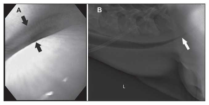
Endoscopic examination of the trachea revealed a severe, dorsoventrally collapsed trachea commencing at the level of the thoracic inlet extending caudal to approximately 10 cm cranial to the tracheal bifurcation (Figure 1A).
Figure 1.
A — Endoscopic view of tracheal collapse and stenosis (black arrows) at the level of the thoracic inlet in a 4-month-old Holstein-Friesian female calf. B — Lateral thoracic radiograph showing severe narrowing of the trachea starting at the level of the fifth cervical vertebra (white arrow).
Lateral thoracic radiographs confirmed a dorsoventrally collapsed trachea commencing at the level of the fifth cervical vertebra extending beyond the thoracic inlet to the second pair of ribs, with the most severe tracheal narrowing at the level of the thoracic inlet (Figure 1B).
After the diagnosis of tracheal collapse, surgical treatment options were discussed with the owners; however, they elected to euthanize the calf 10 d after presentation.
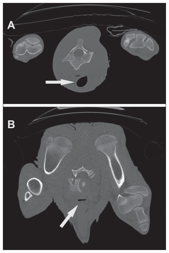
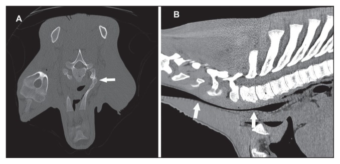
A computed tomography (CT) study performed post-mortem revealed severe narrowing of the trachea from the level of the fifth cervical vertebral body extending caudally to the thoracic inlet (Figure 2). The collapse of the trachea continued caudally and resolved at the level of the second and third pairs of ribs. The first and second ribs showed bilateral callus formation indicative of bone healing following previous rib fractures (Figure 3).
Figure 2.
Computed tomography images showing (A) a normal tracheal diameter at the level of the second cervical vertebra (white arrow) and (B) severe tracheal collapse at the level of the first thoracic vertebra (white arrow).
Figure 3.
Computed tomography images showing (A) callus formation (white arrows) of the first pair of ribs and (B) severe narrowing of the trachea starting at the level of the fifth cervical vertebrae extending caudal to the second thoracic vertebrae (between white arrows).
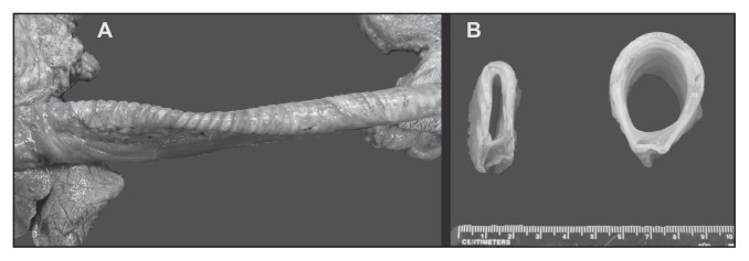
Post-mortem examination revealed subcutaneous edema along the length of the ventral neck, as well as severe edema of the thymus. The collapsed trachea extended for about 15 cm from the level of the thoracic inlet. The luminal diameter of the most severely collapsed segment was 7 mm, compared with 18 mm in the mid-cervical trachea (Figure 4). The first and second pairs of ribs bilaterally, and the third right rib, had prominent bony callus formation from healing rib fractures. There was marked subcutaneous emphysema along the thoracic walls, and mild pulmonary edema.
Figure 4.
A — Segmental tracheal collapse at the level of the thoracic inlet. B — Intraluminal diameter of collapsed segment and a normal tracheal ring at the level of mid-cervical tracheal segment.
Discussion
The present case reflects the most commonly reported etiology for tracheal collapse in calves, namely cranial thoracic trauma or fracture of the first pair of ribs during birth (3–7). Rib fractures are common in neonatal calves and foals, with a prevalence of 20.1% for costochondral dislocation or rib fractures in foals less than 3 d old (8), and 23% for rib fractures in perinatally dying calves (9). When forced extraction is considered, the prevalence of rib fractures in calves has been reported to be up to 40% (10).
Tracheal collapse is common in small dogs, and although the etiology is unknown, the consensus is that tracheal collapse in dogs is caused by congenital weakening of the cartilage rings, with exacerbation of clinical signs due to obesity, airway irritants, or cardiac disease (11). The case presented in this report had a history of dystocia, having been delivered by forced extraction. Forced extraction causes dorsoventral compression and injury to the tracheal rings, which could be exacerbated by bony callus formation during fracture healing, or by the negative inspiratory pressure continuously weakening the affected tracheal segment (6). Most of the calves reported with tracheal collapse were born in the breech position and assisted mechanically during parturition (3–5).
Reported clinical signs of tracheal collapse in calves include depression, dyspnea at rest, open mouth breathing, coughing, cyanosis, inspiratory stridor, and severely reduced tolerance for exercise (3,5,6), as observed in this case. Palpable callus formation or deformation of the ribs has also been reported (3,4). Commonly, affected calves are treated medically for suspected pneumonia or laryngitis, with no response to treatment (2–5). Clinical signs of tracheal collapse can appear within a few days (4) to several weeks after birth (3). The onset of clinical signs in this case reportedly appeared several weeks after birth, which hindered the association between clinical signs and dystocia during the initial assessment.
A diagnosis of tracheal collapse can be confirmed with diagnostic imaging modalities such as radiography, endoscopy, and CT (3,5,6). Tracheoscopy provides an intra-luminal view of the extent and severity of the collapsed segment (1) and correlates well with radiographic findings, as seen in this case. Rib fractures or healed rib fractures can be diagnosed with lateral radiographs (3,5), but overexposure, underexposure, or motion artifacts can preclude adequate interpretation of lateral radiographs (8). Lateral thoracic radiographs will reveal a dorsoventrally collapsed trachea as observed in this case. A latero-lateral collapsed trachea may not be seen on lateral thoracic radiographs (12); however, this type of collapse has not been reported in calves. Computed tomography (CT) allows differentiation between a dorsoventral and latero-lateral tracheal collapse, assessment of rib fractures and effect on the thoracic inlet diameter (12). Additionally, CT offers more benefits in cases suitable for surgical treatment, as three-dimensional reconstructions enable better evaluation of the severity of the rib fractures, collapsed trachea, and pre-surgical planning (5). Radiographs have been shown to underestimate the diameter of the affected collapsed segment compared to CT (1). Ultrasonographic examination of the lung fields is useful to differentiate tracheal collapse from pneumonia (13), and even the use of a linear probe would give an indication of a possible presence of pneumonia. A recent study using thoracic ultrasonography to determine lung consolidation associated with pneumonia in calves reported a sensitivity of 85% and 94% for chronic clinical and acute subclinical cases, respectively, with specificities of 98% and 100%, respectively (14). The same study also documented the presence of rib fractures in 6% of the calves (14), which is lower than previously reported, indicating that rib fractures can easily be missed on thoracic ultrasonographic evaluation.
A classification system has been published for tracheal collapse in dogs, which grades the severity of tracheal collapse from I to IV, whereby a grade I is normal, a grade II is 50% reduction of the tracheal lumen, grade III is 75% reduction, and grade IV is obliteration of the tracheal lumen by flattened tracheal rings (15). No classification system has been published for large animals.
In dogs, medical treatment is often successful in mildly affected individuals, by breaking the cycle of inflammation that triggers coughing and further inflammation (1,11). As the etiology of tracheal collapse in calves is different than that in dogs, medical treatment is unlikely to improve the long-term outcome in calves. Tracheostomy to relieve the respiratory distress is also not effective to treat these calves, due to the location of the stenosis. Intranasal oxygen administration has been shown to increase arterial partial pressure of oxygen and arterial oxygen saturation in neonatal calves with respiratory distress syndrome (16), and could temporarily increase arterial oxygen in calves with hypoxia from tracheal collapse.
The most common surgical treatments of tracheal collapse in dogs are extra-luminal rings and intra-luminal stents (11). A surgical treatment for tracheal collapse in calves with an extraluminal ring prosthesis has been reported (3), with varying degrees of success. The ring prosthesis temporarily restored the intra-luminal diameter of the trachea at the time of placement; however, continued tracheal growth caused varying degrees of tracheal narrowing and stenosis by mucosal infolding of the trachea after surgery (3). Therefore, it has been recommended that the ring-shaped prosthesis be removed 2 to 3 mo after surgery, based on radiographic findings or re-collapse. An alternative recommendation is to place a spiral prosthesis to accommodate tracheal growth without stenosis formation (3). Surgical rib resection to increase the thoracic inlet diameter has also been reported in calves (3). Costectomy of the first and second ribs with associated callus formation in affected calves did not improve the diameter of the collapsed trachea (3). More recently, partial rib resection of the first and second ribs for the treatment of tracheal collapse in 3 calves with rib fractures sustained during difficult deliveries was reported (5). Two out of the three calves recovered well after surgery, whereas 1 had recurrence of wheezing and subsequently was slaughtered. Partial rib resection led to improvement of stridor in 2 cases, indicating that not all cases of tracheal collapse and stenosis may require placement of tracheal prosthesis (5). The optimal surgical intervention to treat tracheal collapse in calves may vary according to age, degree of severity of the collapse, and degree of severity of callus formation after rib fractures, which can be assessed by CT examinations.
Tracheal collapse should be considered as a differential diagnosis in calves presenting with worsening dyspnea and failure to respond to medical treatment, particularly those calves with a history of dystocia and forced delivery. In cases with palpable thoracic wall abnormalities, the suspicion of traumatic tracheal collapse should be considered. The historical and clinical presentation of the calf presented in this report are characteristic of tracheal collapse cases. Dorso-ventral compression of the cranial thorax can occur during assisted delivery and may become a risk factor in the development of tracheal collapse (3). The overall incidence of tracheal collapse in calves is unknown but likely to be low. However, this condition may be underestimated as the final diagnosis may require specialized diagnostic equipment. CVJ
Footnotes
Use of this article is limited to a single copy for personal study. Anyone interested in obtaining reprints should contact the CVMA office (hbroughton@cvma-acmv.org) for additional copies or permission to use this material elsewhere.
References
- 1.Tappin SW. Canine tracheal collapse. J Small Anim Pract. 2016;57:9–17. doi: 10.1111/jsap.12436. [DOI] [PubMed] [Google Scholar]
- 2.Watt BR. Collapse of the trachea in two calves. Aust Vet J. 1983;60:309–310. doi: 10.1111/j.1751-0813.1983.tb02818.x. [DOI] [PubMed] [Google Scholar]
- 3.Fingland RB, Rings DM, Vestweber JG. The etiology and surgical management of tracheal collapse in calves. Vet Surg. 1990;19:371–379. doi: 10.1111/j.1532-950x.1990.tb01211.x. [DOI] [PubMed] [Google Scholar]
- 4.Jelinski M, Vanderkop M. Alberta. Tracheal collapse/stenosis in calves. Can Vet J. 1990;31:780. [PMC free article] [PubMed] [Google Scholar]
- 5.Hidaka Y, Hagio M, Kashiba I, et al. Partial costectomy for tracheal collapse and stenosis associated with perinatal rib fracture in three Japanese Black calves. J Vet Med Sci. 2016;78:451–455. doi: 10.1292/jvms.15-0378. [DOI] [PMC free article] [PubMed] [Google Scholar]
- 6.Rings DM. Tracheal collapse. Vet Clin North Am Food Anim Pract. 1995;11:171–175. doi: 10.1016/s0749-0720(15)30515-6. [DOI] [PubMed] [Google Scholar]
- 7.Vestweber JG, Leipold HW. Tracheal collapse in three calves. J Am Vet Med Assoc. 1984;184:735–736. [PubMed] [Google Scholar]
- 8.Jean D, Laverty S, Halley J, Hannigan D, Leveille R. Thoracic trauma in newborn foals. Equine Vet J. 1999;31:149–152. doi: 10.1111/j.2042-3306.1999.tb03808.x. [DOI] [PubMed] [Google Scholar]
- 9.Schuijt G. Iatrogenic fractures of ribs and vertebrae during delivery in perinatally dying calves: 235 cases (1978–1988) J Am Vet Med Assoc. 1990;197:1196–1202. [PubMed] [Google Scholar]
- 10.Mee JF. Newborn dairy calf management. Vet Clin North Am Food Anim Pract. 2008;24:1–17. doi: 10.1016/j.cvfa.2007.10.002. [DOI] [PubMed] [Google Scholar]
- 11.Tinga S, Thieman Mankin KM, Peycke LE, Cohen ND. Comparison of outcome after use of extra-luminal rings and intra-luminal stents for treatment of tracheal collapse in dogs. Vet Surg. 2015;44:858–865. doi: 10.1111/vsu.12365. [DOI] [PubMed] [Google Scholar]
- 12.Heng HG, Lim CK, Gutierrez-Crespo B, Guptill LF. Radiographic and computed tomographic appearance of tracheal collapse with axial rotation in four dogs. J Small Anim Pract. 2018;59:53–58. doi: 10.1111/jsap.12679. [DOI] [PubMed] [Google Scholar]
- 13.Buczinski S, Forte G, Francoz D, Belanger AM. Comparison of thoracic auscultation, clinical score, and ultrasonography as indicators of bovine respiratory disease in preweaned dairy calves. J Vet Intern Med. 2014;28:234–242. doi: 10.1111/jvim.12251. [DOI] [PMC free article] [PubMed] [Google Scholar]
- 14.Ollivett TL, Leslie KE, Duffield TF, et al. Field trial to evaluate the effect of an intranasal respiratory vaccine protocol on calf health, ultrasonographic lung consolidation, and growth in Holstein dairy calves. J Dairy Sci. 2018;101:8159–8168. doi: 10.3168/jds.2017-14271. [DOI] [PubMed] [Google Scholar]
- 15.Tangner C, Hobson HP. A retrospective study of 20 surgically managed cases of collapsed trachea. Vet Surg. 1982;11:146–149. [Google Scholar]
- 16.Bleul UT, Bircher BM, Kahn WK. Effect of intranasal oxygen administration on blood gas variables and outcome in neonatal calves with respiratory distress syndrome: 20 cases (2004–2006) J Am Vet Med Assoc. 2008;233:289–293. doi: 10.2460/javma.233.2.289. [DOI] [PubMed] [Google Scholar]


