● Cellular/Molecular
Antideath Transcription Factors
Sp1 and Sp3 Are Oxidative Stress-Inducible, Antideath Transcription Factors in Cortical Neurons
Hoon Ryu, Junghee Lee, Khalequz Zaman, James Kubilis, Robert J. Ferrante, Brian D. Ross, Rachael Neve, and Rajiv R. Ratan (see pages 3597–3606)
Cells react to oxidative stress with a complex tapestry of adaptive responses. “Antideath” molecules can counteract this cellular stress by reducing reactive oxygen species and repairing DNA damage. Because oxidative stress can alter the expression of antideath genes, the role of transcription factors in regulating this process has come under increasing scrutiny. A report this week from Ryu et al. shows that the zinc finger transcription factors Sp1 and Sp3 are redox-regulated in neurons. In cultured cortical neurons, oxidative stress dramatically increased the low basal DNA-binding activity of Sp1 and Sp3, an early response that was prevented by antioxidants and appeared to follow cellular increases in Sp1 and Sp3 protein. Furthermore, overexpression of the zinc finger DNA-binding proteins prevented oxidative stress- or DNA damage-induced neuronal death. To investigate the activity of Sp1 proteins in vivo, the authors used a transgenic and a chemical mouse model of Huntington's disease; Sp1 and Sp3 levels were increased in both model systems. Together, these results suggest that the Sp1 and Sp3 transcription factors form one thread of a compensatory neuronal response to oxidative stress.
▴ Development/Plasticity/Repair
Dendritic Patterns in Drosophila
Distinct Developmental Modes and Lesion-Induced Reactions of Dendrites of Two Classes of DrosophilaSensory Neurons
Kaoru Sugimura, Misato Yamamoto, Ryusuke Niwa, Daisuke Satoh, Satoshi Goto, Misako Taniguchi, Shigeo Hayashi, and Tadashi Uemura (see pages 3752–3760)
Many neurons, such as Purkinje cells, can be instantly recognized by their distinct dendritic arbors, yet the mechanisms underlying the extent of dendritic fields and branching patterns is far from clear. Advances in imaging technology are helping to answer such questions. Sugimura et al. generated cell-specific markers and then used time-lapse videomicroscopy to watch dendritic development in class I and class IV sensory neurons in the peripheral nervous system of theDrosophila embryo. They followed dendrite maturation in larva and the response to branch severing. They saw distinct branching behaviors in the dendritic arborization of class I and class IV neurons, which differ in their arbor complexity and morphology. Their analysis was aided by the fact that these neurons form largely two-dimensional dendritic arbors. One interesting property of class IV sensory neurons is the complete but minimal-overlapping innervation of their receptive fields, called “tiling.” Several signaling molecules have been identified recently as important to dendritic arbor formation. The current work, through laser ablation of dendritic branches, seems to confirm that a class-specific intercellular inhibitory communication between class IV neurons is necessary and sufficient for tiling. This system should be a useful model to investigate the underlying molecular mechanisms.
▪ Behavioral/Systems/Cognitive
Neural Correlates of Remembering
Functional Dissociation among Components of Remembering: Control, Perceived Oldness, and Content
Mark E. Wheeler and Randy L. Buckner (see pages3869–3880)
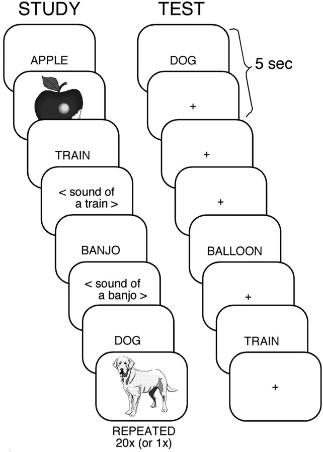
In an investigation of human brain function, Wheeler and Buckner used functional magnetic resonance imaging to examine specific components of that treasured cortical function, memory, and to define the cortical areas that mediate it. The very nature of remembering (the ability to recall events experienced at some time in one's past) requires multiple interdependent elements. Here, Wheeler and Buckner examined three such elements: control, the intentional retrieval of information; perceived oldness, the realization that information is in fact not new; and content, the actual “stuff” of the memory, which can be given context by the modality in which it was experienced. They separated the three components by manipulating the study content, the presentation, and the conditions of the retrieval tasks. They found distinct areas associated with each component. The control element correlated with activity in the left prefrontal cortex, whereas activity in parietal and frontal regions accompanied perceived oldness and activity in inferior temporal regions was content-related.
Word cues paired with pictures or sounds were used to test components of remembering. This image is taken from Figure 1 of this article.


