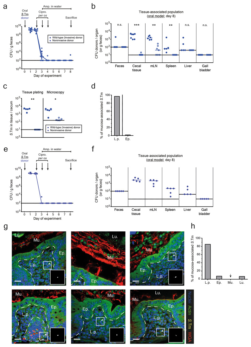
Extended data Fig. 4. Quantification and localization of S.Tm in the host mucosa after antibiotic treatments in the oral model.
a-d) Mice were orally infected with either an invasive (SL1344 P2cat; blue solid circles; n=7) or noninvasive (SL1344noninv P2cat; T3SS-1 negative; blue open circles; n=7) donor and treated with antibiotics. Mice were sacrificed at day 8 after infection (when recipients are normally added) and organs were analyzed. Dotted lines indicate detection limits. a) Fecal populations were monitored daily by selective plating on MacConkey agar. Blue lines connect medians. b) Organ loads were determined by selective plating. mLN: mesenteric lymph nodes. c) Population size of donors in the cecal mucosa determined by selective plating after a gentamycin protection assay or microscopy of tissue sections (same mice for each quantification method). Each data point is the average of 12 sections (10 μm thick). Panel b-c) Statistics are performed using a two-tailed Mann-Whitney U test p>0.05 (ns), p<0.05 (*), p<0.01 (**), p<0.001 (***), p<0.0001 (****) comparing mice infected with invasive or noninvasive donors for each organ. d) Localization of S.Tm detected in the cecal tissue by microscopy, reported as a percentage of bacteria detected in panel c in either the lamina propria (L.p.) or epithelium (Ep.). Bar indicates the median from 5 mice. e-h) Analysis of persister reservoirs in Carnoy-fixed cecal tissue sections. Mice were orally infected with an invasive (SL1344 P2cat; n=5) donor and treated with antibiotics. Mice were sacrificed at day 8 after infection (when recipients are normally added) and organs were analyzed. Dotted lines indicate detection limits. e) Fecal populations were monitored daily by selective plating on MacConkey agar. Blue lines connect medians. f) Organ loads were determined by selective plating. mLN: mesenteric lymph nodes. Line indicates median. g) A Carnoy fixation was performed on ceca of mice to preserve the mucus structure. 10 μm sections were stained to visualize S.Tm (yellow; α-LPS O5), actin (green; phalloidin-FITC), the mucus (red; wheat germ agglutinin (WGA) AF647 conjugate), and nuclei (blue; DAPI). Ep., epithelium; Lu., Lumen; L.p., lamina propria. Mu., mucus. Scale bars represent 20μm. White arrows highlight S.Tm (magnified in inset). Representative images shown from two independent experiments. h) Localization of S.Tm detected in the cecal tissue by microscopy, reported as a percentage of bacteria detected each section (12 sections per mouse cecum) in the lamina propria (L.p.), epithelium (Ep.), mucus (Mu.), or lumen (Lu.). Bar indicates the median from 5 mice.

