Abstract
Background
Schistosomiasis is the second major human parasitic disease next to malaria, in terms of socioeconomic and public health consequences, especially in sub-Saharan Africa. Schistosoma haematobium (S. haematobium) is a trematode and one of the species of Schistosoma that cause urogenital schistosomiasis (urinary schistosomiasis). Although the knowledge of this disease has improved over the years, there are still endemic areas, with most of the reported cases in Africa, including Ghana. Not much has been done in Ghana to investigate cytological abnormalities in individuals within endemic communities, although there are epidemiologic evidences linking S. haematobium infection with carcinoma of the bladder.
Aim
The aim of this study was to identify microscopic and cytological abnormalities in the urine deposits of S. haematobium-infected children.
Methodology
Three hundred and sixty-seven (367) urine samples were collected from school children in Zenu and Weija communities. All the samples were examined microscopically for the presence of S. haematobium eggs, after which the infected samples and controls were processed for cytological investigation.
Results
S. haematobium ova were present in 66 (18.0%) out of the 367 urine samples. Inflammatory cells (82%, 54/66), hyperkeratosis (47%, 31/66), and squamous cell metaplasia (24%, 16/66) were the main observations made during the cytological examination of the S. haematobium-infected urine samples.
Conclusion
Cytological abnormalities in S. haematobium-infected children may play an important role in the severity of the disease, leading to the possible development of bladder cancer in later years, if early attention is not given. Therefore, routine cytological screening for urogenital schistosomiasis patients (especially children) at hospitals in S. haematobium-endemic locations is recommended.
1. Introduction
Schistosomiasis is considered a neglected tropical disease that ranks second after malaria in terms of human suffering, in the tropics and subtropics. The genus Schistosoma contains different species that cause acute and chronic infection in humans: Schistosoma haematobium (S. haematobium), S. mansoni, S. japonicum, S. mekongi, S. intercalatum, S. malayensis, and S. guineensis [1]. S. haematobium causes urogenital schistosomiasis (urinary schistosomiasis) [1–3]. In 2016, it was estimated that globally schistosomiasis affects over 200 million people [2]. It is believed that the majority of people living with urinary schistosomiasis are in Africa, including Ghana, and this is evident from numerous prevalence studies conducted across the country [3–7]. Urogenital schistosomiasis has been reported to be higher in children than in adult [8]. Even though there has been an improvement in knowledge of urogenital schistosomiasis, repeated cases with accompanying morbidities and mortalities are still being reported [9, 10].
The infection of S. haematobium still leads to some form of abnormalities, regardless of whether it was during the early stage or not, there is mixed infection with other schistosomes or not, and the quantity of eggs of S. haematobium present in the tissues [11]. Mortality may occur from granulomatous inflammation complications [12], renal insufficiency [13], possible bladder cancers [14], and ulceration or depletion of the vesicle, as well as the ureteral wall [12]. Emerging evidence shows that immune regulation of inflammatory response, independent of infectious burden, is linked with the risk of some forms of morbidity due to schistosomiasis infection [15]. Therefore, light infection should not be overlooked as a cause for disability; hence, the recommendation that its prevention be integrated as part of the design for population-based schistosomiasis control programmes [15].
Studies in Africa and other parts of the world have linked squamous cell carcinoma of the urinary bladder with S. haematobium infection [14, 16–19]. This association is based on the close correlation of bladder cancer incidence with the prevalence of S. haematobium infection within different geographic areas [16–20], as well as expression of markers such as cyclooxygenase-2 (COX-2) and inducible nitric oxide synthase (iNOS) in urinary schistosomiasis patients [21].
Cytopathological examination of urine samples has been considered a routine noninvasive diagnostic procedure used to detect cancer of the urinary tract, primarily bladder cancer [22, 23]. This diagnostic procedure is also used during the follow-up procedures of the patients previously treated for bladder cancer, in order to detect recurrence as early as possible [23]. In the high-risk population, the cytopathological examination of urine is remarkably used for the screening of urothelial carcinoma [22].
Despite the link between S. haematobium and bladder cancer, only limited data are available on cytopathological findings in schistosomiasis-related tumors. Meanwhile, damage from S. haematobium infection has been attributed to parasite eggs deposited at the affected sites [24]. To the best of our knowledge, there seems not to be a study focusing on cytological examination of urine samples from S. haematobium-infected children within endemic communities in Ghana. Therefore, the connection between these cytological abnormalities and the severity of the disease in these areas has not been demonstrated. The current study is among the first to present finding from cytological and wet mount microscopic observations made in the urine of Schistosoma haematobium-infected children, living in endemic communities in the capital city of Ghana.
2. Materials and Methods
2.1. Approach, Study Design, Study Sites, and Scope of the Research
A cross-sectional study was conducted in two communities (Zenu and Weija) of the Greater Accra Region, the most urbanized region in Ghana. The geographical coordinates of the Zenu community are 5°42′0″ North, 0°20′0″ West [7] while those of Weija are 5°34′ North, 0°20′ West, with Weija occupying a total land area of about 341.838 square kilometres [25]. The major economic activities of these communities are subsistent farming and fishing, with a few inhabitants being civil servants. The presence of a lake in the Zenu community and a river in the Weija community (serving as a great source of water supply for various domestic activities) might have attracted most of the settlers [8].
Earlier studies [7, 26] have considered these two communities (Zenu and Weija) as urogenital schistosomiasis-endemic areas, and thus, they were selected for the current study, in order to increase the probability of obtaining S. haematobium-infected samples.
After obtaining informed consent, fifty-milliliter (50 ml) clean, dry, leak-proof, wide-mouthed containers were given to each child to provide urine, with adequate instructions. Urine sample collection was done between 10:00 am and 12:00 pm for maximum yield [27]. The urine samples were transported on ice immediately to the Parasitology Laboratory of the Medical Microbiology Department, School of Biomedical and Allied Health Sciences, University of Ghana, for analysis.
The scope of this study was to delineate observations (cytological and wet mount microscopic) in the urine of Schistosoma haematobium-infected individual, giving a hint of S. haematobium implication in bladder cancer. The study focused on children of school-going age within the selected communities of the Greater Accra Region of Ghana.
2.2. Urine Chemistry and Wet Mount Examination
The collected urine samples were analyzed for haematuria and proteinuria, alongside other urine chemistry parameters (pH, specific gravity, glucose, ketones, bilirubin, urobilinogen, and leucocytes) using urine chemistry reagent strips (URIT 10V, URIT Medical Electronic Co. Ltd., China). The results were recorded as negative or positive (trace, +, ++, and +++) [28]. Ten milliliters (10 ml) of the urine sample was transferred into 15 ml centrifuge tube and centrifuged at 300 rpm for 5 minutes. The supernatant was decanted until the one milliliter (1 ml) mark, after which fifty microlitres (50 μl) of the sediment was transferred onto a clean glass microscope slide (Biogenix Inc Private Ltd., Uttar Pradesh, India). The presence and intensity of S. haematobium eggs were determined microscopically using the ×10 objective of an optical light microscope (Leica Galen III, catalogue E no. 317506, serial no. ZG6JA4). With the eggs intensity count, the number of eggs present per 10 ml of urine sample was categorized as mild (≤50 ova/10 ml of urine) and heavy (≥50 ova/10 ml of urine) [29].
2.3. Cytological Examination
With the urine cytology, 5 ml of the infected and control samples were centrifuged at 1500 rpm for 10 minutes after which the supernatant was decanted leaving about 1.5 ml. The volume was resuspended and then cytocentrifuged 3–5 drops per slide in order to prepare monolayer smear of urine deposits directly onto the slide [30]. The smear slides were wet fixed with 95% ethanol for 15 minutes before staining with Papanicolaou (PAP) stains. Abnormalities such as squamous metaplastic cells, inflammatory cells, and hyperkeratotic cells were examined and reported.
2.4. Statistical Analysis
Data obtained were stored in Microsoft Excel and analyzed using the Statistical Package for the Social Sciences (IBM® SPSS® version 20.0). Data were summarized by determining frequencies of abnormalities detected in both urine deposit wet mount examination and cytological examination.
2.5. Ethical Statement
This work received ethical clearance from the Ethical and Protocol Review Committee of the College of Health Sciences, University of Ghana (Protocol ID no: MS-Et/M.7-P 3.1/2015-2016). The consent of the students, teachers as well as parents and guardians was sought before sampling.
3. Results
Among the 367 urine samples used, 66 were infected with Schistosoma haematobium, giving a prevalence of 18% for this study. Out of the 66 samples that contained Schistosoma eggs according to the microscopy of the wet mount, when subjected to cytological examination, S. haematobium eggs were detected in 40 (40/66, 61%) of them (Table 1).
Table 1.
Observations made in the urine samples after wet preparation microscopy and cytological examinations.
| Observations | n (%) |
|---|---|
| Cytological abnormality (N = 66) | |
| Squamous metaplasia | 16 (24) |
| Inflammatory cells | 54 (82) |
| Hyperkeratosis | 31 (47) |
| Egg positivity (N = 66) | |
| S. h eggs by wet mount | 66 (100) |
| S. h eggs by cytology | 40 (61) |
| Haematuria (N = 66) | |
| RBC (cytology) | 22 (33.3) |
| RBC (R/E-wet prep) | 29 (44) |
| RBC (urine chemistry) | 37 (56.0) |
| Blood intensity by urine dip stick (N = 37) | |
| Trace | 8 (12.1) |
| + | 5 (7.5) |
| ++ | 10 (15.1) |
| +++ | 14 (21.2) |
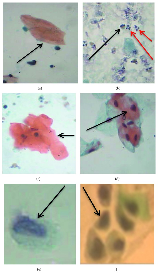
Papanicolaou staining of the urine smears revealed different cytological abnormalities (Figure 1). Squamous metaplastic cells (Figure 1(f)), inflammatory cells (Figure 1(b)), and hyperkeratotic cells (Figure 1(a)) were the main cytological observations made in the samples. Urothelial/transitional cells were also found (Figure 1(e)). Inflammatory cells were observed in most (82%) of the urine samples, followed by hyperkeratosis (Table 1). Haematuria was observed by urine reagent strips, wet mount microscopy examination, and cytological examination in 56%, 44%, and 33.3% of the urine samples, respectively (Table 1).
Figure 1.
Papanicolaou-stained smears showing cytological observations. (a) Anucleated keratinized cell (arrow—hyperkeratotic cell). (b) Inflammatory cells (red arrows point to neutrophils while black arrow points to eosinophils). (c) Keratinized cell (nucleated). (d) Cluster of keratinized and nonkeratinized cells (arrow pointing to keratinized cells). (e) Reactive urothelial/transitional cells. (f) Squamous metaplastic cells (arrow pointing to one of such cells) (source of images: authors' laboratory work).
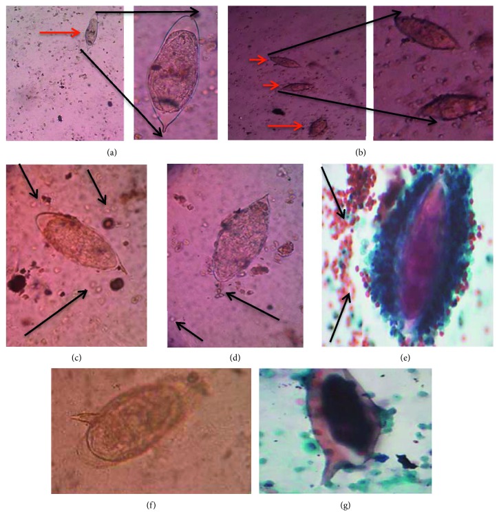
From the microscopic examination of wet preparation, 47% (31/66) of the samples had high egg intensity (above 50 eggs/10 ml urine), as depicted in Figure 2(b) in comparison with the low intensity represented in Figure 2(a). Both wet mount and cytological examinations showed RBCs in the presence of Schistosoma haematobium eggs (Figures 2(c)–2(e)). Out of the sixty-six S. haematobium-infected samples, two contained Schistosoma mansoni eggs (usually found in stool), in wet mount and cytology stained slides, respectively (Figures 2(f) and 2(g)).
Figure 2.
Wet mount and Papanicolaou-stained smears. (a) Low/light intensity of S. haematobium eggs (with a zoom in), (b) high intensity of S. haematobium eggs in the samples (with a zoom in), (c, d) presence of high and low number of red blood cells observed in wet mount (black arrows pointing to some red blood cells), (e) presence of blood cells in Papanicolaou-stained smears (black arrows pointing to cluster of red blood cells around an S. haematobium egg), eggs of S. mansoni in the S. haematobium-infected urine samples (f) in wet mount and (g) in Papanicolaou-stained smears (source of images: authors' laboratory work).
4. Discussions
This study found Schistosoma haematobium infection in 66 out of the 367 children (representing 18%), indicating that urinary schistosomiasis continues to be a disease of great public health importance, especially in children. S. haematobium infection in children has been observed to cause stunting and wasting, which are commonly unacknowledged morbidity [10, 31–33]. In a study by Botelho et al. [32], the proportion of all children and adolescents who had body mass index (BMI) less than 15 kg/m2 increased from 57.78% to 66.67% in S. haematobium-infected children, who were more stunted and wasted than in noninfected children. The mean weight-for-age Z score (WAZ) among the children in that study was significantly reduced in S. haematobium-infected as compared to noninfected [32]. Bustinduy et al. [33] associated wasting with Schistosoma haematobium infection in male children even with light intensity of the infection. Our observation therefore supports their suggestion that this vulnerable population urgently needed to be targeted for implementation of measures for treatment and control [33].
The public health importance of this infection was highlighted in this study where the majority of the S. haematobium-infected children were found to have the different types of cytological abnormalities such as squamous metaplastic cells which could develop into malignant cells in the continuous presence of a carcinogenic agent such as S. haematobium. The International Agency for Research on Cancer classified S. haematobium infection as a group 1 biocarcinogen. Schistosoma worm and egg-derived estrogen-like molecules and their metabolites have been postulated as the main carcinogenic substances implicated in schistosomiasis-linked cancers [34]. A retrospective study done in Accra associated 20.5% of cancer cases with tissue schistosomiasis, where the urinary bladder was the most commonly (93.6%) affected organ [31]. Their study also found 98.7% of all tissue schistosomiasis to be caused by S. haematobium [31]. With the retrospective study in mind, and the findings from the current study, it can be inferred that the possibility of some of the S. haematobium-infected pupils (especially those who had squamous cell metaplasia), having the risk of developing cancer later in life, cannot be overlooked.
While the specimen used for the retrospective study was from clinical cases, seemingly healthy children were used in the current study. Therefore, it is important to note that if this seemingly “benign” Schistosoma infection is left untreated in these children now, there is a probability of some of them having a future (with respect to developing cancer) similar to that of the subjects in the study by Der et al. [31].
Ketabchi and Moshtaghi-Kashanian [16] presented a case study involving a 60-year-old man who briefly lived in a schistosomiasis-endemic area of Khuzestan Province (neighbouring province of Persian Gulf in Iran). Approximately 20 years after his short stay at the schistosomiasis-endemic area, he was referred to the Urology Department of Kerman University of Medical Sciences, with haematuria and dysuria. Sonography revealed a polypoid mass on the bladder floor while cystoscopy and biopsy of the bladder tumor showed simultaneous squamous cell carcinoma and transitional bladder cell carcinoma in the presence of S. haematobium [35]. They indicated that bladder tumors related to schistosomiasis are mainly squamous cell type and there are rare reports of the simultaneous presence of squamous cell bladder carcinomas and transitional cell carcinoma [35].
Even though this study did not set out to ascertain the mechanism of progression from squamous cell metaplasia to carcinomas, it is important to note that these cellular changes, and abnormalities that precede carcinomas, can be used as markers, hinting of a possible squamous cell bladder carcinoma development early enough and possibly treat or manage before they get to an advanced stage. This will help to avoid the situation in some developing countries, including Ghana, where cancers (including squamous cell bladder carcinomas) seem to be diagnosed at the advanced stage. The use of cytology might therefore be very useful in suspecting early, the development of squamous cell bladder carcinomas associated with S. haematobium infection in endemic communities like it was in this study.
Cytopathological examination of urine samples has been described as a routine noninvasive diagnostic procedure used to detect cancer of the urinary tract, primarily, bladder cancer [22, 23]. Thus, it can be applied in the early detection of a developing squamous cell bladder carcinoma, caused by S. haematobium infection in endemic communities, as in the current study. Urine cytology has been identified to have good sensitivity (greater than 90%) for high-grade urothelial tumors and carcinoma in situ, among individuals in the United States [36], and has been considered an important adjunct in the evaluation of patients at high risk of urothelial tumors, because of its positive predictive value in such patients [37]. Ninety-seven (97%) specificity for urine cytology have been observed against 63% for ImmunoCyt and 90% for UroVysion in detecting urothelial carcinoma in patients with a previous history of bladder cancer [38]. Also, a positive predictive value of 100% in the diagnosis of urological malignancies has been reported among urinary cytology specimen requests made at the Department of Urology, Royal Sunderland Hospital [39].
The observation that inflammatory cell was the cytological abnormality in the majority of the samples, followed by hyperkeratosis and squamous cell metaplasia, respectively is similar to an earlier study [40]. In that study [40], the prevalence of inflammation was the highest (39%), followed by metaplasia (33%), hyperkeratosis (30%), and frank atypia (0.4%). Meanwhile, in the current study, frank atypia was not observed in any of the samples, but this observation is not surprising, since in the previous study, just a small percentage (0.4%) of frank atypia was observed, in spite of the very high number (N = 1,014) of participants used [40].
Even though the link between schistosomiasis and bladder cancer is generally accepted, its carcinogenic mechanisms are less clearly defined [16]. In some cases, severe metaplasia in bladder urothelium may represent a precancerous transformation, while in others it may merely serve as a marker for the prolonged inflammation that is associated with high cancer risk [41–44]. Hyperkeratosis as observed in the current study population has been identified to be linked with cancer formation in patients with chronic irritation due to bladder stones, chronic infection, or prolonged catheterization [45, 46], as well as S. haematobium infection [42]. In the capuchin monkey model, Cheever et al. [44] demonstrated that intense S. haematobium infection is associated with the development of significant multifocal proliferative lesions that resemble low-grade carcinomas. In these animals, the natural loss of infectious burden after several years was associated with regression of these lesions, indicating that they were noncancerous in nature. However, this sort of proliferative growth, combined with increased excretion and/or local formation of mutagens in the S. haematobium-inflamed bladder [45], could be a contributory factor for the onset of cancer formation in humans. The risk of cancer formation is said to be greater when chronic inflammation is combined with exposure to urinary carcinogens [46].
The observation of high number of eggs coupled with blood in urine is not strange, since these have always been associated with the infection and the disease severity [28, 29, 47, 48]. It has also been indicated that painless heamaturia is among the most common presenting signs of bladder cancer [49, 50]. Approximately 1.3% of patients with asymptomatic microscopic heamaturia (three or more red blood cells per high-power field, in a properly collected specimen, in the absence of an obvious benign cause) will have bladder cancer, with estimates ranging from 0.4% to 6.5% [49]. In a study conducted in Nigeria, Onile et al. [48] observed a close relationship between the intensity of S. haematobium infection and the presence of bladder abnormalities.
The identification of Schistosoma mansoni eggs in this study might also not be strange, since S. mansoni has been described as more invasive, compared to S. haematobium [47]. Thus, S. mansoni may be found outside where they are frequently observed, which is the superior mesenteric veins in the small intestine [51]. This study did not investigate how coinfection could lead to bladder morbidity as was the case with Lynn et al. [52], where they investigated bladder abnormality in coinfected Schistosoma population in Senegal and observed that the presence of S. mansoni tends to reduce the risk of S. haematobium-associated bladder morbidity. The two cases of mixed infection in this study give a confirmation that S. mansoni can be found in urine and that coinfection of Schistosoma species may be common in endemic communities.
5. Conclusions
Squamous cell metaplasia, inflammatory cells, and hyperkeratosis observed in S. haematobium-infected children may play an important role in the disease condition, leading to a severe form of clinical presentation such as bladder cancer in later years, if early attention is not given.
Considering the urinary tract symptoms and the high incidence of bladder carcinoma in S. haematobium-endemic areas, it is encouraged that additional diagnostic screening for cancer is appropriate for S. haematobium-infected individuals, especially if done at the early stages. Since urine cytology is considered a simple noninvasive procedure used in the diagnosis of urinary tract-associated cancers, its routine use at various healthcare centres on S. haematobium-infected patients, especially children, is recommended.
Acknowledgments
The authors wish to thank all the children who participated in the study as well as their parents. The authors also thank staff of the recruited schools in the Weija and Zenu communities and laboratory staff of the Department of Medical Microbiology, SBAHS, University of Ghana. Special thanks go to Mrs. Esther N. Okine of the Central Laboratory Services, Korle-Bu Teaching Hospital, Korle-Bu, Accra, Ghana, and Dr. John Ahenkorah of the Department of Anatomy, SBAHS, University of Ghana, for all their support.
Data Availability
The hard copies and electronic data used to support the findings of this study are available from the corresponding author upon request via patborket2002@yahoo.com or pbtetteh-quarcoo@ug.edu.gh.
Ethical Approval
This work was conducted in accordance with the Declaration of Helsinki (1964). Approval of the Ethical and Protocol Review Committee of the College of Health Sciences, University of Ghana, was obtained for this study, with Protocol ID no.: MS-Et/M.7-P 3.1/2015-2016.
Consent
All participants provided written informed consent.
Conflicts of Interest
The authors declare that there are no conflicts of interest regarding the publication of this article.
Authors' Contributions
PBT-Q and PFA-K conceived the idea. PBT-Q, PFA-K, BKA, and EA designed the experiments. BKA, IAO, RA, AA, CS, KA-O, SEQ, and RKG participated in the collection, handling, and analyses of the samples and data. PBT-Q, BKA, IAO, SKA, EA, RA, KA-O, SEQ, and RKG contributed to the interpretation of the data. PBT-Q, BKA, and EA drafted the manuscript. PBT-Q, PFA-K, and SKA jointly developed the structure and arguments for the manuscript. PFA-K, PBT-Q, SKA, IAO, EA, AA, SEQ, and RKG made critical revisions and approved the final version. All authors read and approved the final manuscript.
References
- 1.Webster B., Southgate V., Littlewood D. A revision of the interrelationships of schistosoma including the recently described Schistosoma guineensis. International Journal for Parasitology. 2006;36(8):947–955. doi: 10.1016/j.ijpara.2006.03.005. [DOI] [PubMed] [Google Scholar]
- 2.WHO. Weekly Epidemiological Record. Geneva, Switzerland: WHO; 2006. Schistosomiasis and soil-transmitted helminthiases: number of people treated in 2016. [Google Scholar]
- 3.Bosompem K. M., Bentum I. A., Otchere J., et al. Infant schistosomiasis in Ghana: a survey in an irrigation community. Tropical Medicine and International Health. 2004;9(8):917–922. doi: 10.1111/j.1365-3156.2004.01282.x. [DOI] [PubMed] [Google Scholar]
- 4.Aboagye I. F., Edoh D. Investigation of the risk of infection of urinary schistosomiasis at Mahem and Galilea communities in the Greater Accra region of Ghana. West African Journal of Applied Ecology. 2009;15(1) doi: 10.4314/wajae.v15i1.49426. [DOI] [Google Scholar]
- 5.Anto F., Asoala V., Anyorigiya T., et al. Simultaneous administration of praziquantel, ivermectin and albendazole, in a community in rural northern Ghana endemic for schistosomiasis, onchocerciasis and lymphatic filariasis. Tropical Medicine & International Health. 2011;16(9):1112–1119. doi: 10.1111/j.1365-3156.2011.02814.x. [DOI] [PubMed] [Google Scholar]
- 6.Tay S. C. K., Amankwa R., Gbedema S. Y. Prevalence of Schistosoma haematobium infection in Ghana: a retrospective case study in Kumasi. International Journal of Parasitology Research. 2011;3(2):48–52. doi: 10.9735/0975-3702.3.2.48-52. [DOI] [Google Scholar]
- 7.Tetteh-Quarcoo P. B., Attah S. K., Donkor E. S., et al. Urinary schistosomiasis in children-still a concern in part of the Ghanaian capital city. Open Journal of Medical Microbiology. 2013;03(03):151–158. doi: 10.4236/ojmm.2013.33023. [DOI] [Google Scholar]
- 8.Nmorsi O. P., Ukwandu N. C., Ogoinja S., Blackie H. O., Odike M. A. Urinary tract pathology in Schistosoma haematobium infected rural Nigerians. The Southeast Asian Journal of Tropical Medicine and Public Health. 2007;38(1):32–37. [PubMed] [Google Scholar]
- 9.King C. H. Long-term outcomes of school-based treatment for control of urinary schistosomiasis: a re-view of experience in coast province, Kenya. Memórias do Instituto Oswaldo Cruz. 2006;101(1):299–306. doi: 10.1590/s0074-02762006000900047. [DOI] [PubMed] [Google Scholar]
- 10.Koukounari A., Gabrielli A. F., Touré S., et al. Schistosoma haematobium infection and morbidity before and after large-scale administration of praziquantel in Burkina Faso. The Journal of Infectious Diseases. 2007;196(5):659–669. doi: 10.1086/520515. [DOI] [PubMed] [Google Scholar]
- 11.Weerakoon K. G. A. D., Gobert G. N., Cai P., McManus D. P. Advances in the diagnosis of human schistosomiasis. Clinical Microbiology Reviews. 2015;28(4):939–967. doi: 10.1128/cmr.00137-14. [DOI] [PMC free article] [PubMed] [Google Scholar]
- 12.Hams E., Aviello G., Fallon P. G. The schistosoma granuloma: friend or foe. Frontiers in Immunology. 2013;4:p. 89. doi: 10.3389/fimmu.2013.00089. [DOI] [PMC free article] [PubMed] [Google Scholar]
- 13.da Silva G. B., Duarte D. B., Barros E. J. G., De Francesco Daher E. Schistosomiasis-associated kidney disease: a review. Asian Pacific Journal of Tropical Disease. 2013;3(1):79–84. doi: 10.1016/s2222-1808(13)60018-3. [DOI] [Google Scholar]
- 14.Botelho M. C., Alves H., Richter J. Halting Schistosoma haematobium—associated bladder cancer. International Journal of Cancer Management. 2017;10(9):p. 9430. doi: 10.5812/ijcm.9430. [DOI] [PMC free article] [PubMed] [Google Scholar]
- 15.King C. H., Dickman K., Tisch D. J. Reassessment of the cost of chronic helmintic infection: a meta-analysis of disability-related outcomes in endemic schistosomiasis. The Lancet. 2005;365(9470):1561–1569. doi: 10.1016/s0140-6736(05)66457-4. [DOI] [PubMed] [Google Scholar]
- 16.Ketabchi A., Moshtaghi-Kashanian G. Urinary schistosomiasis with simultaneous bladder squamous cell carcinoma and transitional cell carcinoma. Iranian Journal of Parasitology. 2012;7(3):96–98. [PMC free article] [PubMed] [Google Scholar]
- 17.Botelho M. C., Joachim R. Editorial: parasites and cancer. Frontiers in Medicine. 2019;6(55) doi: 10.3389/fmed.2019.00055. [DOI] [PMC free article] [PubMed] [Google Scholar]
- 18.Gouda I., Mokhtar N., Bilal D., El-Bolkainy T., El-Bolkainy N. M. Bilharziasis and bladder cancer: a time trend analysis of 9843 patients. Journal of the Egyptian National Cancer Institute. 2007;19(2):158–162. [PubMed] [Google Scholar]
- 19.Rambau P. F., Chalya P. L., Jackson K. Schistosomiasis and urinary bladder cancer in north western Tanzania: a retrospective review of 185 patients. Infectious Agents and Cancer. 2013;8(1):p. 19. doi: 10.1186/1750-9378-8-19. [DOI] [PMC free article] [PubMed] [Google Scholar]
- 20.Bedwani R., Renganathan E., El Kwhsky F., et al. Schistosomiasis and the risk of bladder cancer in Alexandria, Egypt. British Journal of Cancer. 1998;77(7):1186–1189. doi: 10.1038/bjc.1998.197. [DOI] [PMC free article] [PubMed] [Google Scholar]
- 21.Hassan H. E., Mohamed A. A. B., Bakhiet A. O., Ahmed H. G. Immunohistochemical expression of COX2 and iNOS in bladder cancer and its association with urinary schistosomiasis among Sudanese patients. Infect Agent Cancer. 2013;8(9) doi: 10.1186/1750-9378-8-9. [DOI] [PMC free article] [PubMed] [Google Scholar]
- 22.Flezar M. S. Urine and bladder washing cytology for detection of urothelial carcinoma: standard test with new possibilities. Radiology and Oncology. 2010;44(4):207–214. doi: 10.2478/v10019-010-0042-8. [DOI] [PMC free article] [PubMed] [Google Scholar]
- 23.Grossfeld G. D., Litwin M. S., Wolf J. S., et al. Evaluation of asymptomatic microscopic hematuria in adults: the American urological association best practice policy-part II: patient evaluation, cytology, voided markers, imaging, cystoscopy, nephrology evaluation, and follow-up. Urology. 2001;57(4):604–610. doi: 10.1016/s0090-4295(01)00920-7. [DOI] [PubMed] [Google Scholar]
- 24.Brouwer K. C., Ndhlovu P. D., Wagatsuma Y., Munatsi A., Shiff C. J. Epidemiological assessment of Schistosoma haematobium-induced kidney and bladder pathology in rural Zimbabwe. Acta Tropica. 2003;85(3):339–347. doi: 10.1016/s0001-706x(02)00262-0. [DOI] [PubMed] [Google Scholar]
- 25.Asante K. A., Quarcoopome T., Amevenku F. Y. K. Water quality of the Weija reservoir after 28 years of impoundment. West African Journal of Applied Ecology. 2008;13(1) doi: 10.4314/wajae.v13i1.40588. [DOI] [Google Scholar]
- 26.Anim-Baidoo I., Gadri L., Asmah R., et al. Urinary schistosomiasis and its related anaemia among children in a high risk community in Ghana. International Journal of Tropical Disease & Health. 2017;22(4):1–9. doi: 10.9734/ijtdh/2017/29532. [DOI] [Google Scholar]
- 27.Houmsou R. S., Agere H., Wama B. E., Bingbeng J. B., Amuta E. U., Kela S. L. Urinary schistosomiasis among children in Murbai and Surbai communities of Ardo-Kola local government area, Taraba state, Nigeria. Journal of Tropical Medicine. 2016;2016:7. doi: 10.1155/2016/9831265.9831265 [DOI] [PMC free article] [PubMed] [Google Scholar]
- 28.Yameny A. A. The validity of haematuria and proteinuria by chemical reagent strip for diagnosis of Schistosoma haematobium infection. Journal of Bioscience and Applied Research. 2017;3(4):229–236. [Google Scholar]
- 29.Samie A., Nchachi D. J., Obi C. L., Igumbor E. O. Prevalence and temporal distribution of Schistosoma haematobium infections in the Vhembe district, Limpopo province, South Africa. African Journal of Biotechnology. 2010;42(9):7157–7164. [Google Scholar]
- 30.Kim J. Y., Kim H. J. A comparison between thinprep monolayer and cytospin cytology for the detection of bladder cancer. Korean Journal of Urology. 2014;55(6):390–394. doi: 10.4111/kju.2014.55.6.390. [DOI] [PMC free article] [PubMed] [Google Scholar]
- 31.Der E. M., Quayson S. E., Mensah J. E., Tettey Y. Tissue schistosomiasis in Accra Ghana: a retrospective histopathologic review at the Korle-Bu teaching hospital (2004–2011) Pathology Discovery. 2015;3(1):p. 1. doi: 10.7243/2052-7896-3-1. [DOI] [Google Scholar]
- 32.Botelho M. C., Machado A., Carvalho A., et al. Schistosoma haematobium in Guinea-Bissau: unacknowledged morbidity due to a particularly neglected parasite in a particularly neglected country. Parasitology Research. 2016;115(4):1567–1572. doi: 10.1007/s00436-015-4891-3. [DOI] [PubMed] [Google Scholar]
- 33.Bustinduy A. L., Mutuku F., Parraga I. M., et al. Impact of polyparasitic infections on anemia and undernutrition among Kenyan children living in a Schistosoma haematobium-endemic area. The American Journal of Tropical Medicine and Hygiene. 2013;88(3):433–440. doi: 10.4269/ajtmh.12-0552. [DOI] [PMC free article] [PubMed] [Google Scholar]
- 34.Gouveia M. J., Brindley P. J., Santos L. L., Correia da Costa J. M., Gomes P., Vale N. Mass spectrometry techniques in the survey of steroid metabolites as potential disease biomarkers: a review. Metabolism. 2013;62(9):1206–1217. doi: 10.1016/j.metabol.2013.04.003. [DOI] [PMC free article] [PubMed] [Google Scholar]
- 35.Salem S., Mitchell R. E., El-Alim El-Dorey A., Smith J. A., Barocas D. A. Successful control of schistosomiasis and the changing epidemiology of bladder cancer in Egypt. BJU International. 2011;107(2):206–211. doi: 10.1111/j.1464-410x.2010.09622.x. [DOI] [PubMed] [Google Scholar]
- 36.Davis R., Jones J. S., Barocas D. A., et al. American urological association. diagnosis, evaluation and follow-up of asymptomatic microhematuria (AMH) in adults: AUA guideline. Journal of Urology. 2012;188(6):2473–2481. doi: 10.1016/j.juro.2012.09.078. [DOI] [PubMed] [Google Scholar]
- 37.Witjes J. A., Compérat E., Cowan N. C., et al. EAU guidelines on muscle-invasive and metastatic bladder cancer: summary of the 2013 guidelines. European Urology. 2014;65(4):778–792. doi: 10.1016/j.eururo.2013.11.046. [DOI] [PubMed] [Google Scholar]
- 38.Sullivan P. S., Nooraie F., Sanchez H., et al. Comparison of immunocyt, urovysion, and urine cytology in detection of recurrent urothelial carcinoma. Cancer Cytopathology. 2009;117(3):167–173. doi: 10.1002/cncy.20026. [DOI] [PubMed] [Google Scholar]
- 39.Nabi G., Greene D. R., O’Donnell M. How important is urinary cytology in the diagnosis of urological malignancies? European Urology. 2003;43(6):632–636. doi: 10.1016/s0302-2838(03)00147-7. [DOI] [PubMed] [Google Scholar]
- 40.Hodder S. L., Ouma J. H., Stein R. L., et al. Predisposition to urinary tract epithelial metaplasia in Schistosoma haematobium infection. The American Journal of Tropical Medicine and Hygiene. 2000;63(3):133–138. doi: 10.4269/ajtmh.2000.63.133. [DOI] [PubMed] [Google Scholar]
- 41.Bayissa C., Min-Ho C., Kyung C. M., Hyung K., Cheol K., Sung-Tae H. Development of urinary bladder pre-neoplasia by Schistosoma haematobium eggs and chemical carcinogen in mice. The Korean Journal of Parasitology. 2017;55(1):21–29. doi: 10.3347/kjp.2017.55.1.21. [DOI] [PMC free article] [PubMed] [Google Scholar]
- 42.Santos J., Fernandes E., Ferreira J. A., et al. P53 and cancer-associated sialylated glycans are surrogate markers of cancerization of the bladder associated with Schistosoma haematobium infection. PLoS Neglected Tropical Diseases. 2014;8(12) doi: 10.1371/journal.pntd.0003329.e3329 [DOI] [PMC free article] [PubMed] [Google Scholar]
- 43.Zaghloul M. S. Bladder cancer and schistosomiasis. Journal of the Egyptian National Cancer Institute. 2012;24(4):151–159. doi: 10.1016/j.jnci.2012.08.002. [DOI] [PubMed] [Google Scholar]
- 44.Cheever A. W., Kuntz R. E., Moore J. A., Huang T.-C. Pathology of Schistosoma haematobium infection in the capuchin monkey (Cebus apella) Transactions of the Royal Society of Tropical Medicine and Hygiene. 1988;82(1):107–111. doi: 10.1016/0035-9203(88)90279-9. [DOI] [PubMed] [Google Scholar]
- 45.Weitzman S., Weitberg A., Clark E., Stossel T. Phagocytes as carcinogens: malignant transformation produced by human neutrophils. Science. 1985;227(4691):1231–1233. doi: 10.1126/science.3975611. [DOI] [PubMed] [Google Scholar]
- 46.de Ridder D., van Poppel H., Demonty L., et al. Bladder cancer in patients with multiple sclerosis treated with cyclophosphamide. Journal of Urology. 1998;159(6):1881–1884. doi: 10.1016/s0022-5347(01)63185-9. [DOI] [PubMed] [Google Scholar]
- 47.Ratard R. C., Ndamkou C. N., Kouemeni L. E., Ekani Bessala M. M. Schistosoma mansoni eggs in urine. The Journal of Tropical Medicine and Hygiene. 1991;94(5):348–351. [PubMed] [Google Scholar]
- 48.Onile O. S., Awobode H. O., Oladele V. S., Agunloye A. M., Anumudu C. I. Detection of urinary tract pathology in some Schistosoma haematobium infected Nigerian adults. Journal of Tropical Medicine. 2016;2016:5. doi: 10.1155/2016/5405207.5405207 [DOI] [PMC free article] [PubMed] [Google Scholar]
- 49.Hall M. C., Chang S. S., Dalbagni G., et al. Guideline for the management of nonmuscle invasive bladder cancer (stages Ta, T1, and Tis): 2007 update. Journal of Urology. 2007;178(6):2314–2330. doi: 10.1016/j.juro.2007.09.003. [DOI] [PubMed] [Google Scholar]
- 50.Davis R., Jones J. S., Barocas D. A., et al. Diagnosis, evaluation and follow-up of asymptomatic microhematuria (AMH) in adults: AUA guideline. Journal of Urology. 2012;188(6S):2473–2481. doi: 10.1016/j.juro.2012.09.078. [DOI] [PubMed] [Google Scholar]
- 51.Wu G. Y., Halim M. H. Schistosomiasis: progress and problems. World Journal of Gastroenterology. 2000;6(1):12–19. doi: 10.3748/wjg.v6.i1.12. [DOI] [PMC free article] [PubMed] [Google Scholar]
- 52.Lynn M., Moustaph M., Kim V., Joris M., Souleymane M., Katja P. Bladder morbidity and hepatic fibrosis in mixed Schistosoma haematobium and Schistosoma mansoni infections: a population-wide study in northern Senegal. PLoS Neglected Tropical Diseases. 2012;69:101–111. doi: 10.1371/journal.pntd.0001829. [DOI] [PMC free article] [PubMed] [Google Scholar]
Associated Data
This section collects any data citations, data availability statements, or supplementary materials included in this article.
Data Availability Statement
The hard copies and electronic data used to support the findings of this study are available from the corresponding author upon request via patborket2002@yahoo.com or pbtetteh-quarcoo@ug.edu.gh.


