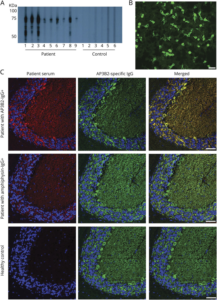
Figure 3. Western blot with recombinant protein and confocal microscopy confirm the target antigen is a cytosolic protein, adaptor protein 3, subunit B2 (AP3B2).
(A) By western blot, immunoglobulin G (IgG) from 9 patient sera, but none of 6 healthy control sera shown, bind to a recombinant AP3B2 C-terminal polypeptide fragment. (B) Positivity by AP3B2-specific cell-binding assay in serum of patient 7. (C) Commercial AP3B2 antibody colocalizes well with immunoreactivity produced on mouse cerebellum by patient AB3B2-IgG (top), but not with amphiphysin-IgG-positive patient (middle) or healthy control (bottom). Left column, patient IgG; middle column, rabbit anti‐AP3B2 IgG; right column, merged images. Scale bars, 50 μm.

