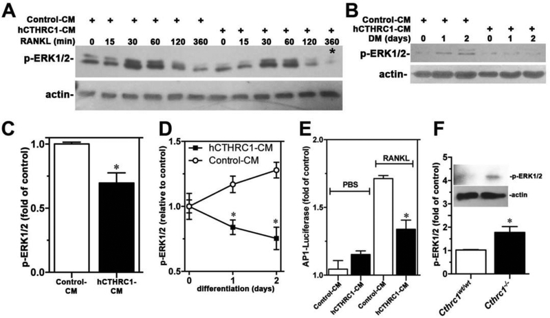
Figure 7.
(A) An immunoblot analysis of a time course of ERK1/2 activation (p-ERK1/2) in RAW264.7 cells stimulated with RANKL in the presence of control-CM or hCTHRC1-CM is shown. The corresponding quantification of the signal marked with * is shown in (C). In (B) p-ERK1/2 levels in RAW264.7 cells growing in differentiation medium (DM) for up to 2 days are shown with corresponding quantification shown in (D). (E) hCTHRC1 inhibits RANKL-induced AP-1 luciferase reporter activity in RAW264.7 cells transfected with the AP-1 promoter reporter plasmid. (F) Western blot analysis of p-ERK1/2 level in femur lysates from wildtype and Cthrc1 null mice. All data represent means±SEM of replicates ≥3 with * = p<0.05.

