Abstract
Introduction:
To compare the efficacy of eccentrically-focused resistance exercise (ECC RT) to concentrically-focused resistance exercise (CNC RT) on knee osteoarthritis (OA) symptoms and strength.
Methods:
90 participants consented. Participants were randomized to CNC RT, ECC RT or a wait-list no-exercise control group (CON). Four-months of supervised exercise training were completed using traditional weight machines (CNC RT), or modified-matched machines that overloaded the eccentric action (ECC RT). Main outcomes included one-repetition maximal strength (1RM; knee extension, leg flexion and leg press), weekly rate of strength gain, Western Ontario McMaster University Osteoarthritis Index (WOMAC) total score and sub-scores.
Results:
54 participants (60–85yr, 61% women) completed the study. Both CNC RT and ECC RT groups showed 16%−28% improvement relative to CON group (p = 0.003 to 0.005) for all leg strength measures. The rate of weekly strength gain was greater for CNC RT than ECC RT for leg press and knee flexion (by 2.9%−4.8%; both p < 0.05), but not knee extension (0.7%; p = 0.38). There were no significant differences in WOMAC total and sub-scores across groups over time. Leg press strength change was the greatest contributor to change in WOMAC Total scores (R2=0.223). The change in knee flexion strength from baseline to month four was a significant predictor of the change in WOMAC pain sub-score (F ratio 4.84, df=45, p=.032). Both modes of strength training were well-tolerated.
Conclusions:
Both resistance training types effectively increased leg strength. Knee flexion and knee extension muscle strength can modify function and pain symptoms irrespective of muscle contraction type. Which mode to pick could be determined by preference, goals, tolerance to the contraction type, and equipment availability.
Keywords: joint pain, strength training, safety, adherence
Introduction
Knee osteoarthritis (OA) is a major source of pain and disability globally.(1) OA is among the top 10 causes of physical disability worldwide.(2) Knee pain impacts multiple facets of quality of life, impedes physical function and is related to muscle loss. Pain itself predicts a trajectory of functional decline in well-functioning adults.(3) Loss of muscle mass and knee extensor-flexor muscle strength are independently associated with symptomatic progression of OA and reduction in overall health status.(4) Isokinetic strength testing indicates that individuals with OA have 11%−56% lower concentric leg extensor strength and 76% lower eccentric strength.(5) Pain inhibits corticospinal and intracortical pathways and causes a central activation deficit and strength reduction in the leg muscles about the knee.(6) While no cure exists for this condition, current management strategies for knee OA target potential modifiable symptoms such as pain, and contributing risk factors like strength.(1)
Resistance exercise can reduce knee pain severity and leg strength in participants with symptomatic knee OA.(7) Exercise interventions using free weights or machines have generally focused on movements with concentric muscle contractions. Previous interventions were developed based on loads lifted during the concentric phase, not the eccentric phase.(8) Hence, the eccentric phase is often relatively under-loaded compared to the muscle capabilities.(8, 9) What remains unknown for the population with knee OA is whether increasing the eccentric loading component of resistance training could enhance training benefits on pain and strength. Limited data show that eccentric contractions increase muscular strength and hypertrophy at a lower metabolic, cardiac and neural cost than concentric contractions. (8, 10) Even among strength trained young men, accentuated eccentric loading during strength training increases isometric torque and muscle activation more than concentric training.(9) While both types of muscle training can induce hypertrophy in healthy adults, the muscle architecture adaptations for eccentric training largely occur with fascicle length and hypertrophy is more evident in the distal ends of the muscle; after concentric training, muscle hypertrophy occurs largely in the muscle belly and induces changes in pennation angle.(11) Moreover, compared to concentric training, eccentric training reduces intracortical inhibition and increases corticospinal excitability by 37%−51%, both of which may promote the documented cross-transfer of strength improvement to opposite limbs.(12) The available literature however, is fraught with methodological variability in training mode, exercise type, unilateral training status, and equipment which compromises the ability to determine comparative effectiveness for clinical populations such as knee OA. Our laboratory has developed and published an eccentric exercise weight machine model of a well-used concentric weight machine counterpart.(13) Until now, there has not been a head-to-head comparison of concentric and eccentric resistance exercise for therapeutic benefit, but our model allows us to perform this comparison.
Recent meta-analysis showed that exercise reduces OA pain severity similarly to traditional analgesic agents.(14) Regimens that maximize strength with relatively less physiological stress are highly attractive for the older adult with debilitating OA. Evidence from the Osteoarthritis Initiative shows that as knee OA progresses and pain increases over time, knee flexor and extensor muscle strength declines linearly in both men and women.(15) This strength loss appears to be independent of radiographic stage of the disease(16) and is minimally explained by comorbidities or depression.(15) Pain reduction and reduction of pain impact on physical functioning are related to strength gains from a variety of exercises.(17, 18) Previous investigations have documented improvements in joint disease-specific instruments such as the Western Ontario and McMaster Universities Osteoarthritis Index (WOMAC), up to 54.%% with resistance exercise, (19) and that changes in knee extensor strength mediate WOMAC scores.(20) What remains unclear is whether eccentric or concentric-based resistance training is more effective for reducing knee OA pain symptoms and reversing the OA-related strength decline. We performed a randomized, controlled study to compare the effectiveness of eccentrically-focused resistance exercise (ECC RT) and concentrically-focused resistance exercise (CNC RT) on knee OA symptoms and leg muscle strength over four months. We hypothesized that ECC RT would elicit superior improvements in knee pain, perceived function and leg maximal strength compared to CNC RT.
Methods
Study Design.
This was a four-month randomized, controlled, single-blinded study of two different resistance exercise training programs on knee OA symptoms. This study followed the Consolidated Standards of Reporting Trials (CONSORT) 2010 guidelines for reporting parallel group randomized trials. The study was registered as a clinical trial NCT00187863.
Participants.
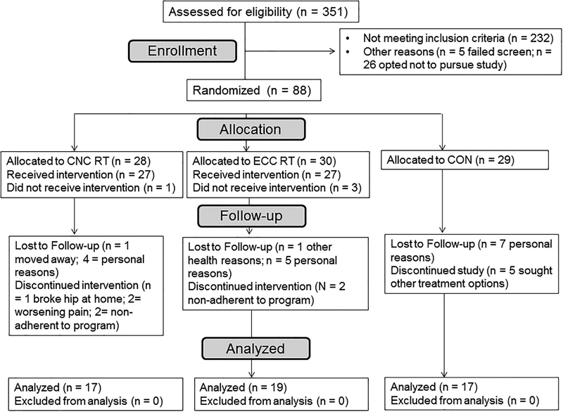
Older adults with knee OA were recruited from study flyers and newspaper advertisements posted in the Gainesville area and surrounding regions using the UF Orthopaedics Clinics, the Clinical Trials Register, and a list of older adults provided by the UF Claude Pepper Aging Center. Recruitment occurred during November 2010 to December 2012. Inclusion criteria: Men and women aged 60–85 years; presence of OA of the knee (using American College of Rheumatology criteria) for ≥6 months;(21) knee pain primarily due to tibiofemoral OA and not from patellofemoral OA; bilateral standing anterior-posterior radiograph demonstrating Kellgren and Lawrence OA grade two or three out of the target knee;(22) willing and able to participate in regular exercise for four months; free from musculoskeletal limitations that would preclude resistance exercise participation (i.e. joint contractures, fractures); free of abnormal cardiovascular responses during the screening graded maximal walk test. Exclusion criteria: Any surgery to either knee within the last 12 months, lumbar radiculopathy, vascular claudication; significant anterior knee pain due to diagnosed isolated patella-femoral syndrome or chondromalacia in either knee; had corticosteroid or hyaluronic acid injections administered within three months of study participation, have added new over the counter or prescription pain medication within two months of study participation. Knee OA eligibility criteria were first reviewed on each potential participant by the study coordinator and the PI (physician) on the study to ensure that the appropriate participants were enrolled. This study was approved by the University of Florida Institutional Review Board, and all procedures on human subjects were conducted in accordance with the Helsinki Declaration of 1975, as revised in 1983. All participants provided written, informed consent to participate. The CONSORT study flow diagram is shown in Figure 1.
Figure 1.
Study flow diagram. CNC RT= concentrically focused resistance exercise group, ECC RT = eccentrically focused resistance exercise group, wait-list control condition (CON).
Additional Screening Measures and Study Visits.
All study measures were collected at the University of Florida Human Dynamics Laboratories. Visit one included an orientation to the laboratory testing area and a familiarization with the machines. Participants completed the WOMAC for knee pain-related quality of life impact. Prior to clearance, the participant’s maximal rate of oxygen consumption was determined using a walking symptom-limited graded exercise test at baseline (incremental treadmill Naughton test). All procedures followed the American College of Sports Medicine (ACSM) guidelines with electrocardiogram heart monitoring and blood pressure measures. Open-circuit spirometry was used to determine the rate of oxygen use and carbon dioxide production using a metabolic cart (VIASYS©, CareFusion Corp. San Diego, CA). The test was stopped at voluntary exhaustion or when knee pain prevented further walking. Rating of perceived exertion values were collected at rest, at each exercise stage and during recovery. If no abnormal cardiovascular responses occurred, the participant continued in the study.
Visit two involved maximal strength testing of major muscle groups to develop a training program. First, participants were familiarized to each exercise machine, and the settings of each machine were individually customized to match the anthropometry of each participant. Once the strength values for each exercise were determined, a training schedule was established. After the training period, participants completed a third visit for post-training measures.
Randomization Procedure.
Participants were randomly assigned to one of three study groups: a concentrically-based resistance exercise training program (CNC RT), an eccentrically-based resistance exercise training program (ECC RT) or a wait-list, non-exercise control group (CON). Randomization was achieved using a computer-generated list and hidden sequencing of the individual assignment. The assignments per participant number were placed in numbered sealed envelopes. Each new enrolled participant opened an envelope to receive the group assignment. One study coordinator issued the assignment and the PI and other investigators were blinded to the allocation sequence. A total of 90 participants were enrolled into the study.
Resistance Exercise Interventions.
CNC RT was performed on traditional commercial dynamic resistance exercise machines (MedX®). ECC RT was performed on modified MedX® machines that contained a novel design that resistance loads during the eccentric phase of the contraction while “assistance” was provided by the machine during the concentric phase. This allowed for each type of contraction to use a load that more appropriately matched its respective force-producing capabilities when compared to CNC RT machines. Exercise training sessions were performed in a supervised laboratory setting over a four-month period. All participants were familiarized with all the testing equipment and performed a light exercise set on each of the exercise machines to configure the machine seat position. Participants in the trained groups reported to the laboratory two times a week for one-on-one training sessions with an experienced exercise physiologist. Work performed during each exercise session was recorded in a personalized training chart.
Blinding to Treatment.
Coordinators and exercise physiologists conducted the testing sessions and the assessments for the study. The physiologists and the physicians who provided coverage and interpretation of the testing were blinded to the randomization, group allocation and related interventions.
Body Composition.
Body composition was tracked using air plethysmography (BODPOD; Life Measurement, Concord, CA) from pre- to post-training. This is a reliable technique of body volume and composition measurement and is highly correlated with the reference standard of underwater weighing.(23) The intraclass coefficient for body density was .996 for a heterogeneous sample. The test–retest correlation for body mass measurement is r = .999.(23)
Strength Testing.
Resistance loads were set using a percentage of the one repetition maximum (1RM) technique for each exercise. The 1RM were determined using the following protocol: for each exercise, a warm up of five repetitions at a low weight was followed by three repetitions at a higher weight of each dynamic exercise. One lift was performed at progressively higher loads until the dynamic exercise could not be performed or performed with good form. 1RM values were secondary outcomes. Recovery periods between each lift were 60 seconds. Specific training appointment times were established for each participant during the week to avoid exposure to other participants and contamination of data. All adverse events and unanticipated events were tracked during the study.
Concentrically-Focused Resistance Training (CNC RT).
For clarity, we define here that the phase of the motion that involves pushing the weight away from the body is the concentric phase, and the phase of the motion that involves controlled weight return back toward the body is the eccentric phase. We modified our exercise protocol that was successfully used on our older adult population of a similar age range using MedX® machines and is in accord with the guidelines prescribed by the ACSM. Participants randomized to this group performed two resistance exercise sessions per week, and one set of each exercise was completed in each session: leg press, knee flexion, knee extension, chest press, seated row, overhead press, biceps curl, and calf press. A description of the exercise details can be found in Supplemental Table 1 (see Table, Supplemental Digital Content 1, Exercise start and end positions, directions and cues for MedX® resistance exercise machines). Each set contained 12 repetitions performed at a resistance load of 60% of the concentric 1RM for that exercise. Participants subjectively rated the effort of the exercise set using a 6–20 point Borg scale. As the participant adapted, the effort was less, and the resistance load was increased for the set to keep the RPE value at approximately 17–18 for the exercise over the study duration.
Eccentrically-Focused Resistance Training (ECC RT).
The resistance strategy for enhanced eccentric training is therefore to continually perform the eccentric muscle action or weight return with the equivalent of the concentric 1RM. During the pushing or concentric phase of each lift here, the resistance was set at 60% of 1RM. Each set consisted of eight repetitions and the participants subjectively rated the effort of the exercise set using a 6–20 point Borg scale. Progression of loading over time was identical to that of the CNC RT group described above. The repetition structure on the eccentric exercise machine and comparative concentric exercise machine were adjusted to equalize the work performed on a given exercise between the study groups.
Wait-List Control Condition (CON).
Participants continued participating in their normal activities during the four month study period if assigned to this group. This group was offered the opportunity to complete either the CNC RT or ECC RT program after the control period. Telephone contact was made weekly to help encourage adherence to the knee symptom management guidelines and to provide attention to this group. Finally, all participants wore dual-axis accelerometers for seven days before and after the training period (StepWatch activity monitors; SAM, Cyma, Seattle, WA) to track whether habitual activity changed.
Main Outcomes.
The main study outcomes included patient-reported knee symptoms and function, dynamic muscle strength and ambulatory knee pain severity.
Western Ontario and McMaster Universities Osteoarthritis Index (WOMAC).
The WOMAC is a disease-specific, reliable and valid measure of patient-reported knee or hip status. A total of 24 items were grouped into three subscales, including pain (five items), stiffness two items) and physical function (17 items). Questions were asked in four-point Likert scale format. The point range for this format was 0 points (no difficulty, pain or stiffness) to 100 points (worst pain, stiffness and worst function). Internal consistency for this instrument is high with Chronbach’s α values for pain, stiffness and subscale scores ranging from 0.86–0.55.(24) WOMAC test-retest reliability intraclass correlation coefficient (ICC) for these WOMAC subscales ranges from 0.90–0.95. (25) The minimum clinically important improvement (MCII) in the WOMAC global with treatment is −39.0%. MCII for pain and physical function are −40.8% and −26.0%, respectively.(26) The study was powered based on the WOMAC as the primary study outcome.
Muscle Strength.
The 1RM values for knee extension, knee flexion and leg press on the MedX® machines represented the dynamic muscle strength values. Strength of muscles around the knee joint is clinically important because lower strength is associated with higher WOMAC pain score.(15) The test-retest reliability ICC for 1RM measures in the lower extremity range from to 0.94–0.99 in other older populations(27) and persons with mobility limitations.(28) The knee flexor-to-extensor ratio was calculated before and after the training period. This ratio has been used to estimate knee function and muscle balance; higher flexor-to-extensor rations indicate lower quadricep strength.(29) Muscle strength values for other muscle groups are reported in Supplementary table 2 (see Table, Supplemental Digital Content 2, Maximal strength measurements across intervention groups for other muscle groups).
Feasibility and Safety.
The feasibility of the training interventions was examined by the weekly rate of strength gain. Safety was tracked by adverse events related to the intervention, and included but was not limited to: worsening of knee pain, falls, knee joint swelling, onset of other joint pain. Adverse events were documented from the time of enrollment to completion of the four month study for each participant, and were reviewed as they occurred and on a monthly basis with the study team.
Statistics.
All analyses were conducted in JMP Pro 12.0 (SAS Institute, Inc., Cary, NC). Differences in baseline categorical measures across concentric (CNC RT), eccentric (ECC RT), and control (CON) groups was assessed using chi-square tests. Differences in baseline continuous measures across CNC RT, and ECC RT groups were assessed with ANOVA, using the Tukey-Kramer test for pairwise comparisons, which also adjusted for multiple comparisons using the Bonferroni method. Non-normal measures were log transformed prior to analyses. For primary outcomes, data were analyzed by intent-to-treat approach, using linear mixed models were used. These models included time (pre or post) and study group as main effects, with an interaction model between time and group. A significant time × group interaction would indicate that change in outcome from pre to post differed among groups. Mixed models use maximum likelihood estimation to handle missing data, including full information from all individuals randomized in the study.
General linear models were also run and compared to assess which gains in strength (calculated as pre to post improvements in leg press, knee flexion, and knee extension) best explained improvements in WOMAC Total scores. The first model (Model 1) included pre-intervention WOMAC total score as the independent variable and post-intervention WOMAC total score as dependent variable to model change in total WOMAC score. Subsequent models (Model 2a-2c) added each strength measure (leg press change, knee flexion change, knee extension change) separately and then all together (Model 3). Model fit and parsimony was assessed with R2 (% variance explained in WOMAC total score) and Akaike’s Information Criteria (AIC). A reduction in AIC would indicate greater model parsimony and better model fit.
Sample Size Estimation.
Power analysis was calculated using previously published data regarding differences elicited for the WOMAC pain subscale.(30–33) This variable was chosen because it is a primary reason for people with knee OA to undergo a total knee arthroplasty for OA.(31–33) The minimum clinically relevant decrease in the WOMAC pain subscale is 1.2 cm on a 10 cm scale.(34) For this investigation, 1.5 cm represents a 30% difference between the two exercise interventions. Sample size estimation using a mean of five cm with a standard deviation of two cm and a desired effect size of 1.5 cm indicated that 20 participants per group was needed to have a power of 0.80 at an alpha level of 0.05. With our past experience, we anticipated a 30% dropout rate, and increased the number of participants in each group to 30.
Results
Participant Characteristics.
Figure 1 shows the study flow diagram. A total of 351 people were screened by phone, and 237 candidates did not meet all the inclusion criteria or met one or more exclusion criteria. A total of 114 candidates were offered initial appointments, and 90 were enrolled. Table 1 provides the baseline characteristics of the three study groups. The proportion of participants who used pain medications for knee pain or the mean number of pain medications used by the three groups was not different from pre- to post-training. Mean pain medication numbers post-training were 0.8 ± 0.5 (CNC RT), 0.8 ± 0.6 (ECC RT) and 1.2 ± 1.1 (CON; p=0.347). Mean step counts from the activity monitors were not different across groups over time, indicating no change in habitual activity levels (CNC RT 3600 to 3496 steps/day; ECC RT 4652 to 4643 steps/day and CON 5002 to 4851 steps per day; p=0.962).
Table 1.
Baseline characteristics of older adults with knee osteoarthritis (OA). Values are means ± SD or % of the group.
| CNC RT (n =28) |
ECC RT (n =30) |
CON (n =32) |
p (sig) | |
|---|---|---|---|---|
| Age (yr) | 69.5 ± 6.5 | 66.8 ± 5.4 | 68.6 ± 7.2 | 0.287 |
| Body mass (kg) | 92.7 ± 19.7 | 79.5 ± 20.7 | 86.8 ± 19.7 | 0.203 |
| BMI (kg/m2) | 32.8 ± 7.4 | 28.7 ± 6.6 | 30.1 ± 6.2 | 0.069 |
| Fat-free mass (%) | 57.3 ± 11.8 | 61.0 ± 9.6 | 61.7 ± 10.8 | 0.486 |
| Sex (%) | ||||
| Female | 67.0 | 70.0 | 66.0 | |
| Male | 33.0 | 30.0 | 34.0 | 0.381 |
| Race/Ethnicity (#, %) | ||||
| White | 85.0 | 93.0 | 81.0 | |
| African-American | 11.0 | 7.0 | 6.0 | |
| Other | 4.0 | 0.0 | 13.0 | 0.439 |
| Work status (%) | ||||
| Working | 40.0 | 23.0 | 34.0 | |
| Not working | 4.0 | 7.0 | 13.0 | |
| Retired | 52.0 | 70.0 | 50.0 | |
| Disabled | 4.0 | 0.0 | 3.0 | 0.981 |
| Duration of pain, median [Q1–Q3] | 4.5 [2–11] | 10 [2.75–20] | 5 [2–10] | 0.150 |
| Location of knee pain (%) | ||||
| Left | 18.0 | 20.0 | 9.0 | |
| Right | 15.0 | 17.0 | 28.0 | |
| Both | 67.0 | 63.0 | 63.0 | |
| Pain Medications (#) | 1.0 ± 0.5 | 0.9 ± 0.5 | 1.1 ± 1.1 | 0.863 |
Adherence to the Intervention.
From the initial 90 who consented to participate, 88 were randomized into a study group and 54 completed the study. Figure 1 provides the details for dropouts. For the two groups who trained, the percentage of exercise training sessions completed was 94.8% and 96.4% in the CNC RT and ECC RT, respectively (p=0.533). The average training duration, or days to complete the training sessions were 126 ± 21 days (114 to 137 days 95% CI) for the CNC RT and 130 ±12 days (123 to 135 days 95% CI) for the ECC RT (p=0.628).
Body Composition.
There were no significant changes in body mass or composition among the three groups from pre- to post-training. Post-training body weight for the CNC RT, ECC RT and CON were 93.2 kg ± 20.0kg, 79.5 kg ±0.5 kg and 87.7 kg ± 19.2 kg, respectively (p=0.509). The percent fat-free mass values were also not different among the three groups (57.0% ± 11.4% [CNC RT], 59.6% ± 9.8% [ECC RT], and 61.5% ± 11.2% [CON]; p=0.715).
WOMAC Responses.
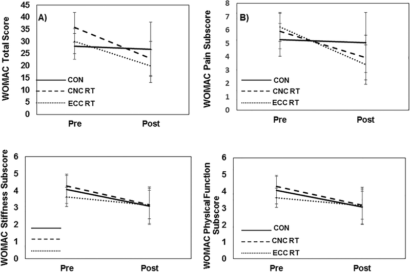
There were no statistically significant differences in WOMAC measures across groups (Table 2). Fifty-four (n=54) patients had post-intervention assessments. Figure 2 shows change in WOMAC scores from pre to post-intervention. As shown in Figure 2a–2d, there were no statistically significant group differences in this change for WOMAC total scores (F(2,50) = 2.1, p = 0.13), WOMAC stiffness scores (F(2,51) = 0.01, p = 0.98), and WOMAC function scores (F(2,51) = 1.0, p = 0.37), though there was a trend for differences for WOMAC pain scores (F(2,51) = 2.6, p = 0.08). A total of 50% and 68.4% of the CNC RT and ECC RT achieved the minimum clinically relevant reduction in the WOMAC pain sub-score, respectively. There were no main effects (p > 0.050 of age nor sex on any WOMAC measure.
Table 2.
Baseline WOMAC and maximal leg strength measurements across intervention groups. Values are mean ± SD. WOMAC scores are expressed in points and strength values are expressed in Nm.
| CNC RT (n =28) |
ECC RT (n =30) |
CON (n =32) |
p (sig) | |
|---|---|---|---|---|
| WOMAC Pain | 5.9 ± 3.2 | 6.2 ± 2.8 | 5.3 ± 3.2 | 0.495 |
| WOMAC Stiffness | 4.3 ± 1.6 | 3.6 ± 1.5 | 4.1 ± 1.5 | 0.383 |
| WOMAC Physical Function | 25.6 ± 11.1 | 20.2 ± 10.0 | 19.2 ± 10.2 | 0.061 |
| WOMAC Total | 35.9 ± 14.7 | 30.1 ± 13.2 | 28.0 ± 13.7 | 0.115 |
| Leg press | 528.9 ± 207.9 | 597.4 ± 207.7 | 672.4 ± 179.6 | 0.701 |
| Knee flexion | 250.6 ± 85.3 | 281.9 ± 110.2 | 303.6 ± 98.2 | 0.211 |
| Knee extension | 232.8 ± 106.9 | 314.1 ± 178.8 | 332.9± 172.8 | 0.091 |
Figure 2.
Change (pre- to post-intervention) in WOMAC scores for CNC RT group (dashed line), ECC RT group (dotted line) and CON group (solid grey line). Error bars represent 95% confidence intervals. A) WOMAC total scores, B) Pain sub-scores, C) Stiffness sub-scores, and D) Physical Function sub-scores.
Muscle Strength.
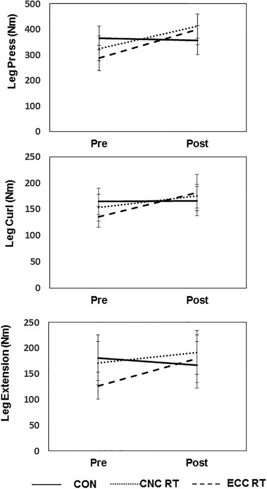
There were statistically significant group differences for pre- to post-intervention change in leg press, knee flexion, and knee extension (Figure 3; all p<0.05). Specifically, for all leg strength measures, both CNC RT and ECC RT groups showed greater improvement relative to CON group (p = 0.003 to 0.005), but there were no statistically significant differences between CNC RT and ECC RT groups. The percent change in the leg press strength from pre to post-training in the CON, CNC RT and ECC RT were −2.2% (95%CI: −18.7%; 14.2%), 33.5% (95%CI: 16.1%; 50.9%) and 32.8% (95%CI: 17.2%; 48.4%), respectively. The percent change in the knee extension strength from pre to post-training in the CON, CNC RT and ECC RT were-7.4% (95%CI: −19.8%; 5.1%), 29.2% (95%CI: 7.1%; 51.2%) and 20.2% (95%CI: 4.1%; 36.4%). Finally, the percent change in the knee flexion strength values in these same three groups were −0.5% (95%CI: −10.2%; 9.3%), 20.8% (95%CI: 10.7%; 30.9%) and 13.3% (95%CI: 10.2%; 28.2%), respectively. Maximal strength values for all other muscle groups are reported in Supplementary table 2 (see Table, Supplemental Digital Content 2, Maximal strength measurements across intervention groups for other muscle groups). There were no statistically significant group by time interactions for strength for chest press, shoulder press and seated row. There were significant main effects (p < 0.05) of both age and sex on strength measures.
Figure 3.
Change (pre- to post-intervention) in leg strength for concentric group (dashed line), eccentric group (dotted line) and control group (solid grey line). Error bars represent 95% confidence intervals.
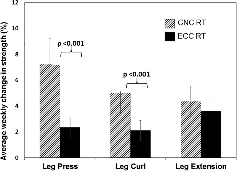
There were significant differences in weekly strength gains between the CNC RT and ECC RT groups (Figure 4). Specifically, the CNC RT had greater mean weekly gains compared to the ECC RT for leg press (7.2±2.0% versus 2.3±0.7%; p < 0.001) and knee flexion (5.0±1.5% versus 2.1±0.7%; p < 0.001), but not for knee extension (4.3±1.2% versus 3.6±1.2%; p = 0.38). Finally, the knee flexor-to-knee-extensor strength ratio was not different across groups over time (p=.109). The mean strength flexor-to-knee extensor ratios for the three groups from pre and post-training were CNC RT (from 1.11 to 1.00), ECC RT (from 1.01 to 1.00) and CON (from 1.03 to 1.14).
Figure 4.
Mean percent (%) weekly improvement in leg strength for CNC RT group (white bars) and ECC RT group (black bars). Error bars represent 95% confidence intervals. There were statistically significant group differences for leg press and knee flexion weekly improvements.
Relationship Between Muscle Strength and WOMAC Pain Scores.
Model fitting procedures examined the relationship between the individual strength leg gains on the change in WOMAC pain score, the results of which are shown in Supplemental Table 2 (see Table, Supplemental Digital Content 2, Maximal strength measurements across intervention groups for other muscle groups). Models accounted for age, sex, baseline strength value and study group. Among the three leg strength measures, knee flexion was a significant predictor of pain reduction (p=.033). Model fitting procedures also examined the relationship between gains in leg strength (for overall sample of patients) and improvements in WOMAC total scores (see Table, Supplemental Digital Content 3, Fixed effects results for the relationship between gains in leg strength and change in WOMAC Pain scores). The best fitting, most parsimonious model (in terms of combined R2 and AIC) resulted from the inclusion of the leg press strength as an independent predictor of improvement in WOMAC total scores. This model explained nearly 30% of the variance in the outcome (see Table, Supplemental Digital Content 4, Model fitting results for the relationship between gains in strength and improvement in total WOMAC scores, Model 2a). Greater gains in the leg press exercise results in greater decreases (improvements) in WOMAC total score (β = −0.29, SE = 0.13, p = 0.027).
Safety and Feasibility.
The dropout rates in the CNC RT, ECC RT and CON groups were 39%, 36% and 45%, respectively. The proportion of patients experiencing a non-serious adverse event was 10.7% in the CNC RT (arm ligament strain, fall unrelated to study), 3.3% in the ECC RT group (increased hip pain) and 0% in the CON group. The proportion of patients experiencing a severe adverse event was 1.7% (1 out of 58 total exercisers) and this was not related to the study (broken hip due to fall in the home). A total of 13.7% of participants were lost to follow-up due to “personal reasons” (lost interest, assumed care for family member or spouse). Among the others lost to follow-up in the two exercising groups, 6.8% (n=4) were non-adherent to the exercise program, one participant moved away, and one was unexpectedly diagnosed with cancer. In the CON group, seven participants cited personal reasons for stopping the study and decided not to wait for their opportunity to train, and five sought other treatment options for the knee pain.
Discussion
We compared the efficacy of ECC RT to traditional CNC RT on knee pain, perceived function and leg maximal strength over four months compared to a control group. Maximal strength improved with both resistance exercise programs, but the rate of strength gain was higher in the CNC RT group. The ECC RT was well tolerated and safe. These findings indicate that ECC RT provides comparable strength benefits to strength or pain reduction compared to CNC RT over four months.
We found wide variability in the pain and strength responsiveness to resistance exercise among these participants, with individual knee pain changes ranging from −65% to 78%, and strength improvements ranging from 4% to 54%. Because neither training program appeared superior to the other with respect to mean strength gains and perceived function and pain, there is flexibility for the patient with OA and the care provider to determine which training program best matches the patient’s goals in light of other health considerations.
Data from the Osteoarthritis Initiative show that with disease progression, for each increase in WOMAC pain, knee extensor and flexor strength linearly decrease by1.6–1.9% to 1.6–2.5%, respectively.(15) We anticipated that pain severity would be reduced in parallel with training-induced strength gains. The mean group changes in the WOMAC pain scores over the four month period were not statistically different. However, we did find that there was a subset of individuals who responded better to the training than others. Specifically, about 50% of the exercising participants in the ECC RT and CNC RT groups achieved clinically significant pain reduction represented by a 30% reduction in WOMAC pain sub-score from baseline (2–2.8 point reduction). A recent systematic review and meta-regression from 45 varied exercise trials in knee osteoarthritis revealed that detection of pain improvement or knee function is unlikely unless leg muscle strength gains were ≥30% from pre-training values.(18) In our present study, both resistance training groups made gains in leg strength ranging from 13.3% (knee flexion) to 33.5% (leg press). Variability existed in the pain responsiveness to training, where some patients achieved clinically meaningful improvements to pain whereas others did not. These collective findings suggest that the overall gains made here may not have been sufficient for all exercising participants to obtain clinically meaningful pain relief. Alternatively, the findings could mean that: 1) strength gain is but one part of the exercise benefit on pain relief in knee OA, or 2) that contraction type may differentially impact pain depending on individual variations in OA grade, location and size of cartilage deficits. Recent evidence indicates that eccentric quadriceps strength compared to concentric strength is lower in persons with focal cartilage lesions.(35) These areas warrant further study to identify which patients would respond best to which contraction mode depending on OA joint morphology.
Published studies of total body strength training and WOMAC scores in knee OA are limited. One study that used weight machines and free weights reported that a 26–43% increase in knee flexors/extensors strength paralleled a WOMAC pain score reduction of 9–4.9 points.(36) Another study that used Keiser pneumatic machines as the training stimulus did not find training group by time interactions for total WOMAC scores and subscores.(37) Pain reductions in the sham and training groups were 1.2–1.8 WOMAC points, respectively (21%−32% pain reductions). The authors explained these findings in part due the fact that their control group (‘sham exercise’) was more than a sham as it consisted of low volume knee extensions.(37) Hence, resistance exercise even in low volumes may positively improve WOMAC scores.
Despite a significantly higher rate of weekly strength gain in the CNC RT group compared to ECC RT, we found that the maximal leg strength gains were not different between training groups. Our findings are similar to studies showing no difference in strength gain and torque between eccentrically and concentrically-focused protocols after five weeks of training,(9) but disagree with other evidence that superior strength gains are achieved after 5–12 weeks of eccentric resistance training than concentric training.(42) One possibility to explain this finding is that the regular resistance exercise, irrespective of type of contraction, may prepare an individual to perform maximal lifts during post-testing. Familiarity with the machines and the sensations experienced during maximal lifts could have mitigated adverse perceptions of the strength testing (e.g., temporary knee pain, fatigue, elevated heart rate) so that maximal loads were better tolerated and improved in both groups after four months. Alternatively, ECC RT may have improved volitional drive by reducing corticospinal inhibition to muscle more than CNC RT,(10) enabling participants to achieve similar maximal lifts during strength testing as those in the CNC RT group. The therapeutic application of these findings lies in the ability to individualize strength protocols depending on the goals of the patient as both were equally effective in terms of improving function while decreasing pain. There is the possibility that there are sex differences in the strength gains and muscle adaptations to different forms of resistance exercise, and thus individualizing strength protocols may be important. While data to support this point are limited, Miller et al.(40) reported sex differences in the mitochondrial content and improved contractility of muscle fibers after training in men than women and believed that much of the strength and functional adaptation in women was related to neural mechanisms. Potentially the eccentric and concentric stimulus affects strength and function differently in women and men. Future studies should consider sex as a factor in responsiveness to training protocols. The relative contribution of the gains in leg press, knee extension and knee flexion on OA symptom changes is not clear. Previous intervention studies showed that strengthening exercise involving both knee extensors and flexors produces better WOMAC pain symptom relief than knee extension alone.(41) Here, knee flexor strength gain was a significant predictor of pain reduction. This is in contrast to most of the published cross sectional work of the relationships between leg muscle strength and pain symptoms.(42) The hamstring-to-quadriceps ratio (knee flexor: knee extensor) has not been found to be associated with pain severity during daily tasks including stair climb, lying down, standing up and sitting cross-legged on the floor.(42) Our pre-training knee flexor: extensor strength ratios were close to 1, indicating that there was not a relative quadriceps to hamstrings strength deficit in the in our cohort, as has been shown in other knee OA groups. The potential significance of the role in hamstrings to pain reduction could be in part due to favorable shifting of the femoral and tibial contact points relative to the location of the cartilage lesions. Regression modeling revealed that greater gains in the leg press exercise resulted in better WOMAC total scores (β = −0.29, SE = 0.13, p = 0.027). The importance of this finding may lie in the translation of leg press strength to the functionality of the knee. Tevald et al(43) and Aalund et al.(44) showed that leg press power contributed to the ability to perform physical function tasks such as 10m fast walks, stair climb, get up and go tests and chair rise time. Hence, leg press may be more functionally relevant than knee extension or curl in this group. Achievement of strength gain did not appear to be superior in the ECC RT to the CONC RT. This suggests that either mode of training would provide benefit to leg muscles in knee OA. The choice of which strengthening machines to use in the clinical setting can thus be decided by the patient and practitioner depending on the goals of the treatment plan. For those patients who have OA and issues with hypertension or cardiovascular disease, a program that emphasizes the eccentric phase may prove to be the preferable option as the cardiovascular cost of this type of contraction is lower compared to the concentric phase.
Limitations and Strengths.
There are several limitations to the present study. In contrast to our expectations, our control group was very healthy and demonstrated similar patterns of WOMAC physical function and stiffness improvements (Figure 2). Also, our CNC RT group tended to have higher BMIs, lesser fat-free mass and more were working; it is possible that at baseline, WOMAC functional scores were greater as a consequence. Only N=54 patients completed post-intervention measurements. There was also greater variability in the pain responsiveness to the training than initially expected. Thus, a greater sample size may have been required to detect statistical significance in both the pain and strength outcomes; follow-up calculations show that N=66 patients with completed post-intervention data would be needed to detect the observed differences in WOMAC pain scores at statistically significant at an alpha=0.05. We used maximum likelihood estimation to handle missing data and uses full information for all patients, and this is in line with an intent-to-treat approach. The number of dropouts to the final number of 54 participants was due to a combination of factors including inability to commit to an intensive four month schedule, inability to follow study procedures, personal reasons and sought other procedures for knee pain. The percent dropouts were not different among the three study groups, ranging from 36.6% to 41.3%, which was 6.6–11.3% higher than we anticipated at the outset. We were unable to obtain radiographic images of joint space changes and differences in tibiofemoral contact points which would have provided insight on mechanisms of exercise effects. Study strengths included a rigorous RCT design, matched exercise equipment models for head-to head comparison of muscle contraction type(13) and use of validated, reliable instruments.(24, 25) A strong finding from this study is that overall, both exercise programs were well-tolerated and there were no adverse events related to the exercise that required medical intervention. The machines for the CNC RT are readily available in most fitness and therapy centers, and the protocol is highly feasible. As more companies develop eccentrically-focused machines, the ECC RT will also become more feasible for the general public.
Conclusion.
ECC RT and CONC RT were both effective in increasing leg muscle strength. WOMAC pain reduction was related to knee flexion strength gains in people with knee osteoarthritis, and overall WOMAC scores were predicted in part by leg press strength gains. These data indicate that programs that either use traditional modes of resistance training or those that emphasize the eccentric component of the contraction cycle can be well-tolerated in this population. Neither proved to be advantageous compared to the other allowing the provider and the patient to determine which method is best tolerated, fits the patient’s goals and other health considerations most appropriately. The reduced cardiovascular stress associated with eccentric contractions may make this mode more appropriate for patients with cardiovascular disease allowing strength and functional gains while minimizing cardiovascular stress.
Supplementary Material
SDC 1: Exercise start and end positions, directions and cues for MedX® resistance exercise machines. These same details applied to both traditional machines and the modified machines that provided eccentric overloading stimulus. All exercises were executed with a three-second concentric phase and three-second eccentric phase.
SDC 2: Maximal strength measurements across intervention groups for other muscle groups. Values are mean ± SD and expressed in Nm
SDC 3: Fixed effects results for the relationship between gains in leg strength and change in WOMAC Pain scores.
SDC 4: Model fitting results for the relationship between gains in strength and improvement in total WOMAC scores.
Acknowledgments.
Research reported in this publication was supported by the National Institute of Arthritis and Musculoskeletal and Skin Diseases of the National Institutes of Health under Award Number AR059786. The content is solely the responsibility of the authors and does not necessarily represent the official views of the National Institutes of Health. The results of this study do not constitute endorsement by the American College of Sports Medicine.
Conflict of interest and Source of Funding: Kevin Vincent and Heather Vincent are current associate editors of MSSE. Dr. Kevin Vincent received support from National Institute of Arthritis and Musculoskeletal and Skin Diseases of the National Institutes of Health (NIH) under Award Number AR059786. For the remaining authors none were declared. The results of the present study do not constitute endorsement by ACSM. The results of the study are presented clearly, honestly, and without fabrication, falsification, or inappropriate data manipulation.
References
- 1.Munukka M, Waller B, Rantalainen T, et al. Efficacy of progressive aquatic resistance training for tibiofemoral cartilage in postmenopausal women with mild knee osteoarthritis: a randomised controlled trial. Osteoarthritis Cartilage. 2016;24(10):1708–17. [DOI] [PubMed] [Google Scholar]
- 2.Neogi T The epidemiology and impact of pain in osteoarthritis. Osteoarthritis Cartilage. 2013;21(9):1145–53. [DOI] [PMC free article] [PubMed] [Google Scholar]
- 3.White DK, Neogi T, Nguyen U-SDT, Niu J, Zhang Y. Trajectories of functional decline in knee osteoarthritis: the Osteoarthritis Initiative. Rheumatol Oxf Engl. 2016;55(5):801–8. [DOI] [PMC free article] [PubMed] [Google Scholar]
- 4.Gonçalves RS, Pinheiro JP, Cabri J. Evaluation of potentially modifiable physical factors as predictors of health status in knee osteoarthritis patients referred for physical therapy. The Knee. 2012;19(4):373–9. [DOI] [PubMed] [Google Scholar]
- 5.Alnahdi AH, Zeni JA, Snyder-Mackler L. Muscle impairments in patients with knee osteoarthritis. Sports Health. 2012;4(4):284–92. [DOI] [PMC free article] [PubMed] [Google Scholar]
- 6.Kittelson AJ, Thomas AC, Kluger BM, Stevens-Lapsley JE. Corticospinal and intracortical excitability of the quadriceps in patients with knee osteoarthritis. Exp Brain Res. 2014;232(12):3991–9. [DOI] [PMC free article] [PubMed] [Google Scholar]
- 7.Gür H, Cakin N, Akova B, Okay E, Küçükoğlu S. Concentric versus combined concentric-eccentric isokinetic training: effects on functional capacity and symptoms in patients with osteoarthrosis of the knee. Arch Phys Med Rehabil. 2002;83(3):308–16. [DOI] [PubMed] [Google Scholar]
- 8.Reeves ND, Maganaris CN, Longo S, Narici MV. Differential adaptations to eccentric versus conventional resistance training in older humans. Exp Physiol. 2009;94(7):825–33. [DOI] [PubMed] [Google Scholar]
- 9.Walker S, Blazevich AJ, Haff GG, Tufano JJ, Newton RU, Häkkinen K. Greater strength gains after training with accentuated eccentric than traditional isoinertial loads in already strength-trained men. Front Physiol. 2016;7:149. [DOI] [PMC free article] [PubMed] [Google Scholar]
- 10.Li Y, Su Y, Chen S, et al. The effects of resistance exercise in patients with knee osteoarthritis: a systematic review and meta-analysis. Clin Rehabil. 2016;30(10):947–59. [DOI] [PubMed] [Google Scholar]
- 11.Franchi MV, Reeves ND, Narici MV. Skeletal muscle remodeling in response to eccentric vs. concentric loading: morphological, molecular, and metabolic adaptations. Front Physiol. 2017;8:447. [DOI] [PMC free article] [PubMed] [Google Scholar]
- 12.Kidgell DJ, Frazer AK, Daly RM, et al. Increased cross-education of muscle strength and reduced corticospinal inhibition following eccentric strength training. Neuroscience. 2015;300:566–75. [DOI] [PubMed] [Google Scholar]
- 13.Vincent HK, Percival S, Creasy R, et al. Acute Effects of Enhanced Eccentric and Concentric Resistance Exercise on Metabolism and Inflammation [Internet]. J Nov Physiother. 2014;4(2) doi: 10.4172/2165-7025.1000200. [DOI] [PMC free article] [PubMed] [Google Scholar]
- 14.Henriksen M, Hansen JB, Klokker L, Bliddal H, Christensen R. Comparable effects of exercise and analgesics for pain secondary to knee osteoarthritis: a meta-analysis of trials included in Cochrane systematic reviews. J Comp Eff Res. 2016;5(4):417–31. [DOI] [PubMed] [Google Scholar]
- 15.Ruhdorfer A, Wirth W, Eckstein F. Association of knee pain with a reduction in thigh muscle strength - a cross-sectional analysis including 4553 osteoarthritis initiative participants [Internet]. Osteoarthritis Cartilage. 2016; doi: 10.1016/j.joca.2016.10.026. [DOI] [PMC free article] [PubMed] [Google Scholar]
- 16.Ruhdorfer A, Wirth W, Hitzl W, Nevitt M, Eckstein F, Osteoarthritis Initiative Investigators. Association of thigh muscle strength with knee symptoms and radiographic disease stage of osteoarthritis: data from the Osteoarthritis Initiative. Arthritis Care Res. 2014;66(9):1344–53. [DOI] [PubMed] [Google Scholar]
- 17.Knoop J, Steultjens MPM, Roorda LD, et al. Improvement in upper leg muscle strength underlies beneficial effects of exercise therapy in knee osteoarthritis: secondary analysis from a randomised controlled trial. Physiotherapy. 2015;101(2):171–7. [DOI] [PubMed] [Google Scholar]
- 18.Bartholdy C, Juhl C, Christensen R, Lund H, Zhang W, Henriksen M. The role of muscle strengthening in exercise therapy for knee osteoarthritis: A systematic review and meta-regression analysis of randomized trials [Internet]. Semin Arthritis Rheum. 2017; doi: 10.1016/j.semarthrit.2017.03.007. [DOI] [PubMed] [Google Scholar]
- 19.DeVita P, Aaboe J, Bartholdy C, Leonardis JM, Bliddal H, Henriksen M. Quadriceps-strengthening exercise and quadriceps and knee biomechanics during walking in knee osteoarthritis: A two-centre randomized controlled trial. Clin Biomech Bristol Avon. 2018;59:199–206. [DOI] [PubMed] [Google Scholar]
- 20.Hall M, Hinman RS, Wrigley TV, Kasza J, Lim B-W, Bennell KL. Knee extensor strength gains mediate symptom improvement in knee osteoarthritis: secondary analysis of a randomised controlled trial. Osteoarthritis Cartilage. 2018;26(4):495–500. [DOI] [PubMed] [Google Scholar]
- 21.Altman R, Asch E, Bloch D, et al. Development of criteria for the classification and reporting of osteoarthritis. Classification of osteoarthritis of the knee. Diagnostic and Therapeutic Criteria Committee of the American Rheumatism Association. Arthritis Rheum. 1986;29(8):1039–49. [DOI] [PubMed] [Google Scholar]
- 22.Kellgren JH, Lawrence JS. Radiological assessment of osteo-arthrosis. Ann Rheum Dis. 1957;16(4):494–502. [DOI] [PMC free article] [PubMed] [Google Scholar]
- 23.Noreen EE, Lemon PWR. Reliability of air displacement plethysmography in a large, heterogeneous sample. Med Sci Sports Exerc. 2006;38(8):1505–9. [DOI] [PubMed] [Google Scholar]
- 24.Bellamy N, Buchanan WW, Goldsmith CH, Campbell J, Stitt LW. Validation study of WOMAC: a health status instrument for measuring clinically important patient relevant outcomes to antirheumatic drug therapy in patients with osteoarthritis of the hip or knee. J Rheumatol. 1988;15(12):1833–40. [PubMed] [Google Scholar]
- 25.Dunbar MJ, Robertsson O, Ryd L, Lidgren L. Appropriate questionnaires for knee arthroplasty. Results of a survey of 3600 patients from The Swedish Knee Arthroplasty Registry. J Bone Joint Surg Br. 2001;83(3):339–44. [DOI] [PubMed] [Google Scholar]
- 26.Tubach F, Ravaud P, Baron G, et al. Evaluation of clinically relevant changes in patient reported outcomes in knee and hip osteoarthritis: the minimal clinically important improvement. Ann Rheum Dis. 2005;64(1):29–33. [DOI] [PMC free article] [PubMed] [Google Scholar]
- 27.Abdul-Hameed U, Rangra P, Shareef MY, Hussain ME. Reliability of 1-repetition maximum estimation for upper and lower body muscular strength measurement in untrained middle aged type 2 diabetic patients. Asian J Sports Med. 2012;3(4):267–73. [DOI] [PMC free article] [PubMed] [Google Scholar]
- 28.LeBrasseur NK, Bhasin S, Miciek R, Storer TW. Tests of muscle strength and physical function: reliability and discrimination of performance in younger and older men and older men with mobility limitations. J Am Geriatr Soc. 2008;56(11):2118–23. [DOI] [PMC free article] [PubMed] [Google Scholar]
- 29.Suh MJ, Kim BR, Kim SR, Han EY, Lee SY. Effects of early combined eccentric-concentric versus concentric resistance training following total knee arthroplasty. Ann Rehabil Med. 2017;41(5):816–27. [DOI] [PMC free article] [PubMed] [Google Scholar]
- 30.Altman RD, Moskowitz R. Intraarticular sodium hyaluronate (Hyalgan) in the treatment of patients with osteoarthritis of the knee: a randomized clinical trial. Hyalgan Study Group. J Rheumatol. 1998;25(11):2203–12. [PubMed] [Google Scholar]
- 31.Altman RD, Rosen JE, Bloch DA, Hatoum HT, Korner P. A double-blind, randomized, saline-controlled study of the efficacy and safety of EUFLEXXA for treatment of painful osteoarthritis of the knee, with an open-label safety extension (the FLEXX trial). Semin Arthritis Rheum. 2009;39(1):1–9. [DOI] [PubMed] [Google Scholar]
- 32.Jinks C, Jordan K, Croft P. Osteoarthritis as a public health problem: the impact of developing knee pain on physical function in adults living in the community: (KNEST 3). Rheumatol Oxf Engl. 2007;46(5):877–81. [DOI] [PubMed] [Google Scholar]
- 33.Jordan JM, Helmick CG, Renner JB, et al. Prevalence of knee symptoms and radiographic and symptomatic knee osteoarthritis in African Americans and Caucasians: the Johnston County Osteoarthritis Project. J Rheumatol. 2007;34(1):172–80. [PubMed] [Google Scholar]
- 34.Ehrich EW, Davies GM, Watson DJ, Bolognese JA, Seidenberg BC, Bellamy N. Minimal perceptible clinical improvement with the Western Ontario and McMaster Universities osteoarthritis index questionnaire and global assessments in patients with osteoarthritis. J Rheumatol. 2000;27(11):2635–41. [PubMed] [Google Scholar]
- 35.Hirschmüller A, Andres T, Schoch W, et al. Quadriceps strength in patients with isolated cartilage defects of the knee: results of isokinetic strength measurements and their correlation with clinical and functional results. Orthop J Sports Med. 2017;5(5):2325967117703726. [DOI] [PMC free article] [PubMed] [Google Scholar]
- 36.Jorge RTB, de Souza MC, Chiari A, et al. Progressive resistance exercise in women with osteoarthritis of the knee: a randomized controlled trial. Clin Rehabil. 2015;29(3):234–43. [DOI] [PubMed] [Google Scholar]
- 37.Foroughi N, Smith RM, Lange AK, Baker MK, Fiatarone Singh MA, Vanwanseele B. Lower limb muscle strengthening does not change frontal plane moments in women with knee osteoarthritis: A randomized controlled trial. Clin Biomech Bristol Avon. 2011;26(2):167–74. [DOI] [PubMed] [Google Scholar]
- 38.Hilliard-Robertson PC, Schneider SM, Bishop SL, Guilliams ME. Strength gains following different combined concentric and eccentric exercise regimens. Aviat Space Environ Med. 2003;74(4):342–7. [PubMed] [Google Scholar]
- 39.Dibble LE, Hale TF, Marcus RL, Gerber JP, LaStayo PC. High intensity eccentric resistance training decreases bradykinesia and improves Quality Of Life in persons with Parkinson’s disease: a preliminary study. Parkinsonism Relat Disord. 2009;15(10):752–7. [DOI] [PubMed] [Google Scholar]
- 40.Miller MS, Callahan DM, Tourville TW, et al. Moderate intensity resistance exercise alters skeletal muscle molecular and cellular structure and function in inactive, older adults with knee osteoarthritis. J Appl Physiol Bethesda Md 1985. 2017;jap.00830.2016. [DOI] [PMC free article] [PubMed] [Google Scholar]
- 41.Al-Johani AH, Kachanathu SJ, Ramadan Hafez A, et al. Comparative study of hamstring and quadriceps strengthening treatments in the management of knee osteoarthritis. J Phys Ther Sci. 2014;26(6):817–20. [DOI] [PMC free article] [PubMed] [Google Scholar]
- 42.Fujita R, Matsui Y, Harada A, et al. Does the Q - H index show a stronger relationship than the H:Q ratio in regard to knee pain during daily activities in patients with knee osteoarthritis? J Phys Ther Sci. 2016;28(12):3320–4. [DOI] [PMC free article] [PubMed] [Google Scholar]
- 43.Tevald MA, Murray AM, Luc B, Lai K, Sohn D, Pietrosimone B. The contribution of leg press and knee extension strength and power to physical function in people with knee osteoarthritis: A cross-sectional study. The Knee. 2016;23(6):942–9. [DOI] [PubMed] [Google Scholar]
- 44.Aalund PK, Larsen K, Hansen TB, Bandholm T. Normalized knee-extension strength or leg-press power after fast-track total knee arthroplasty: which measure is most closely associated with performance-based and self-reported function? Arch Phys Med Rehabil. 2013;94(2):384–90. [DOI] [PubMed] [Google Scholar]
Associated Data
This section collects any data citations, data availability statements, or supplementary materials included in this article.
Supplementary Materials
SDC 1: Exercise start and end positions, directions and cues for MedX® resistance exercise machines. These same details applied to both traditional machines and the modified machines that provided eccentric overloading stimulus. All exercises were executed with a three-second concentric phase and three-second eccentric phase.
SDC 2: Maximal strength measurements across intervention groups for other muscle groups. Values are mean ± SD and expressed in Nm
SDC 3: Fixed effects results for the relationship between gains in leg strength and change in WOMAC Pain scores.
SDC 4: Model fitting results for the relationship between gains in strength and improvement in total WOMAC scores.


