Abstract
Introduction
Knowledge about the mechanisms leading to the establishment of uteroplacental vascularisation is inadequate, and some of what has been thought to be known for decades has recently been challenged by showing that the intervillous space, the major area of maternal-fetal exchange, appears to be perfused by maternal blood at as early as 6 weeks of gestation. The vascular flow then seems relatively constant until 13 weeks when it appears to increase suddenly.
Objectives
The principal objective is to quantify the perfusion of the intervillous space by contrast-enhanced ultrasonography (CEUS) during the first-trimester at three different gestational ages (8, 11 and 13 weeks). The secondary objectives are to: (1) describe the indicators of vascularisation of the placenta (intervillous space) and the myometrium at the three gestational ages, measured by CEUS and three-dimensional power Doppler (3DPD) angiography; (2) compare the diagnostic performance of CEUS and 3DPD for the demonstration and quantification of uteroplacental vascularisation and (3) establish a biological collection of placentas to increase knowledge about placental development and functions during pregnancy.
Methods and analysis
This is a prospective, cross-sectional, multicentre and non-randomised open study. We will include 42 women with ongoing pregnancy and divided into three groups of gestational ages (ie, 14 women by per group): 8, 11 and 13 weeks of gestation. 3DPD and then CEUS will be performed and the data about the perfusion kinetics and the 3DPD indices will be calculated and then compared with each other and for each gestational age.
Ethics and dissemination
The appropriate French Ethics Committee Est III approved this study and the related consent forms on 5 April 2016, and the competent authority (Agence Nationale de Sécurité du Médicament et des Produits de Santé) authorised the study on 21 June 2016. The results of this study will be published in a peer-reviewed journal and will be presented at relevant conferences.
Trial registration numbers
ClinicalTrials.gov registry (NCT02884297); EudraCT registry (2015-005655-27).
Keywords: ultrasonography, prenatal diagnosis, ultrasound, physiology, pathology
Strengths and limitations of this study.
The recent data will be confirmed in our study with a larger number of women.
To our knowledge, this will be the first study comparing three-dimensional power Doppler (3DPD) with contrast-enhanced ultrasonography. Should the results demonstrate the value of 3DPD, studies with a longitudinal follow-up, that is, of ongoing pregnancies, could be conducted and would enable imaging data to be correlated with final pregnancy outcomes.
The groups are selected by gestational age.
The pathophysiological interpretation of the data will be limited by the absence of information about pregnancy outcome.
Introduction
Preeclampsia and intrauterine growth restriction (IUGR) are two principal complications of pregnancy that account for more than 30% of maternal and fetal morbidity and mortality. These diseases, which affect from 4% to 7% of pregnancies, are related to chronic uteroplacental hypoperfusion, and knowledge of its pathophysiological mechanisms remains inadequate. Currently, the major hypothesis is that defective trophoblastic invasion during the first trimester leads to chronic uteroplacental hypoperfusion.1
The mechanisms that establish uteroplacental vascularisation during the first trimester were described several decades ago, based on pathology studies, in the absence of functional imaging tools applicable in vivo in pregnant women.2 3 The presence of trophoblastic (also called endovascular) plugs in the spiral arteries was thought to prevent perfusion of the intervillous space until approximately 10 weeks of gestation, to enable the trophoblast to develop in a favourable hypoxic situation. More recently, ultrasound (US) exploration seemingly confirming the absence of a Doppler signal before 12 weeks strengthened this hypothesis.4 5 Other work, notably histological exploration of early-pregnancy hysterectomy specimens suggesting the absence of vascular ‘connection’ before 8 weeks of gestation between the maternal network and the intervillous space and physiological explorations showing the maintenance of a hypoxic environment within the placenta up to 10 weeks of gestation also pointed in this direction at the beginning of this century.2 6
On the other hand, the chronology and conditions leading to the disappearance of these plugs remained unknown. Based on this knowledge, it was suggested the origin of the chronic placental hypoperfusion phenomena observed in preeclampsia and IUGR might be the premature disappearance of these plugs and therefore the loss of the hypoxic environment.7–11
This entire set of pathophysiological hypotheses was called into question in 2017. Specifically, Roberts et al 12 applied a modern technique of functional imaging—contrast-enhanced ultrasonography (CEUS)—to the placenta to show that the intervillous space was perfused by 6 weeks of gestation.12 They suggested in particular that the plugs disappear between 6 and 8 weeks.
This exploratory work thus presents major interest in terms of the physiological understanding of uteroplacental perfusion in the first trimester. Nonetheless, the study design and the number of pregnancies studied did not make it possible to go farther in understanding the quantitative course of this perfusion of the intervillous space. Accordingly, the authors observed quantitative modification of vascular flow within the intervillous space only starting at 13 weeks; they accordingly hypothesised that late remodelling of the radial arteries led to the reduction of vascular resistance. It is nonetheless also possible that the study lacked the power to show quantitative differences in vascular flow between 6 and 13 weeks.
These new data must imperatively be confirmed by other studies with more subjects, designed in principle with the objective of quantifying flow as a function of gestational age. The demonstration of blood within the intervillous space at 6 weeks of gestation was possible through the use of a US contrast product, known to be strictly intravascular.
Three-dimensional power Doppler (3DPD) angiography is another innovative technique for functional placental imaging. First described in 2004, it has the major interest of being usable in pregnant women because it has no teratogenic risk.13 It appears to present a major potential interest for the study of pathophysiological phenomena associated with uteroplacental vascularisation.14 15 This technique, however, has only been assessed at a gestational age of 11 weeks.16 Its principal limitations are the absence of absolute specificity of the Doppler signal for blood flow and the impossibility of differentiating maternal and fetal flow. Accordingly, it would be interesting to assess whether 3DPD angiography might make it possible to quantify early perfusion of the intervillous space, by comparing it with CEUS, which is specific for maternal blood flow. If this is the case, contrast product would no longer be necessary for exploring uteroplacental vascularisation during pregnancy.
Objective
Primary objective
The principal objective is to quantify the perfusion of the intervillous space by CEUS during the first trimester at three different gestational ages: 8, 11 and 13 weeks.
Secondary objectives
Describe the indicators of vascularisation of the placenta (intervillous space) and the myometrium at the three gestational ages, measured by CEUS and 3DPD.
Compare the diagnostic performance of CEUS and 3DPD for the demonstration and quantification of uteroplacental vascularisation.
Establish a biological collection of placentas to increase knowledge about placental development and functions during pregnancy.
Methods
Trial design
Prospective, physiological, cross-sectional, multicentre and non-randomised open study. This is a multicentre study including a level III university hospital centre (CHRU Nancy) and a level II regional hospital centre (CHR Metz).
Study population
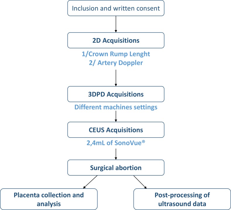
Table 1 presents the inclusion and non-inclusion criteria and figure 1 presents the flowchart of the study. The participants in this study are women with ongoing pregnancy at the first-trimester. The analysis of uteroplacental vascularisation by CEUS can only be performed in contexts of elective abortion, as the contrast product is not authorised for use in ongoing pregnancies.
Table 1.
Inclusion and non-inclusion criteria
| Inclusion criteria | Non-inclusion criteria |
|
|
IUGR, intrauterine growth restriction; PE, preeclampsia; WG, Weeks of Gestation.
Figure 1.
Flowchart.
Before inclusion, the women will be informed of the aim, the procedures and the predictable risks of the study (no identified clinical risk) by the principal investigator.
US acquisition
The US acquisition will begin in 2D mode by measurement of the crown-rump length to verify gestational age and study group. The position of the placenta will be noted and the Doppler spectrum of the uterine arteries, spiral arteries and radial arteries will be recorded, with measurements of the pulsatility and resistance indices.
First, the 3D Doppler acquisitions will be performed at four different predetermined settings (impact of the settings currently underway: NCT03342014). The instrument used will be a Voluson S8 (General Electric Healthcare) with a volumetric convex abdominal transducer (4–8 MHz).
Next, the CEUS acquisitions will be performed with a Logiq E9 (General Electric Healthcare) and an abdominal transducer (1–5 MHz). The US contrast product used will be Sonovue (Bracco Imaging, Italy), administered by bolus injection. A volume of 2.4 mL of contrast product will be injected per patient, and repeated once if necessary.
Settings and equipments are standardised between the two centres.
Image analysis
3DPD angiography
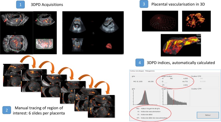
The images will be analysed with VOCAL software, which makes it possible to define a volume of interest and to quantify the Doppler signals to calculate the vascularization indices vasculazisation index (VI), flow index (FI) and vascularization flow index (VFI) automatically (figure 2). Each volume will be recorded and analysed independently. Two regions of interest will be traced: one focused on the placenta and the other on the myometrium (placental bed).
Figure 2.
Three-dimensional power Doppler (3DPD) analysis.
CEUS
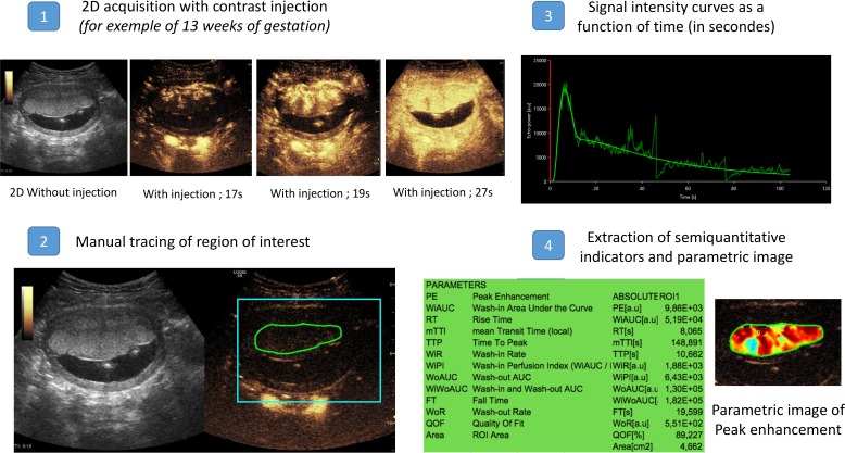
The images will be analysed with specific software that makes it possible to trace the regions of interest and to view the perfusion curves and to extract semiquantitative perfusion indicators from them (figure 3).
Figure 3.
Contrast-enhanced ultrasound analysis.
Placenta collection and analyses
The placentas will be collected and transferred fresh for preparation. The placental villi will be isolated and rinsed in saline solution. A portion of the villi (approximately 1–2 cm3 of tissue) will be frozen at −80°C for the subsequent extraction of RNA and specific proteins. For an immunohistochemical analysis, a second portion will be fixed in buffered formalin for inclusion in paraffin, and serial sections of 5 µm will be made on superfrost slides. The markers of oxidative stress will be specifically studied. The third portion will be fixed in formalin, diluted to 4% and kept at 4°C in order to perform a confocal microscopic analysis.
All the villi will be collected, and we consider that it is representative sampling of the placenta. However, it will be not possible to perform a spatial localisation of the villi because placenta if surgically collected and as multiple parts.
Outcomes
The principal endpoint is the measurement (in arbitrary units) of signal intensity in the intervillous space during the first trimester (at 8, 11 and 13 weeks), obtained by CEUS.
The secondary outcome measures are:
Measurement of indicators of vascularisation in the intervillous space and the myometrium: signal intensity and perfusion kinetics.
Comparison of the quantitative data about uteroplacental vascularisation obtained with each technique.
Procurement of placental villi that can be analysed to study human placental development and functions.
Participant timeline
The enrolment of women began in October 2016. In view of the recruitment capacity of our institutions, the recruitment should be completed by November 2019.
Patient and public involvement
Patients and public were not involved.
Premature ending of patient participation
Participants will be excluded from the study in the following situations:
Lack of CEUS acquisition.
Withdrawal of consent before the end of the study.
Patients will be immediately excluded from the study and replaced with other new participants. Any decision to withdraw consent will not affect the patient’s routine medical care. In the case of an adverse event related to the study, the patient will be informed and excluded. She will also receive what additional medical care is necessary or appropriate.
Follow-Up
No specific follow-up has been planned for participants except for standard routine healthcare. Any adverse events will be noted and reported.
Sample size consideration
In view of recent data from the literature,12 to show a difference in signal intensity between the three groups (8, 11 and 13 weeks) with α (corrected for the multiple comparison) and β risks set, respectively, at 0.017% and 10%, we should have 14 women in each group at analysis, that is, 42 women in all.
Data collection and management
An electronic case report file (e-CRF) will be created for each woman. The women's anonymity will be ensured by mentioning to the maximum extent possible their research code number, followed by the first letters of the last name and first name of the participant on all necessary documents or by deleting their names by appropriate means (white-out) from the copies of source documents intended to document the study.
The US data will be anonymised and transferred via a secure server for storage and archiving directly in the ArchiMed database, reported to the Commission Nationale de l'Informatique et des Libertés (CNIL report number: 1410005) because they cannot be transcribed in the e-CRF. The clinical data concerning the woman collected are shown in table 2.
Table 2.
Data collected
| Family history | Preeclampsia, intrauterine growth restriction, venous thromboembolic disease, high blood pressure and diabetes |
| Obstetrical history | Parity, history of postpartum haemorrhage, history of pregnancy loss or intrauterine fetal death Fibroma or uterine malformation. |
| Clinical data | Body mass index Proteinuria Blood pressure (systolic and diastolic) Smoking |
| Current treatments | |
| Ultrasound and pregnancy data | Date of pregnancy; Crown-rump length (CRL), placental position, depth from the maternal abdomen, blood pressure and heart rate at the beginning of CEUS acquisition |
CEUS, contrast-enhanced ultrasonography.
Statistical analysis
The quantitative indicators will be described by their means±SD, medians, and maximum and minimum values, the qualitative indicators by the number of individuals and percentages. The mean values will be compared between the groups by Student's t-test or the Mann-Whitney test, matched or not, depending on the type of data. The comparison of imaging techniques will be completed by Bland-Altman plots.
The analyses will be performed with R software.
No interim analysis is planned.
No statistical criterion for stopping the study is planned.
Quality control
Right of access to data and source documents
The medical data of each patient will only be transmitted to the sponsor (Metz-Thionville Regional Hospital Centre, CHR Metz-Thionville) or any person duly authorised by the sponsor and, where applicable, to the authorised health authorities, under conditions guaranteeing their confidentiality.
The sponsor and the regulatory authorities may request direct access to the medical file for verification of the procedures and/or data of the clinical trial, without violating confidentiality and within the limits permitted by laws and regulations.
For research purposes, the processing of personal data relating to persons undergoing research will be carried out.
These data are collected and processed solely on the basis of the legal grounds provided for by statute and regulations in the context of the performance of the public interest missions of the Metz-Thionville CHR, in particular those relating to ensuring and contributing to research and innovation (Article 6.1.e of the General Data Protection Regulation (GDPR)). The processing of personal data of persons participating in research is permitted by the exception provided for in Article 9.2(i) and (j).
This data processing is part of the MR001 reference methodology that the Metz-Thionville CHR has undertaken to respect.
In accordance with the GDPR, persons participating in research have a right of access to their data (Article 15), a right of rectification (Article 16), a right to erase their data (right to forget) under the conditions provided for in Article 17, a right to limit the processing provided for in Article 18 and a right to object to the processing of their personal data (Article 21). These rights are exercised with the investigators, who will inform the research sponsor as soon as possible.
The persons participating in the research also have a right to complain to the supervisory authority in France, namely, CNIL.
Study monitoring
The monitoring will be performed by the appropriate department of the Support Platform for Clinical Research of CHR Metz-Thionville, throughout the study.
A Clinical Research Associate (CRA) will travel regularly to each centre to perform the quality control of the e-CRF data.
The CRA will verify that the research is conducted according to the protocol provided in accordance with regulations and will ensure that every e-CRF contains all the information requested.
Each patient's e-CRF must be consistent with the source documents.
The investigators will allocate adequate time for such monitoring activities. The investigators will also make sure that the monitor or other compliance or quality assurance reviewer is given access to all the above noted study-related documents and study-related facilities (eg, US, diagnostic laboratory and so on), and has adequate space to conduct the monitoring visits.
Data management and quality control
Data management will be carried out by the clinical research team CIC-IT of Nancy (INSERM CIC 1433). The data images (US) will be automatically transferred to Nancy CIC-IT and stored after verification in the ArchiMed database declared to the French authorities (CNIL declaration number: 1410005).
Patient data protection
Each patient must be identified on the e-CRF, US data and placenta collection with her initials and identification number indicating her order of inclusion into the study. The investigators must keep the list of all the patients, including identification numbers, full names and last known addresses.
Patients must be informed in writing about the possibility of audits by authorised representatives of the sponsor and/or regulatory authorities, in which case the relevant parts of study-related hospital records may be required.
Patients must also be informed that the results obtained will be computerised and analysed, that local laws will be applied, that their confidentiality will be preserved, and that they are entitled to obtain any information concerning the data stored and analysed by the computerised system.
Potential risks related to the study
This study may expose the women participating in it to rare, transient and mild side effects. It will comply at all times with the Good Clinical Practices defined by the Ministry of Health.
The only constraint associated with the study is the addition of 3D Doppler acquisitions after the standard 2D US; these acquisitions will prolong the procedure by 10 min. The medical devices (Voluson and Logiq E9, GE Healthcare) are CE-marked and used routinely in clinical practice.
The contrast product (SonoVue) is authorised for use in exploring lesions of the liver, breasts and great vessels. It will be used according to the guidelines described in the summary of product characteristics in its French marketing authorisation. In the current state of knowledge about the safety of contrast products (which have not yet been authorised for use during pregnancy), adequate evidence exists to affirm their absence of permanent or serious adverse effects among women.
Ethical permission
The sponsor and all investigators undertake to conduct this study in accordance with the Declaration of Helsinki (Ethical Principles for Medical Research Involving Human Subjects, Tokyo 2004) and its updates, the provisions of European Directive 2001/20-CE as transposed into French law by L. 2004-806 dated 9 August 2004, on public health policy and 2004-800 dated 6 August 2004, on bioethics and their implementation decrees, and to comply with the guidelines of Good Clinical Practices (I.C.H. version 4 of 1 May 1996 and Decision of 24 November 2006).
They undertake to adhere to all legislative and regulatory provisions that may concern the research. In accordance with Article L. 1123–6 of the Public Health Code, the sponsor has submitted the research protocol the sponsor to the appropriate Committee and the related consent forms on 5 April 2018 (Number 16.03.02). The competent authority authorised the study on 21 June 2018 (Number 160 187A 12).
The research is to be conducted in accordance with the present Protocol. The investigators undertake to respect the protocol in all respects, especially with regard to obtaining consent and the notification and follow-up of serious adverse events.
Information letter and informed consent
Research participants will be informed of the objectives and constraints of the study, their rights to refuse to participate in the study or to withdraw from the study at any time. When all essential information has been conveyed to the subject and the investigators have ensured that the patient has understood the implications of participating in the trial, the patient's written consent shall be obtained by an investigator in two original copies. A copy of the information forms and signed consents will be given to the patient (online supplementary annexe 1).
bmjopen-2019-030353supp001.pdf (1.1MB, pdf)
The investigator will retain the second copy for a minimum of 15 years.
Protocol amendment
The sponsor must be informed of any proposed amendment to the protocol by the coordinating investigator. The changes must be described, substantive or not.
A substantial change is a change which is susceptible, in one way or another, to modify the assurances made to participants who consent to biomedical research (modification of an inclusion criterion, extending the inclusion period, participation of new centre, so on).
Once the research has begun, any substantial modification thereof at the initiative of the sponsor must obtain, prior to its implementation, a favourable approval of the committee and an authorisation from the competent authority. In this case, if necessary, the committee ensures that a new consent from individuals participating in research is obtained.
Any substantial change requires that an authorisation request be made by the sponsor to the ANSM and/or a notification request by the CPP in accordance with legislative regulation no. 2004-806 of 9 August 2004.
Final research report
The coordinator and the mandated biostatistician will collaboratively write the final research report. This report will be submitted to each of the investigators for review. Once consensus has been reached, the final version must be endorsed with the signature of each of the investigators and sent to the sponsor as early as possible after the effective end of the research. A report prepared according to the reference plan of the competent authority must be forwarded to the competent authority and the CPP within a year after the end of the research.
Discussion
This study is a follow-up to that recently published by Roberts et al. 12
Our first objective is to confirm their data, that is, the existence of early perfusion of the intervillous space (from 8 weeks of gestation) and an increase in vascular flow starting at 13 weeks. We also seek to explore the course of vascular flow rates with gestational age to test both of the hypotheses advanced about the absence of modification of flow before 13 weeks: lack of power or external regulatory factors (remodelling of the radial arteries)?
To test the first hypothesis, we chose to increase the number of individuals in each group and to select three gestational age targets: 8, 11 and 13 weeks of gestation. A significant result between 8 and 13 weeks and between 11 and 13 weeks is expected and would thus confirm the data of Roberts et al. The intermediate group at 11 weeks was chosen arbitrarily and its size was calculated to be able to show a difference between 8 and 11 weeks.
To test the second hypothesis, and as Roberts and Frias have suggested, we will examine vascular resistance of the radial arteries at these different gestational ages and assess the level of correlation between the changes in resistance and in blood flow within the intervillous space.5 17 18
The major difference between their study and ours is the choice of contrast product, a choice linked solely to the product available by geographic zone. They used Definity while in Europe; we use Sonovue. In principle, this difference will have no effect on the comparison of our results, as their properties and performance are similar.19
Moreover, we have chosen to perform 3DPD angiography at the same time to assess the applicability of this technique for the detection of blood flow before 11 weeks. The use of Doppler in the first trimester of pregnancy is a problem mainly when used with regard to the fetus. Indeed, the main hypothesis is the rise in temperature that can alter organogenesis and induce spontaneous malformations or miscarriages. In this study protocol, the acquisitions are focused on the placenta and on sections where the fetus is not visible. There is, therefore, no priori any fetal risk. Obviously, a safety assessment study will be needed before it is used in clinical practice. Should there be a high correlation between the 3DPD indices and the indicators of perfusion by CEUS, future studies will be able to choose only the 3DPD, which will make it possible to envision a longitudinal follow-up.
For placental analysis, we expect to perform a confocal microscopic analysis, immunohistochemichal analysis and extraction of RNA and specific protein. We will consider to perform also a histomorfometric analysis as Rizzo et al. 20
Trial status
This is an ongoing trial. Recruitment began in October 2016. We expect to complete recruitment by November 2019. We plan to publish final results in 2020.
Supplementary Material
Footnotes
Contributors: CB, M-LE and OM designed the study and are the principal investigators; AC and NM are project managers; CB wrote the manuscript; CB and MT will carry out recruitment, ultrasound acquisition, placental analysis and will collect the data; GH is in charge of statistical analysis and all authors reviewed and contributed to the manuscript. All authors have read, approved the paper and meet the criteria for authorship as established by the International Committee of Medical Journals Editors.
Funding: This study won the regional award for clinical research (2016).
Competing interests: None declared.
Patient consent for publication: Not required.
Ethics approval: ANSM (the French National Agency for Medicines and Health Products Safety) and the Committee for the Protection of Persons (Comité de Protection des Personnes, CPP) approved this study.
Provenance and peer review: Not commissioned; externally peer reviewed.
References
- 1. Burton GJ, Jauniaux E. Pathophysiology of placental-derived fetal growth restriction. Am J Obstet Gynecol 2018;218:S745–S761. 10.1016/j.ajog.2017.11.577 [DOI] [PubMed] [Google Scholar]
- 2. Jauniaux E, Watson A, Burton G. Evaluation of respiratory gases and acid-base gradients in human fetal fluids and uteroplacental tissue between 7 and 16 weeks’ gestation. Am J Obstet Gynecol 2001;184:998–1003. 10.1067/mob.2001.111935 [DOI] [PubMed] [Google Scholar]
- 3. Kurjak A, Kupesic S. Doppler assessment of the intervillous blood flow in normal and abnormal early pregnancy. Obstetrics & Gynecology 1997;89:252–6. 10.1016/S0029-7844(96)00422-X [DOI] [PubMed] [Google Scholar]
- 4. Carbillon L, Challier JC, Alouini S, et al. Uteroplacental circulation development: Doppler assessment and clinical importance. Placenta 2001;22:795–9. 10.1053/plac.2001.0732 [DOI] [PubMed] [Google Scholar]
- 5. Coppens M, Loquet P, Kollen M, et al. Longitudinal evaluation of uteroplacental and umbilical blood flow changes in normal early pregnancy. Ultrasound Obstet Gynecol 1996;7:114–21. 10.1046/j.1469-0705.1996.07020114.x [DOI] [PubMed] [Google Scholar]
- 6. Burton GJ, Jauniaux E, Watson AL. Maternal arterial connections to the placental intervillous space during the first trimester of human pregnancy: the Boyd collection revisited. Am J Obstet Gynecol 1999;181:718–24. 10.1016/S0002-9378(99)70518-1 [DOI] [PubMed] [Google Scholar]
- 7. Greenwold N, Jauniaux E, Gulbis B, et al. Relationship among maternal serum endocrinology, placental karyotype, and intervillous circulation in early pregnancy failure. Fertil Steril 2003;79:1373–9. 10.1016/S0015-0282(03)00364-9 [DOI] [PubMed] [Google Scholar]
- 8. Hempstock J, Jauniaux E, Greenwold N, et al. The contribution of placental oxidative stress to early pregnancy failure. Hum Pathol 2003;34:1265–75. 10.1016/j.humpath.2003.08.006 [DOI] [PubMed] [Google Scholar]
- 9. Jauniaux E, Watson AL, Hempstock J, et al. Onset of maternal arterial blood flow and placental oxidative stress. A possible factor in human early pregnancy failure. Am J Pathol 2000;157:2111–22. [DOI] [PMC free article] [PubMed] [Google Scholar]
- 10. Jauniaux E, Burton GJ. The role of oxidative stress in placental-related diseases of pregnancy]. J Gynecol Obstet Biol Reprod 2016;45:775–85. [DOI] [PubMed] [Google Scholar]
- 11. Trophoblastic oxidative stress in relation to temporal and regional differences in maternal placental blood flow in normal and abnormal early pregn. - PubMed - NCBI [Internet]. Available: https://www.ncbi.nlm.nih.gov/pubmed/12507895 [Accessed 10 Mar 2019].
- 12. Roberts VHJ, Morgan TK, Bednarek P, et al. Early first trimester uteroplacental flow and the progressive disintegration of spiral artery plugs: new insights from contrast-enhanced ultrasound and tissue histopathology. Hum Reprod 2017;32:2382–93. 10.1093/humrep/dex301 [DOI] [PMC free article] [PubMed] [Google Scholar]
- 13. Shih JC, Ko TL, Lin MC, et al. Quantitative three-dimensional power Doppler ultrasound predicts the outcome of placental chorioangioma. Ultrasound Obstet Gynecol 2004;24:202–6. 10.1002/uog.1081 [DOI] [PubMed] [Google Scholar]
- 14. Duan J, Perdriolle-Galet E, Chabot-Lecoanet A-C, et al. Placental 3D Doppler angiography: current and upcoming applications]. J Gynecol Obstet Biol Reprod 2015;44:107–18. [DOI] [PubMed] [Google Scholar]
- 15. Duan J, Chabot-Lecoanet A-C, Perdriolle-Galet E, et al. Utero-Placental vascularisation in normal and preeclamptic and intra-uterine growth restriction pregnancies: third trimester quantification using 3D power Doppler with comparison to placental vascular morphology (EVUPA): a prospective controlled study. BMJ Open 2016;6:e009909 10.1136/bmjopen-2015-009909 [DOI] [PMC free article] [PubMed] [Google Scholar]
- 16. Hafner T, Kurjak A, Funduk-Kurjak B, et al. Assessment of early chorionic circulation by three-dimensional power Doppler. J Perinat Med 2002;30:33–9. 10.1515/JPM.2002.005 [DOI] [PubMed] [Google Scholar]
- 17. Burton GJ, Woods AW, Jauniaux E, et al. Rheological and physiological consequences of conversion of the maternal spiral arteries for uteroplacental blood flow during human pregnancy. Placenta 2009;30:473–82. 10.1016/j.placenta.2009.02.009 [DOI] [PMC free article] [PubMed] [Google Scholar]
- 18. Kurjak A, Zudenigo D, Predanic M, et al. Assessment of the fetomaternal circulation in threatened abortion by transvaginal color Doppler. Fetal Diagn Ther 1994;9:341–7. 10.1159/000263959 [DOI] [PubMed] [Google Scholar]
- 19. Hyvelin J-M, Gaud E, Costa M, et al. Characteristics and echogenicity of clinical ultrasound contrast agents: an in vitro and in vivo comparison study. J Ultrasound Med 2017;36:941–53. 10.7863/ultra.16.04059 [DOI] [PubMed] [Google Scholar]
- 20. Rizzo G, Silvestri E, Capponi A, et al. Histomorphometric characteristics of first trimester chorionic villi in pregnancies with low serum pregnancy-associated plasma protein-A levels: relationship with placental three-dimensional power Doppler ultrasonographic vascularization. J Matern Fetal Neonatal Med 2011;24:253–7. 10.3109/14767058.2010.482627 [DOI] [PubMed] [Google Scholar]
Associated Data
This section collects any data citations, data availability statements, or supplementary materials included in this article.
Supplementary Materials
bmjopen-2019-030353supp001.pdf (1.1MB, pdf)


