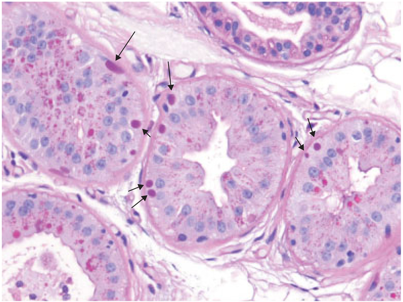
Fig. 3.
Skin biopsy from the same patient as above with periodic acid-Schiff staining. Arrows indicate pathognomonic Lafora bodies (LB) in the myoepithelia (at the bases) of apocrine sweat glands. Note, similarly stained structures near gland lumens(e.g., in the gland at the bottom left corner of the image). Those are the normal secretory products of these glands and are not LB. They are a common source of obviously highly undesired false-positive diagnosis in patients undergoing skin biopsy as part of a workup for progressive myoclonus epilepsy.

