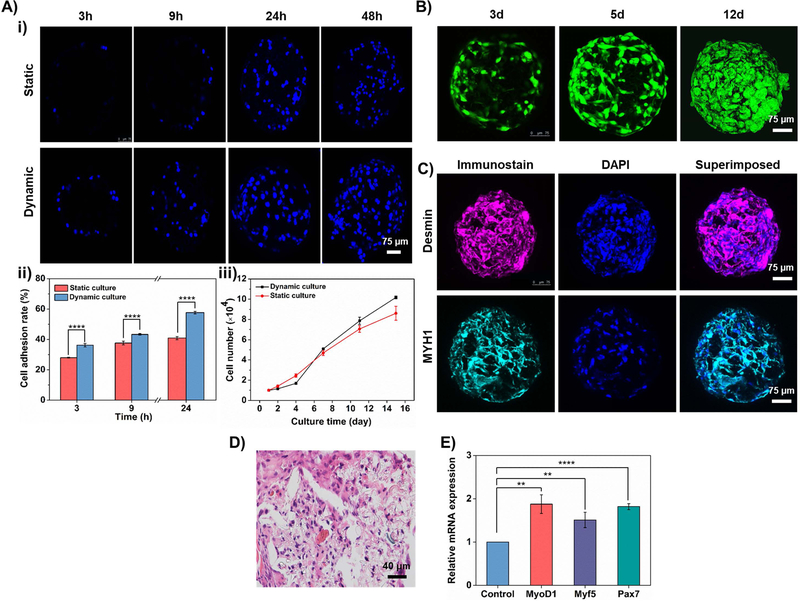
Figure 6. Construction of cell-laden PLGA HOPMs in vitro.
A) i) CLSM images showing the time-dependent growth of myoblasts cultured in the PLGA HOPMs for various time periods (3, 9, 24, and 48 h). ii, iii) Graphical representations showing ii) adhesion of the cells (3, 9, and 24 h) as well as iii) the number of myoblasts, under static and dynamic cultures. B) CLSM images showing the proliferation of myoblasts on the PLGA HOPMs for 12 days. C) Immunohistological analysis of myoblasts in the PLGA HOPMs by staining them against desmin and MYH1 (counterstained by DAPI for nuclei) for 7 days. D) H&E staining of myoblasts in the PLGA HOPMs. E) Gene expression profiles of myoblasts in the PLGA HOPMs (MyoD1, Myf 5, and Pax 7). **P 0.01, ****P<0.0001.

