Abstract
OBJECTIVES
To assess and monitor lateral semicircular canal (LSC) function over time in patients affected by chronic otitis media with cholesteatoma (CHO) complicated by fistula of LSC (LSC-F) before and after surgery using video Head Impulse Test (vHIT).
MATERIALS and METHODS
Eight patients aged 18–67 years affected by CHO with imaging-ascertained LSC-F were included in this preliminary prospective study. The following protocol has been applied: oto-microscopic diagnosis with patient’s history; computed tomography scan of the temporal bone; surgery with concomitant resurfacing of LSF-F; audiological and vestibular evaluation before surgery (T0) and at 30 days (T1), 6 months (T2), and 1 year after surgery (T3). vHIT was used to assess vestibulo-ocular reflex (VOR) in LSC.
RESULTS
None of the patients showed deterioration of bone conduction hearing levels during the different time of evaluation. Three patients showed a reduced VOR gain and catch-up saccades at T0, with VOR gain normalization at T2. This finding remained stable at the 1-year follow-up. The VOR gain in the nonaffected side generally experienced an increase, paralleled by the normalization on the affected side, with statistically significant correlation. The subjects with normal vHIT before surgery did not show any variation following surgery.
CONCLUSION
vHIT allows the assessment of LSC function in case of fistula. The adopted surgical fistula repair did not induce deterioration of the auditory or LSC function, but indeed, it could prevent worsening and help promoting recovery to the normal function.
Keywords: Lateral semicircular canal, cholesteatoma, video Head Impulse Test, vestibulo-ocular reflex, labyrinthine fistula
INTRODUCTION
Labyrinthine fistula (LF) is a possible complication from chronic otitis media with cholesteatoma (CHO), with an estimated incidence rate between 2.4% and 16.7%, which in about 90% of cases, involves the lateral semicircular canal (LSC) [1–3]. The surgical management of LF is mandatory to avoid complications, including possible hearing and vestibular impairments, the former up to deafness. In fact, the surgical procedure includes both removal of the pathological lesion and the resurfacing of the eroded canal. However, some authors are in favor of a more conservative approach that encompasses leaving the CHO matrix over the canal opening at the end of the surgical procedure, thereafter planning a second-look for the final eradication, when few reports even indicate disappearance of the residual tissue at the revision surgery [4–6].
The evaluation of the LSC function is classically performed by caloric testing, with the stimulus impacting at distance on this structure via the external auditory canal and the eardrum [7, 8]. Under this pathological condition, the Head Impulse Test (HIT), as described by Halmagyi and Curthoys [9], shows to be less invasive and feasible to become very popular in the clinical setting as the video Head Impulse Test (vHIT) [10, 11].
The aim of this preliminary, prospective study was to assess the LSC function in a consecutive series of subjects affected by CHO with an imaging-ascertained fistula of LSC (LSC-F) who underwent surgery with simultaneous resurfacing of the eroded canal. The assessment was performed with vHIT and allowed us to evaluate on the role played by this diagnostic tool for investigating the LSC function pre- and postoperatively, as well as for assessing the effectiveness of the adopted surgical procedure on the vestibular function of the operated side.
MATERIALS AND METHODS
The study was designed and performed according to Declaration of Helsinki [12]. Ethics committee approval was received.
From January 2014 to April 2016, 76 patients (48 male and 28 female) affected by chronic otitis media with CHO were admitted for surgery at the Otolaryngologic clinic of a tertiary referral university hospital. Informed consent was obtained from all subjects. Based on the aim of this prospective study, only primary and unilateral cases were included, as confirmed by high-resolution computed tomography (HRCT) (Philips Brilliance iCT 128, Amsterdam, The Netherlands) of the temporal bone. Revision cases (19 patients) and cases with different or multiple sites of LF (9 cases), concomitant or previous vestibular diseases on the affected (2 cases) or contralateral side (14 cases), and absence of LSC-F at HRCT scan (22 cases) were excluded. Ten patients (six males, four females) were included, but two of them were lost to follow-up due to transfer to another city, so that only eight patients successfully completed the 1-year follow-up examination and were therefore included.
The following protocol has been applied: oto-microscopic diagnosis with patient’s history, with particular reference to reported episodes of vertigo spells, either spontaneous or provoked by ear canal occlusion, together with appearance of nystagmus (Hennebert sign), and to hearing impairment; HRCT scan of the temporal bone; pure tone audiometry; vHIT before surgical treatment (T0); surgery with concomitant resurfacing of the eroded LSC; pure tone audiometry and vHIT at 30 days (T1), 6 months (T2), and 1 year after surgery (T3). vHIT was performed by a professional having experienced in vestibular assessment at our center.
Physiological artifacts were evaluated in the present analysis. All v-HITs collected and accepted by the algorithm of the device software were stored and traces with artifacts were manually filtered by processing raw quantitative data exported from the ICS Impulse device, using Matlab R2017a (Mathworks, Natick, Mass., USA) according to the coding manual (online suppl. Appendix B) for the classification of HIT artifacts. [13]
The surgical report allowed classifying the LSC-F according to Dornhoffer and Milewski [1].
Preoperative Assessment (T0)
Audiometric Data
All patients underwent pure tone audiometry to measure air and bone conduction hearing levels. A cochlear involvement was defined when bone conduction (BC) thresholds between 0.5 and 4 kHz (four-tone pure tone average, PTA-4) were worse than 25 dB HL [14].
Vestibular Testing
The Hennebert sign (eye movements and vertigo induced by pressure on the external auditory canal) was searched using Frenzel’s glasses. The vestibulo-ocular reflex (VOR) in LSC was evaluated using the vHIT system (ICS Impulse System, GN Otometrics, Taastrup, Denmark). The evaluated parameters included the VOR mean gain [ratio of eye velocity to head velocity for every head rotation; normal value (gain)>0.79] [15] and the occurrence of saccades (covert or overt catch-up saccades) [10, 11].
Imaging
It included HRCT of the temporal bone (images are displayed in the axial and coronal plane) for detection of LSC-F, as evaluated by the senior surgeon.
Surgical Technique
All the procedures were performed by the senior surgeon (MB). The surgical procedures included an intact canal wall up or closed technique (three subjects) and an “in-to-out” open technique (five subjects), in all cases with a good exposure of the site of the LSC-F. During surgery, part of the CHO tissue was temporarily left over the fistula until the bulky CHO mass was completely eradicated. Then, a three-layer sealing procedure was performed and consisted the following:
gentle dissection of the matrix over the bony defect, helped by fine suction tip and sharp dissector;
immediate covering of the exposed defect with temporalis fascia;
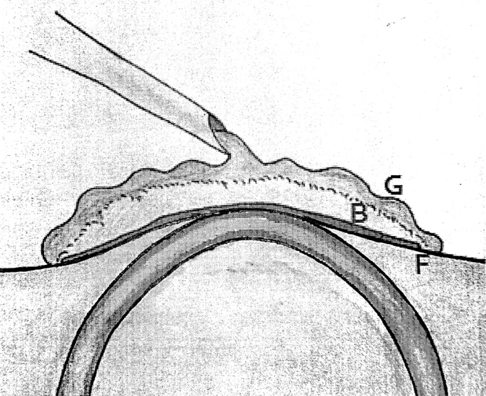
covering of the fascia with bone pâté collected during the bone drilling work and final stabilization with fibrin glue (Figure 1) [16].
Figure 1.

Artist’s drawing of the surgical repair of the lateral semicircular canal erosion.
F: temporal fascia; B: bone pâté; G: Fibrin glue.
Statistical Analysis
A statistical analysis was performed using Statistical Package for the Social Sciences (SPSS), version 22.0 (IBM Corp.; Armonk, NY, USA). A nonparametric, paired test, Wilcoxon-signed rank test (p<0.05) was used for statistical analysis to evaluate the difference between unfiltered and filtered data of VOR gain. The Pearson correlation coefficient test was used to index the correlation between VOR gain values of the affected and non-affected ear, separately, for each exam, with high correlation values defined by p>0.7.
RESULTS
Eight patients (five males, three females; aged between 18 and 71 years) affected by CHO complicated by LSC-F (five on the right ear, three in the left ear) were studied. Demographic data, including type of fistula according to Dornhoffer and Milewski classification [1], are reported in Table 1. Six patients showed conductive hearing loss, while in two of them, a mixed hearing loss was found. Tinnitus was reported in three patients (37.5 %), episodes of vertigo in two (25 %), and a positive Hennebert sign in one (12.5 %), characterized by left-beating horizontal nystagmus. The intraoperative findings identified four patients (50 %) with a stage I fistula, three (37.5 %) with stage II, and one (12.5 %) with stage III.
Table 1.
Demographics of the study group and auditory features
| PTS | Age | Gender | Affected side | Fistula type | Type of hearing loss | Pre BC | Post 1 BC | Post 6 BC | Post 12 BC | Tinnitus |
|---|---|---|---|---|---|---|---|---|---|---|
| 1 | 63 | M | R | I | MHL | 24 | 25 | 26 | 29 | N |
| 2 | 18 | F | R | III | CHL | 6 | 10 | 9 | 9 | Y |
| 3 | 71 | F | L | II | CHL | 19 | 24 | 19 | 19 | Y |
| 4 | 44 | M | R | II | MHL | 26 | 18 | 16 | 16 | N |
| 5 | 54 | M | L | I | CHL | 8 | 13 | 10 | 10 | N |
| 6 | 50 | M | R | I | CHL | 20 | 18 | 21 | 21 | Y |
| 7 | 27 | M | R | I | CHL | 16 | 9 | 10 | 11 | N |
| 8 | 38 | F | L | II | CHL | 11 | 14 | 16 | 16 | N |
Fistula type, according to Dornhoffer and Milewski classification (1995). MHL: mixed hearing loss; CHL: conductive hearing loss; BC: mean pure tone average at 0.5, 1, 2, and 4 kHz (PTA4) bone conduction threshold.
Postoperatively, the next day, none of the patients showed spontaneous nystagmus or dizziness. No postoperative facial palsy or intracranial complications occurred.
All the patients underwent vHIT before surgery (T0) and then 1 (T1), 6 (T2), and 12 months (T3) after surgery. No significant gain difference between filtered and unfiltered data was found (p>0.05). The results are shown in Table 2.
Table 2.
Demographics of the study group and vestibular features
| PTS | Side | Fistula Type | D (T0) | HS (T0) | Pre vHIT (T0) | Post 1 vHIT (T1) | Post 6 vHIT (T2) | Post 12 vHIT (T3) | D (T3) | HS (T3) | ||||
|---|---|---|---|---|---|---|---|---|---|---|---|---|---|---|
| L | R | L | R | L | R | L | R | |||||||
| 1 | R | I | N | N | 0.77 | 0.79 | 0.75 | 0.80 | 0.86 | 0.92 | 0.84 | 0.92 | N | N |
| 2 | R | III | Y | Y | 0.83 | 0.47 | 0.83 | 0.47 | 0.92 | 0.88 | 0.86 | 0.91 | N | N |
| 3 | L | II | N | N | 0.55 | 0.94 | 0.65 | 0.90 | 1.25 | 1.42 | 1.36 | 1.51 | N | N |
| 4 | R | II | N | N | 0.88 | 0.43 | 1.01 | 0.56 | 1.03 | 0.80 | 1.06 | 0.82 | N | N |
| 5 | L | I | N | N | 1.11 | 1.18 | 1.08 | 1.15 | 0.94 | 0.97 | 0.96 | 0.94 | N | N |
| 6 | R | I | N | N | 0.95 | 0.95 | 0.94 | 0.95 | 0.90 | 0.91 | 0.91 | 0.90 | N | N |
| 7 | R | I | N | N | 0.82 | 0.94 | 0.94 | 1.04 | 0.90 | 0.98 | 0.92 | 1.01 | N | N |
| 8 | L | II | Y | N | 0.84 | 0.97 | 0.91 | 0.98 | 0.91 | 0.96 | 0.92 | 0.96 | N | N |
Fistula type, according to Dornhoffer and Milewski classification (1995). D: Dizziness; HS: Hennebert sign; vHIT: video Head Impulse Test; T0: basal value; T1: one-month control; T2: six months control; T3: 1 year control. In bold: values of the affected side.
T0
Before surgery, in three patients (no. 2, 3, and 4) (37.5%), a VOR gain reduction was shown in the affected side, with compensatory overt and covert saccades. Patient no. 2, in particular, showed reduced lateral VOR gain with compensatory, overt and covert saccades, and high asymmetry, without spontaneous nystagmus. Patient no. 3 displayed clusters of overt saccades with high asymmetry in addition to the reduced VOR gain. In patient no. 4, vHIT revealed a reduced VOR gain in association with saccades (mostly covert) and high asymmetry (Tab. II).
The remaining five patients (no. 1, 5, 6, 7, and 8) (62,5%) did not show VOR gain reduction on the affected side or presence of saccades.
T1
One month postoperatively, none of the patients showed deterioration of BC hearing levels. In the five patients without pathological findings at Stage T0, vHIT remained normal. Within the group of patients that displayed abnormalities at vHIT, two (no. 2 and 3) showed similar results to the preoperative ones. Patient no. 4 had a slight VOR gain improvement, still below the normal value, with reduction in asymmetry and evidence of more clustered mostly covert saccades.
T2
Six months postoperatively, no patients showed reduction in BC hearing levels. The patients without preoperative vHIT alterations maintained the same pattern as T0. All patients (no. 2, 3, and 4) with a reduced VOR gain at T0, displayed VOR gain values within normal limits:
Patient no. 2 showed an appropriate VOR gain with still evidence of saccades either overt or covert, but without asymmetry.
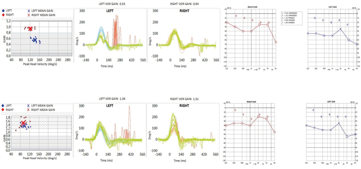
Patient no. 3 showed a high bilateral VOR gain with overt, nonclustered saccades with reduced velocity in the affected side and no asymmetry (Figure 2).
Patient no. 4 showed VOR gain normalization in association with clustered covert saccades and reduced asymmetry.
Figure 2. a–c.
Trend line of vestibulo-ocular reflex (VOR) gain in the affected and nonaffected contralateral side relative to the subjects no. 2 a), no. 3 b), and no. 4 c) with decreased VOR gain at T0 (basal value).
T3
All the evaluated patients showed a normal VOR gain. Only patient no. 2 still showed almost exclusively overt saccades. Patient no. 3 still had high bilateral VOR gain, with nonclustered overt saccades, whose amplitude was reduced. Patient no. 4 showed substantially similar findings.
The VOR gain trend line for each patient is shown in Figure 3 and compared, in the pathological subjects, with data from the contralateral, unaffected side.
Figure 3.
Hearing and video Head Impulse Test (vHIT) testing of patient no. 3. Upper: vHIT graphs performed preoperatively show a reduced VOR gain on the left affected side, with clusters of overt saccades. The preoperative audiogram shows a mixed type of hearing loss on the same side. Lower: vHIT graphs performed at 1 year after surgery shows normalization of VOR gain in the affected side, with nonclustered overt saccades. The postoperative audiogram shows an unaltered bone conduction in the affected ear.
Contralateral Ear
The VOR assessment in the non-affected side, in cases with decreased VOR, showed changes in value during the different time of evaluation (Table 3). In particular, VOR gain in the non-affected side, generally experienced an increase, paralleled by the normalization on the affected side, as confirmed by high Pearson correlation coefficient, calculated for patient no. 2, 3, and 4.
Table 3.
Pearson test on the three subjects with decreased VOR gain at T0. Data from the nonaffected side well correlates with those of the fistula side, with increased VOR gain values over time
| Pt 2 | Pt 3 | Pt 4 | ||||
|---|---|---|---|---|---|---|
| AS | CS | AS | CS | AS | CS | |
| T0 | 0.47 | 0.83 | 0.55 | 0.94 | 0.43 | 0.88 |
| T1 | 0.47 | 0.83 | 0.65 | 0.9 | 0.56 | 1.01 |
| T2 | 0.88 | 0.92 | 1.25 | 1.42 | 0.8 | 1.03 |
| T3 | 0.91 | 0.86 | 1.36 | 1.51 | 0.82 | 1.06 |
| CC | 0.79 | 0.99 | 0.89 | |||
CC: correlation coefficient; AS: affected side; CS: contralateral side. T0: basal value; T1: one-month control; T2: six months control; T3: 1-year control.
DISCUSSION
Labyrinthine fistula is a common severe complication of aggressive CHO and occurs most frequently at the LSC level. The HRCT scan represents the only objective tool to ascertain it since the affected subjects may lack reliable symptoms evocative of this labyrinthine complication. However, HRCT scan has some limitations, being unable to determine the invasion boundaries between bone and membranous labyrinth, an important feature that can only be provided by intraoperative exploration. According to this finding, three types of LF can be distinguished [1]: a) Type 1: bone erosion without penetration of the endosteal layer of the canal; b) Type 2a: bone erosion with penetration of the endosteal layer of the canal; c) Type 2b: same as type 2a, with spontaneous perilymphatic leakage; d) Type 3: complete invasion of the canal. It is likely to assume that an inner ear impairment, both for the auditory and the vestibular compartments, would mostly depend upon the labyrinthine invasion, i.e., being more likely to occur in stage 3 than in stage 1.
Knowing how threatening the presence of an external communication produced by pathological, infectious tissue, such as CHO, could be for the inner ear structures, it would seem of utmost importance to evaluate both auditory and vestibular functions, regardless of the preoperative findings. For the auditory function, it is possible to fully rely on the standard audiometric tests, in particular looking for signs of cochlear involvement, basically shown by deterioration of the BC threshold. In the majority of our study sample, a conductive hearing loss compatible with the middle ear lesion was evidenced, and only in two of them, a mixed hearing loss for a slight decrease of the BC threshold at a high frequency level was found. The same methodology has also allowed monitoring possible noxious effects produced by the surgical procedure, in the immediate as well as the delayed postoperative period. As far as the vestibular impairment is concerned, when an LF is suspected, at first, this pathology can be investigated by applying finger pressure to the external ear canal, performing a fistula test. This test has not been found to be very sensitive, with only 20% of positivity reported in patients with a surgically confirmed fistula [17, 18]. If one considers an instrumental vestibular workout, then it is important to remind that LSC-F represents a unique example of clear-cut LSC lesion. Caloric testing, apart from the general consideration of not using a physiological type of stimulation, is obviously contraindicated in case of CHO, due to the presence of eardrum perforation. So, the search of assessment of the VOR by the HIT, as first proposed by Halmagyi [19], would seem very appropriate in this regard. The integration of this test on software-based video systems, vHIT, has improved its analytical process along with the clinical importance. With vHIT, it is possible to evaluate the peripheral vestibular function using a proper stimulation (high head velocity) in the plane of the canal that houses the receptor of interest [10]. Moreover, VOR is not influenced by pathological changes to the external or middle ear [20].
The assessment of LSC function by vHIT has already been applied in the clinical practice for studying, for example, the effect of plugging and resurfacing of superior semicircular canal dehiscence [21]. In CHO, a recent report on three cases with LSC-F, showed abnormal VOR findings despite a negative fistula test [22].
To our knowledge, this is one of the first studies reporting on the systematic use of vHIT to evaluate LSC function in patients affected by chronic otitis media with CHO complicated by fistula. Although within a limited cohort of subjects, this study has allowed us to retrieve interesting data otherwise not provided by symptomatology or noninstrumental vestibular tests. During surgery, it was always planned to proceed with the complete removal of the pathological tissue and the immediate repair of the bone erosion of the LSC-F, performed as a single surgical step, with a three-layer resurfacing of the eroded bone by temporal fascia, bone pâté, and fibrin glue (Figure 1). It has always been possible to precisely document the extent of the fistula, with its typology, and to proceed with its repair. In 50% of our study sample, a type I fistula was found, with complete bone erosion up to the membranous structures that remained unviolated. Whether this condition would be enough to avoid the noxious effect of CHO tissue via toxic agents is still to be elucidated, although the present findings would be of support. This finding may explain the absence of spontaneous or provoked vertiginous symptomatology at fistula test, for example.
Our results showed a normal VOR gain in 62.5 % of the cases, including all type I and one type II fistulas. One may then assume that the simple bone labyrinthine exposure due to bone erosion is unable to induce a functional impairment if an additional cause, such as a toxic effect, is not concomitant. When considering the three subjects with a preoperative reduced VOR gain, two of them presented a type II fistula and one a type III. Every patient affected by type I fistula (no. 1, 5, 6, and 7) showed, instead, a normal preoperative VOR gain. Also, in type II fistula, one case of VOR normality was found, so that it could be assumable that factors different from the mere labyrinthine exposure, such as the toxic effect of the pathological tissue in contact, could play a role.
Some interesting findings worth to be mentioned and commented were observed when monitoring the vestibular function at distance from surgery. In fact, all the patients with a reduced VOR gain at T0 (no. 2, 3, and 4), showed VOR gain normalization at 6-month follow-up (T2). This finding, which remained stable at the 1-year follow-up, could allow us to assume that fistula repair, along with CHO removal, not only prevents worsening of the canal function but would also be able to promote its functional recovery in case of its impairment.
The increased VOR gain in the healthy side also needs to be elucidated For instance, the disappearance of the clustered overt saccades pattern could be due to the progressive reduction of the compensatory mechanisms. The other patient affected by type II fistula with abnormal vHIT at T0 (no. 4) showed a progressive VOR gain normalization during the follow-up, with changes in saccades pattern, being initially (T0) both overt and covert, and at T2 only overt, as a sign of LSC functional recovery.
Although drawn by a limited number of subjects, it could be likely to assume that the progressive impairment of canal function occurs mostly in type II fistula, while the saccadic compensatory mechanism could be responsible for the referred scarce symptomatology. The appropriateness of the surgical technique used for fistula repair seems also confirmed by the fact that the only patient with a type III fistula (no. 2), with a reduced VOR gain at T0, showed a progressive normalization at the follow-up along with reduction in number and amplitude of overt and covert saccades that represents a true canal function recovery.
It is remarkable to observe that all the patients with a reduced VOR gain at the preoperative evaluation presented on the non-affected side VOR gain values that increased in a fashion like the affected side, with statistically significant correlation. The two data series (VOR gain on affected side and VOR gain on the healthy side), in fact, resulted to be highly correlated (Table 3).
The subjects with normal vHIT before surgery did not show any variation following surgery as a proof that CHO removal with the adopted method of fistula repair were not producing damage to the vestibular system. These findings are also in agreement with audiometric data that showed lack of hearing deterioration in all the operated subjects.
CONCLUSION
In this preliminary report, vHIT appears to be a useful objective method for the assessment of vestibular function in case of LSC fistula, since it can help obtain preoperative information on the canal function, particularly useful when the symptomatology is scarce and noninstrumental signs are negative. Chronic otitis media with CHO, when complicated by LSC fistula, can induce a reduced canal function in a way proportional to the type of the fistula, and patients affected by type I fistula never showed LSC impairment, although no statistical validation has been obtained. Finally, the surgical procedure with the described fistula repair did not induce deterioration of the auditory or LSC function, but indeed, for the latter it could help promote the recovery to the normal function, as definitely observed 6 months postoperatively. Further studies with analysis numerous cohorts will be needed for supporting this preliminary observation.
Acknowledgements
The authors thank Dr Ferrante Daniele for the supporting of the work.
Footnotes
This study was presented at the Vestibular Academy International, 2–4 March 2017, Mumbai, India.
Ethics Committee Approval: Ethics committee approval was received for this study from the ethic committee of Sapienza University.
Informed Consent: Written informed consent was obtained from the patients who participated in this study.
Peer-review: Externally peer-reviewed.
Author Contributions: Concept – M.B., E.C., S.M. Design - M.B., E.C. Supervision – S.M., M.B.; Resource –A.T.B, R.T., E.C.; Materials – A.T.B., R.T.; Data Collection and/or Processing – S.T., V.M., A.T.B., R.T.; Analysis and/or Interpretation – M.B., S.M., E.C.; Literature Search - A.T.B., R.T.; Writing - M.B., S.M., E.C.; Critical Reviews – M.B., S.M.
Conflict of Interest: The authors have no conflict of interest to declare.
Financial Disclosure: The authors declared that this study has received no financial support.
REFERENCES
- 1.Dornhoffer JL, Milewski C. Management of the open labyrinth. Otolaryngol Head Neck Surg. 1995;112:410–4. doi: 10.1016/S0194-5998(95)70275-X. [DOI] [PubMed] [Google Scholar]
- 2.Moon IS, Knon MO, Park CY, Hong SJ, Shim DB, Kim J, et al. Surgical management of labyrinthine fistula in chronic otitis media with cholesteatoma. Auris Nasus Larynx. 2012;39:261–4. doi: 10.1016/j.anl.2011.06.002. [DOI] [PubMed] [Google Scholar]
- 3.Palva T, Ramsay H. Treatment of labyrinthine fistula. Arch Otolaryngol Head Neck Surg. 1989;115:804–6. doi: 10.1001/archotol.1989.01860310042019. [DOI] [PubMed] [Google Scholar]
- 4.Sanna M, Zini C, Gamoletti R, Taibah AK, Russo A, Scandellari R. Closed versus open technique in the management of labyrinthine fistulae. Am J Otol. 1988;9:470–5. [PubMed] [Google Scholar]
- 5.Jang CH, Jo SY, Cho YB. Matrix removal of labyrinthine fistulae by non-suction technique with intraoperative dexamethasone injection. Acta Otolaryngol. 2013;133:910–5. doi: 10.3109/00016489.2013.782105. [DOI] [PubMed] [Google Scholar]
- 6.Hakuba N, Hato N, Shinomori Y, Sato H, Gyo K. Labyrinthine fistula as a late complication of middle ear surgery using the canal wall down. Otol Neurotol. 2002;23:832–5. doi: 10.1097/00129492-200211000-00003. [DOI] [PubMed] [Google Scholar]
- 7.Hullar TE, Della Santina CC, Hirvonen T, Lasker DM, Carey JP, Minor LB. Responses of irregularly discharging chinchilla semicircular canal vestibular nerve afferents during high-frequency head rotations. J Neurophysiol. 2005;93:2777–86. doi: 10.1152/jn.01002.2004. [DOI] [PubMed] [Google Scholar]
- 8.Hallpike CS. The caloric tests. J Laryngol Otol. 1956;70:15–28. doi: 10.1017/S0022215100052610. [DOI] [PubMed] [Google Scholar]
- 9.Halmagyi GM, Curthoys IS. A clinical sign of canal paresis. Arch Neurol. 1988;45:737–9. doi: 10.1001/archneur.1988.00520310043015. [DOI] [PubMed] [Google Scholar]
- 10.MacDougall HG, Weber KP, McGarvie LA, Halmagyi GM, Curthoys IS. The video head impulse test: diagnostic accuracy in peripheral vestibulopathy. Neurology. 2009;73:1134–41. doi: 10.1212/WNL.0b013e3181bacf85. [DOI] [PMC free article] [PubMed] [Google Scholar]
- 11.MacDougall HG, McGarvie LA, Halmagyi GM, Rogers SJ, Manzari L, Burgess AM, et al. A new saccadic indicator of peripheral vestibular function based on the video head impulse test. Neurology. 2016;87:410–8. doi: 10.1212/WNL.0000000000002827. [DOI] [PMC free article] [PubMed] [Google Scholar]
- 12.Williams JR. The Declaration of Helsinki and public health. Bull World Health Organ. 2008;86:650–2. doi: 10.2471/BLT.08.050955. [DOI] [PMC free article] [PubMed] [Google Scholar]
- 13.Mantokoudis G, Saber Tehrani AS, Wozniak A, Eibenberger K, Kattah JC, Guede CI, et al. Impact of artifacts on VOR gain measures by video-oculography in the acute vestibular syndrome. J Vestib Res. 2016;26:375–85. doi: 10.3233/VES-160587. [DOI] [PMC free article] [PubMed] [Google Scholar]
- 14.Committee on Hearing and Equilibrium guidelines for the evaluation of results of treatment of conductive hearing loss. American Academy of Otolaryngology-Head and Neck Surgery Foundation. Inc Otolaryngol Head Neck Surg. 1995;113:186–7. doi: 10.1016/S0194-5998(95)70103-6. [DOI] [PubMed] [Google Scholar]
- 15.McGarvie LA, MacDougall HG, Halmagyi GM, Burgess AM, Weber KP, Curthoys IS. The video Head Impulse Test (vHIT) of semicircular canal function - Age-dependent normative values of VOR gain in healthy subjects. Front Neurol. 2015;6:154. doi: 10.3389/fneur.2015.00154. [DOI] [PMC free article] [PubMed] [Google Scholar]
- 16.Barbara M, Iacolucci C, Atturo F, Bucci MG. Treatment of cholesteatoma-induced labyrinthine fistula. Cholesteatoma and Ear Surgery – An Update; Proceedings of the 9th International Conference on Cholesteatoma and Ear Surgery; June 37, 2012; Nagasaki, Japan. pp. 103–05. [Google Scholar]
- 17.Casale M, Errante Y, Sabatino L, Incammisa A, Salvinelli F, Quattrocchi CC. Perilimphatic fistula test: a video clip demonstration. Eur Rev Med Pharmacol Sci. 2014;18:3549–50. [PubMed] [Google Scholar]
- 18.Kvestad E, Kværner KJ, Mair IW. Labyrinthine fistula detection: the predictive value of vestibular symptoms and computerized tomography. Acta Otolaryngol. 2001;121:622–6. doi: 10.1080/000164801316878935. [DOI] [PubMed] [Google Scholar]
- 19.Halmagyi GM, Curthoys IS, Cremer PD, Henderson CJ, Todd MJ, Staples MJ, et al. The human horizontal vestibulo-ocular reflex in response to high-acceleration stimulation before and after unilateral vestibular neurectomy. Exp Brain Res. 1990;81:479–90. doi: 10.1007/BF02423496. [DOI] [PubMed] [Google Scholar]
- 20.Curthoys IS. The interpretation of clinical tests of peripheral vestibular function. Laryngoscope. 2012;122:1342–52. doi: 10.1002/lary.23258. [DOI] [PubMed] [Google Scholar]
- 21.Mantokoudis G, Saber Tehrani AS, Wong AL, Agrawal Y, Wenzel A, Carey JP. Adaptation and Compensation of Vestibular Responses Following Superior Canal Dehiscence Surgery. Otol Neurotol. 2016;37:1399–405. doi: 10.1097/MAO.0000000000001196. [DOI] [PMC free article] [PubMed] [Google Scholar]
- 22.D’Albora R, Silveira L, Carmona S, Perez-Fernandez N. Diagnostic Bedside Vestibuloocular Reflex Evaluation in the Setting of a False Negative Fistula Test in Cholesteatoma of the Middle Ear. Case Rep Otolaryngol. 2017 doi: 10.1155/2017/2919463. doi: 10.1155/2017/2919463. Epub 2017 Mar 13. [DOI] [PMC free article] [PubMed] [Google Scholar]


