Fig. 2.
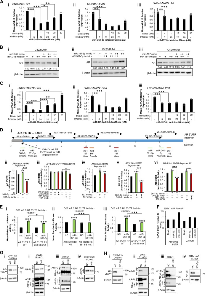
MiR-346, -361-3p and -197-3p alter wild-type and variant androgen receptor activity in prostate cancer (PC) cells partially through association with AR 3′UTR. a qRT-PCR analysis of AR mRNA levels in C42/MAR4 cells transfected with (i) miR-346, (ii) miR-361-3p or (iii) miR-197-3p mimic and/or inhibitor for 24 h. b Western blot analysis of AR protein levels in C42/MAR4 transfected with (i) miR-346, (ii) miR-361-3p or (iii) miR-197-3p mimic and/or inhibitor for 24 h. β-actin was used as a control for loading. Representative blots of three independent experiments are shown. Additional biological replicates and densitometry for three independent experiments are shown (Fig. S1c–g). c qRT-PCR analysis of PSA mRNA levels in LNCaP/MAR4 cells transfected with (i) miR-346, (ii) miR-361-3p or (iii) miR-197-3p mimic and/or inhibitor for 24 h. L19 was used as a normalisation gene. d, e Luciferase assay analysis of 6.9 kb AR 3′UTR activity in HEK293T (d) or C42 (e) cells transfected with pMiR-Report vector containing WT (d) or miR binding site-mutant (e) regions of the AR 6.9 kb 3′UTR as depicted (Fig. S2Di) ± miR mimics or inhibitors (20 nM) for 48 h (d) or 72 h (e) Luciferase activity was normalised to β-galactosidase activity to correct for transfection efficiency. Columns: mean normalised AR reporter luciferase activity from three independent experiments performed in duplicate ± SEM. f AGO2/biotin-miR RNA-IP analysis of miR-197 and miR-346 association with AR 6.9 kb 3′UTR. 22RV1 cells transfected with biotin-labelled miR (200 pmol) for 24 h, followed by two-step immunoprecipitation with AGO2 antibody- and streptavidin-coated beads. RNA was extracted from input and IP samples and qRT-PCR performed for 6.9 kb AR 3′UTR. Data are presented relative to input values. g, h Western blot analysis of wild-type AR (110 kDa) and variant-AR (65–90 kDa) in (i) CWR-R1-D567es (lacking WT-AR but overexpressing v567es), (ii) CWR-R1-AD1 (parental cells expressing WT- and variant-AR), (iii) 22RV1 (expressing WT- and variant-AR) and (iv) 22RV1/AR-FL-KO cells (expressing AR-variants but with WT-AR knocked out) cells transfected with (a) miR-346 or (b) miR-361-3p inhibitor (both 20 nM) for 24 h. β-actin was used as a control for loading. Representative western blot images are shown. See Fig. S5 for independent biological replicates. *P ≤ 0.05, **P ≤ 0.005, ***P ≤ 0.0001. See also Figs. S1, S2, S3, S4 and S5

