Fig. 2.
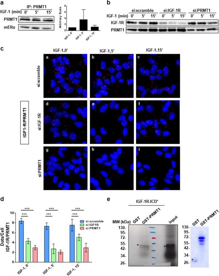
IGF-1R interacts with PRMT1. a MCF-7 cells were treated with IGF-1 for the indicated times, cell lysates were then immunoprecipitated with anti-PRMT1 antibody and its enzymatic activity was evaluated by performing an in vitro methylation assay using the GST-hinge of ERα as a substrate, detected by western blot using the anti-mERα antibody. Quantification of the signal was performed by computer-assisted analysis (right-hand panel). This result is representative of two independent experiments. b MCF-7 cells were transfected with si:scramble or siRNAs targeting IGF-1R or PRMT1 for 72 h, then treated with IGF-1 for different times. The efficacy of protein inhibition was verified by western blot using the corresponding antibodies. c After siRNA transfection and fixation, proximity ligation assay experiments were performed to evaluate IGF-1R/PRMT1 interaction using IGF-1R- and PRMT1-specific antibodies. The detected dimers are represented by red dots. The nuclei were counterstained with mounting medium containing DAPI (blue) (Obj: ×60). d Quantification of the number of dots per cell was performed by computer-assisted analysis as reported in the Materials and Methods section. The mean ± s.e.m. of one experiment representative of three experiments is shown. The P value was determined using the Student t test. ***P < 0.001. e Radioactive GST pull-down assay was performed by incubating the in vitro 35S-labeled intracellular domain of IGF-1R (IGF-1R-ICD*) with GST and GST-PRMT1. The corresponding Coomassie-stained gel is shown in the right-hand panel. *Indicates the full-length fusion proteins. IGF-1R insulin-like growth factor 1 receptor

