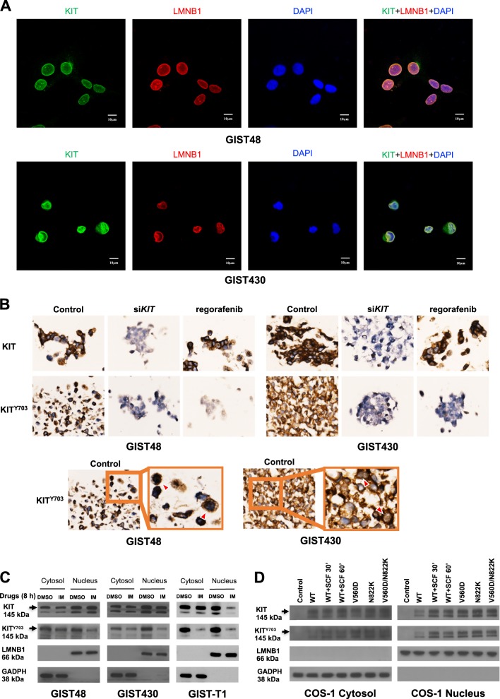
Fig. 1.
Distribution of KIT in the cytoplasm and nucleus of GIST cells. a GIST48 and GIST430 cells were stained using antibodies against KIT and LMNB1. After the cells were immunostained, they were visualized by confocal microscopy, and images were acquired through the Cy2, rhodamine, and DAPI channels (×1000). The data were derived from representative images of five fields/picture for each sample. b Cells were transfected with 150 nM siRNA targeting KIT for 72 h or treated with 1 μM regorafenib for 8 h. The cell blocks were analyzed by immunohistochemistry staining against phosphorylated KIT (KITY703) and total KIT. GIST cells were treated with 1 μM IM for 8 h (c), and COS-1 cells were transfected with various KIT mutants and treated with or without SCF for indicated time (d). The cells were fractionated to separate the cytoplasmic and nuclear proteins and analyzed by immunoblotting for KITY703 and total KIT. LMNB1 and GAPDH were used as nuclear and cytoplasmic markers, respectively. All experiments were repeated at least three times

