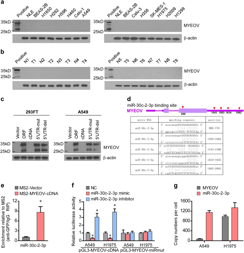
Fig. 2.
Characterization of MYEOV transcript as a ceRNA in NSCLC. a The protein expression levels of MYEOV in indicated cells examined by WB analysis. b The protein expression levels of MYEOV in NSCLC versus the paired adjacent noncancerous tissue examined by WB analysis. c The protein expression levels of MYEOV in indicated cells assessed by WB analysis. d Schematic outlining of the predicted binding sites of miR-30c-2-3p in MYEOV transcript. e MS2-RIP followed by miRNA qRT-PCR to detect miR-30c-2-3p endogenously associated with MYEOV transcript (each bar represents the mean ± SD derived from three independent experiments, two-tailed Student’s t test. *P < 0.05). f The reporters containing WT or mutant MYEOV transcript were co-transfected with miR-30c-2-3p mimic or inhibitor, and luciferase activities were assessed after 48 h (each bar represents the mean ± SD derived from three independent experiments, one-way ANOVA followed by Dunnett’s multiple comparison test. *P < 0.05). g Copy numbers of the MYEOV transcript and miR-30c-2-3p in NSCLC cell lines quantified by absolute quantitative PCR (each bar represents the mean ± SD derived from three independent experiments)

