Abstract
Neuronal nicotinic acetylcholine receptors (nAChRs) expressed on mesostriatal dopaminergic neurons are thought to mediate several behavioral effects of nicotine, including locomotion, habit learning, and reinforcement. Using immunoprecipitation and ligand-binding techniques, we have shown that both α6β2* and α4(nonα6)β2* nAChRs are expressed in the caudate–putamen and that only α6* nAChRs can bind α-conotoxin MII and methyllycaconitine with affinities of 1.3 and 40 nm, respectively. Further studies performed on 6-hydroxydopamine-lesioned striatum led to the identification of nAChR subtypes selectively expressed on dopaminergic terminals [α4α5β2, α4α6β2(β3), and α6β2(β3)], nondopaminergic neuronal structures (α2α4β2), or both structures (α4β2). The identification of the nAChRs expressed on striatal dopaminergic terminals opens up the possibility of developing selective nAChR ligands active on dopaminergic systems and associated diseases, such as Parkinson's disease.
Keywords: nicotinic acetylcholine receptor, mesostriatal dopamine pathway, striatum, immunoprecipitation, 6-hydroxydopamine, α-conotoxin MII
The mesostriatal dopamine (DA) pathway is a major brain target for nicotinic agonists. Its ventral (the mesolimbic DA pathway) and dorsal (the nigrostriatal DA pathway) components both express high levels of nicotinic acetylcholine receptors (nAChRs), which are thought to mediate several behavioral effects of nicotinic agonists (including the modulation of locomotor activity, reinforcement, and habit learning) (Di Chiara, 2000).
Neuronal nAChRs comprise a heterogeneous family of pentameric oligomers made up of combinations of subunits encoded by at least 11 different genes in mammals. They have been grouped into two subfamilies based on their phylogenetic, functional, and pharmacological properties (Le Novére and Changeux, 1995; Corringer et al., 2000), namely the α-bungarotoxin (α-Bgtx)-sensitive or homomeric nAChRs (α7 subunit), and the α-Bgtx-insensitive or heteromeric nAChRs (α2-α6 and β2-β4 subunits). These latter subunits can combine to form a number of functionally and pharmacologically different heteropentamers consisting of two, three, or four different subunits.
In situ hybridization and single-cell PCR studies have shown that 80–100% of midbrain DA neurons express α4, α5, α6, β2, and β3 subunits, 40–60% express α3 and α7, and a few of them express β4 (Le Novère et al., 1996; Klink et al., 2001; Azam et al., 2002). A large number of heteromeric nAChR subtypes are therefore potentially present in these neurons. Previous studies using α-conotoxin MII (α–CntxMII, an antagonist selective for α3β2 or α6β2 interfaces) (Cartier et al., 1996; Champtiaux et al., 2002;Kuryatov et al., 2002) and knock-out (KO) mice lacking specific nAChR subunits have suggested the existence of at least two main receptor populations containing α4β2 or α6β2 subunits (Picciotto et al., 1998; Zoli et al., 1998; Klink et al., 2001;Champtiaux et al., 2002).
Using a combination of techniques (immunoprecipitation and purification of native nAChRs, followed by their pharmacological characterization in intact or DA denervated striatum), we have established the composition of nAChRs expressed in striatal DA projections and in nondopaminergic neuronal structures.
MATERIALS AND METHODS
Animals and materials. Adult male pathogen-free Sprague Dawley rats (Harlan-Nossan, Milan, Italy) were used. All animal experimentation was conducted in accordance with the European Communities Council Directive of 24 November 1986 (86/609/EEC). (+/−)3Η-epibatidine (Epi; specific activity, 50–66 Ci/mmol) was purchased from Amersham Biosciences (Arlington Heights, IL), 125I-Epi (s.a. 2200 Ci/mmol) and 3H-WIN35,428 (s.a. 86 Ci/mmol) from NEN (Boston, MA), and nonradioactive ligands were purchased from Sigma (St. Louis, MO). α-CntxMII was synthesized as described previously (Cartier et al., 1996).
Antibody production and characterization. The polyclonal antibodies against the α2, α3, α4, α5, α6, β2, β3, and β4 nAChR subunits were produced in rabbit as previously described (Vailati et al., 1999) and affinity purified. The peptides obtained from rat or human sequences were located in the putative cytoplasmic loop between M3 and M4 and/or at the COOH terminal. For almost all of the subunits we raised antisera directed against two separate peptides of the same subunit, and the immunoprecipitation values reported are the mean of results obtained using both antisera. The affinity-purified antisera were bound to cyanogen bromide-activated Sepharose at a concentration of 1 mg/ml, and the columns were used for subtype immunopurification.
Characterization of antibody specificity. The antisera were tested by quantitative immunoprecipitation experiments on 2 nm3H-Epi-labeled nAChRs present in 2% Triton X-100 extracts prepared from brain membranes and/or immunopurified nAChRs. Because3H-Epi binds α7* nAChRs, albeit with nanomolar affinity, we always preincubated the membranes and 2% Triton X-100 extracts with 2 μm α-Bgtx. Only the receptors labeled with 3H-Epi were immunoprecipitated, which assured the specificity of the quantification. The antisera were tested in available wild-type (WT) and KO mice (immunoprecipitation expressed as percentage of3H-Epi-labeled receptors in total brain): 60 and 1% (anti-α4 antisera), 11 and 0% (anti-α5 antiserum), 84 and 2% (anti-β2 antisera). Anti-α6 and anti-β3 antisera immunoprecipitated 25 ± 1 versus 1 ± 0.3% and 8 ± 2 versus 2 ± 1%, respectively, of3H-Epi-labeled striatal receptors in α6 WT versus KO mice (N. Champtiaux and C. Gotti et al., unpublished observations). Anti-α2, -α4, and -β2 antisera immunoprecipitated at ∼0, 80, and 90%, respectively, of α4β2 or α4α5β2 receptors immunopurified from rat cortex, whereas anti-α5 antisera immunoprecipitated 1% of the α4β2 receptors but 75% of the α4α5β2 receptors. Anti-α3 and -β4 antisera immunoprecipitated only 1–2% of cortical α4β2 and α4α5β2 receptors but immunoprecipitated 74 and 70%, respectively, of3H-Epi-labeled receptors from rat superior cervical ganglion. Finally, anti-α2 antisera immunoprecipitated up to 27% of α2α5β2 purified from postnatal rat retina (M. Moretti, unpublished observations).
Binding assay and pharmacological experiments. Binding techniques for solubilized or immunoimmobilized nAChRs, receptor immobilization by anti-subunit-specific antisera, and immunoprecipitation of 3H-Epi-labeled receptors by anti-subunit specific antisera were performed as inVailati et al. (1999). The affinity-purified anti-α6 or anti-β2 antisera were bound to microwells (Maxi-Sorp; Nunc, Roskilde, Denmark) and then incubated overnight at 4°C with 200 μl of 2% Triton X-100 total (α6 microwells) or α6 subunit-depleted (β2 microwells) striatal extract containing 10–30 fmol of receptors. We ascertained that 84 ± 2% of 3H-Epi binding could be solubilized from striatal membranes using 2% Triton X-100.
Receptor subtype immunopurification. For each purification experiment the caudate–putamen from 20–30 animals was dissected, immediately frozen at −70°C, and processed as described in Del Signore et al. (2002). The extract was incubated three times with 5 ml of Sepharose-4B bound anti-α6 antisera to remove the α6 receptors. The flow-through of the α6 column was reincubated with 5 ml of Sepharose-4B with bound anti-α4 or β2 antisera. The bound receptors were eluted by competition with 100 μm of the corresponding α6, α4, or β2 peptide used for antiserum production.
6-hydroxydopamine lesion and 3H-WIN 35,428 binding. Unilateral DA denervation of striatum was performed by injecting the selective DA neurotoxin 6-hydroxydopamine (6-OHDA) in the medial forebrain bundle. The animals were deeply anesthetized with halothane, and 6-OHDA (10 μg/4 μl) was injected (coordinates: anterior, −4 mm; lateral, 1.8 mm, dorsal, −7.5 mm) using a 10 μl Hamilton syringe (26G) during 4 min, waiting 2 min before withdrawal of the needle. The animals were killed 14 d after the lesion. The extent of DA denervation was assessed by WIN35,428 binding, a ligand for DA transporter that is selectively localized on DA terminals. In preliminary experiments the affinity of3H-WIN35,428 was determined using established protocols (Kimmel et al., 2000).3H-WIN35,428 binding was determined individually in striata from 30 control and 30 6-OHDA-lesioned rats using a saturating concentration of 100 nm3H-WIN35,428 in the presence or absence of 10 μm GBR 12935. 6-OHDA-lesioned striata with a decrease of 3H-WIN35,428 <80% were discarded.
RESULTS
Overall subunit composition of nicotinic receptors in striatum
Because the contribution of α7* nAChR to nicotine effects on striatum is still debated (Kaiser and Wonnacott, 2000), we first determined the amount of α7* versus (nonα7)* nAChRs in striatal homogenates. We found that 125I-αΒgtx binding is <3% of 3H-Epi binding (4.7 ± 1.6 fmol/mg of protein vs 153.7 ± 15.0 fmol/mg of protein, respectively).
We next determined the overall subunit composition of striatal nAChRs by studying 3H-Epi-labeled receptors immunoprecipitated by subunit-specific antisera. Almost all of the receptors (90.7%) contained the β2 subunit, whereas α4 (69.0%) and α6 (19.3%) appeared to be the most represented α subunits. We also found that a considerable percentage of3H-Epi-labeled receptors contain α5 (18.7) or β3 (8.9%) subunits. Instead, the level of α2, α3, and β4 subunits was low (3.9, 3.3, and 1.3%, respectively).
These results show that α6β2* and α4β2* are the main nAChR populations present in rat striatum, whereas putative α3β2* nAChRs, previously proposed as a major striatal subtype (Kulak et al., 1997;Kaiser et al., 1998), are almost absent from this region.
Subunit composition of striatal α6β2* subtypes
To isolate α6β2* receptors, we immunodepleted the striatal extract of α6* receptors by using an affinity column bearing anti-α6 antisera. Selective α6-containing nAChR immunodepletion was confirmed by the fact that immunoprecipitated α6-containing3H-Epi-labeled receptors decreased from 19.3% in the total striatal extract to 2.9% in the flow-through of the α6 column. In addition, α4-containing and α5-containing receptors increased (from 69.0 to 87.6% and from 18.7 to 21.8%, respectively), β2-containing receptors remained unchanged, and β3- containing receptors markedly decreased (from 8.9 to 1.2%). Indeed, the increase in α4 subunit in the flow-through demonstrates that the majority of the α4 subunit pool is not assembled with α6 subunit.
To identify the subunit composition of the α6-containing receptors, we eluted them from the affinity column with the α6 peptide, and then labeled with 3H-Epi and immunoprecipitated the eluate with subunit specific antisera (Fig.1). The anti-α4, β2 and β3 antisera immunoprecipitated 37.8, 87.9, and 19.7%, respectively, of the purified 3H-Epi-labeled α6-containing receptors. The anti-α2, α3, α5, and β4 antisera immunoprecipitated only 0.1, 0, 2.1, and 2.6%, respectively, of the purified α6-containing receptors.
Fig. 1.
Immunoprecipitation analysis of the subunit content of α6β2* and α4(nonα6)β2* nAChR subtypes immunopurified through affinity column from striatal extracts and labeled with 2 nm3H-Epi. The results are expressed as percentage of total 3H-Epi binding present in the solution before immunoprecipitation. Each data point is the mean ± SEM of five determinations performed in triplicate.
These immunoprecipitation results indicate that purified α6β2* receptor population is a mixture of two main subtypes, namely α6β2 and α4α6β2 nAChRs, some of which also contain the β3 subunit.
Subunit composition of striatal α4(nonα6)β2* subtypes
To determine the subunit composition of α4β2* receptor population that do not contain the α6 subunit (α4(nonα6)β2*), we immunopurified nAChRs present in the flow-through of the α6 column over an anti-α4 column, eluted using the α4 peptide, and performed an immunoprecipitation with subunit-specific antisera (Fig. 1). The anti-α4, -α5, and -β2 antisera immunoprecipitated 84, 21, and 82%, respectively, of 3H-Epi-labeled receptors recovered using this method, whereas the anti-α2, -α3, -α6, -β3, and -β4 immunoprecipitated 7.4, 2.5, 0.9, 2.5, and 1.6%, respectively, of the purified eluate (Fig. 1). The subunit content of these α4* nAChRs was identical to that obtained by passing the α6 flow-through over an anti-β2 column to immunopurify nAChRs (data not shown) and very similar to that determined in the flow-through of the α6 column (see above), indicating that no other main nAChR receptor populations are present in striatum besides α6β2* and α4(nonα6)β2*.
These immunoprecipitation results show that α4(nonα6)β2* nAChRs comprise α4β2 and α4α5β2 subtypes with a minor proportion of the α2α4β2 subtype.
Pharmacological profile of striatal α6β2* and α4(nonα6)β2* nAChRs
To explore the pharmacology of the two receptor populations, we immunoimmobilized the α6β2* receptors using an anti-α6 column and compared their pharmacological profile with that of the α4(nonα6)β2* receptors immobilized over an anti-β2 column.
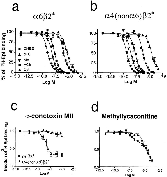
Equilibrium binding assays revealed no significant differences in the affinity for 3H-Epi of the α6β2* and α4nonα6β2* receptor populations [apparentKd value of 34 pm (coefficient of variation, 34%) and 41 pm (coefficient of variation, 25%) for α6β2* and α4(nonα6)β2* receptors, respectively]. We then performed competition binding studies using a number of nicotinic ligands. Although no significant difference was detected for the agonists acetylcholine, nicotine, and cytisine and the antagonists dihydro-β-erythroidine and d-tubocurarine (Fig.2a,b), significant differences were observed for α-CntxMII and methyllycaconitine (MLA). Both ligands showed a statistically significant better fit for a two-site model with a high- and a low-affinity site when tested on the α6β2* nAChRs. α-CntxMII had a high affinity site for α6-containing nAChRs with a Ki of 1.3 nm and a site with no or low affinity with aKi >10 μm(Fig. 2c, Table 1), whereas MLA had a high-affinity site with a Kiof 40 nm and a low-affinity site with aKi of 20.8 μm(Fig. 2d, Table 1). On the other hand, for α4(nonα6)β2* receptors, both ligands showed the presence of only a single class of low-affinity sites with aKi of >10 μmfor α-CntxMII and a Ki of 25 μm for MLA.
Fig. 2.
Inhibition of 125I-Epi binding to native immunoimmobilized α6β2* (a) and α4(nonα6)β2* (b) nAChRs by several nicotinic ligands, including nicotine (Nic), acetylcholine (ACh), cytisine (Cyt), dihydro-β-erythroidine (DHBE),d-tubocurarine (dTC) (a, b), α-CntxMII (c), and MLA (d). The curves were obtained by fitting three or four separate experiments using the LIGAND program (Munson and Rodbard, 1980).
Table 1.
Affinity of nicotinic agonists and antagonists for immunoimmobilized nAChR subtypes
| α6β2* | α4(non α6)β2* | |
|---|---|---|
| Kd (nm) | ||
| 125I-Epibatidine | 0.034 (34) | 0.041 (25) |
| Ki (nm) | ||
| Cytisine | 0.65 (18) | 0.19 (16) |
| Nicotine | 2.5 (23) | 1.75 (23) |
| Acetylcholine | 8.0 (34) | 8.6 (23) |
| Dihydro-β-erythroidine | 524 (25) | 274 (21) |
| d-Tubocurarine | 3,110 (20) | 16,100 (28) |
| α-Conotoxin MII | 1.3 (45) | – |
| >10,000 | >10,000 | |
| Methyllycaconitine | 40 (47) | – |
| 20,800 (18) | 25,000 (23) |
Kd and Ki values were derived from curves of 125I-Epi saturation and competition binding, respectively, to α6β2* or α4(nonα6)β2
immunoimmobilized receptors. Curves obtained from three or four separate experiments were fitted using a nonlinear least-squares analysis program. For both α-conotoxin MII and methyllycaconitine, a two-site model was statistically significant (F test), whereas for d-tubocurarine and cytisine the data were better fitted with a one-site model. Numbers in parentheses represent percentage of coefficient of variation.
Nicotinic receptor subtypes expressed on striatal dopaminergic terminals
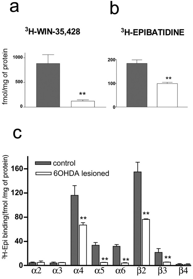
Several neuronal structures in striatum in addition to nigrostriatal dopaminergic terminals express nAChRs (Kaiser and Wonnacott, 2000). To distinguish nAChR subtypes expressed by dopaminergic and nondopaminergic structures, we performed striatal DA denervation using the neurotoxin 6-OHDA. In view of the very low density of noradrenergic terminals in striatum, this technique allows a selective destruction of DA terminals. The extent of the denervation was ∼85%, as assessed by binding to3H-WIN35,428 (Fig.3a).
Fig. 3.
a, b,3H-WIN-35,428 (a) and3H-Epi binding (b) in rat striatal membranes obtained from control and 6-OHDA lesioned rats.c, Immunoprecipitation of nAChR subunits in 2% Triton X-100 extracts from control and 6-OHDA lesioned striata. Each value represents the mean ± SEM of three separate experiments. Statistical analysis according to Mann–Whitney U test, **p < 0.01.
We first examined the effect of DA denervation on the amount of3H-Epi binding, showing a decrease by ∼50% in 6-OHDA-lesioned striata (183 ± 10 and 99 ± 6 fmol/mg protein in intact vs lesioned striatum) (Fig.3b).
We then assessed the nAChR subunit composition of3H-Epi-labeled receptors of control and 6-OHDA-lesioned striata in quantitative immunoprecipitation experiments (Fig. 3c). These experiments revealed an almost complete disappearance of nAChRs containing the α5 (84%), α6 (87%), or β3 (73%) subunits, which matches very closely the reduction in DA innervation, a marked but partial reduction of the receptors containing α4 (42%) and β2 (50%) subunits, whereas the other subunits were unchanged. These results demonstrate that α6, α5, and β3 subunits are selectively enriched in DA terminals, α4 and β2 subunits are present in both dopaminergic and nondopaminergic cells, and α2 subunit is only present in nondopaminergic cells.
Combining the results obtained on DA-denervated striata with those obtained on immunopurified receptors, it can be concluded that striatal DA terminals express α6β2 and α6α4β2 (with or without β3 subunit) as well α4α5β2 and α4β2 nAChR subtypes, whereas nondopaminergic striatal structures express α4β2 and α2α4β2 nAChR subtypes.
DISCUSSION
In this study, we identified the major nAChR subtypes expressed in dopaminergic terminals and nondopaminergic neuronal structures in the caudate–putamen at the molecular and pharmacological level. Much information about native nAChRs in the brain and ganglia has been obtained using immunopurification and immunoprecipitation techniques (for review, see Lindstrom 2000). Our identification of striatal nAChR subtypes relied on the use of a series of antisera raised against unique amino acid sequences of the different subunits. To obtain a quantitative evaluation of the subunit composition of a receptor subtype, it is necessary to evaluate the efficiency of the immunoprecipitation of antigens by their respective antisera. This was assessed for the α3, α4, α5, α6, β2, and β4 subunits, and ranged from 75 to 90%, thus suggesting that the values obtained in this study are probably slightly underestimated. A second caveat concerns the detection limits of the immunoprecipitation and immunopurification techniques and so, in the following discussion, we will not consider the contribution to receptor composition of subunits that were immunodetected in amounts <3%; therefore, this means that the existence of minor nAChR subtypes (<3–5%) may be overlooked. Finally, it must be considered that possible changes in nAChRs expressed on DAceptive neurons induced by DA denervation cannot be presently excluded.
In defining the striatal nAChR subtypes, we followed the current hypothesis that heteromeric nAChRs have at least two subunits bearing the principal amino acid loops for ACh binding interfaces (i.e., α2, α3, α4, or α6 subunits) and two subunits bearing the complementary amino acid loops for ACh binding interfaces (i.e., β2 or β4 subunits), whereas the fifth subunit can be either a complementary subunit or a purely structural subunit (α5 or β3 subunits) (Corringer et al., 2000).
Striatal α6β2* and α4(nonα6)β2* nAChRs have a partially different pharmacology
Present immunopurification approach allowed to isolate two populations of striatal nAChRs: one contains α4β2*, but not α6, subunits and accounts for ∼70% of the nAChRs; the other contains α6β2* subunits and accounts for ∼20%. Furthermore, whereas α6β2* nAChRs are selectively expressed on dopaminergic terminals (see below), α4(nonα6)β2* nAChRs are expressed on both dopaminergic terminals and nondopaminergic cells. These two populations have indistinguishable binding affinity for several classical nicotinic agonists and antagonists, including acetylcholine, nicotine, cytisine, dihydro-β-erythroidine and d-tubo-curarine. However, the antagonists α-CntxMII and MLA could discriminate the two receptor populations by showing low (micromolar) affinity for the α4(nonα6)β2*, but both low (micromolar) and high (nanomolar) affinity for the α6β2* receptors. Because a subset of ∼40% of the α6β2* nAChRs also contain the α4 subunit (Fig. 1a) (see below for discussion), we hypothesize that both compounds bind an α6β2 interface (exclusively present in α6β2*) with nanomolar affinity (Vailati et al., 1999; Barabino et al., 2001; Champtiaux et al., 2002) and an α4β2 interface [present in both α6β2* and α4(nonα6)β2* nAChRs] with micromolar affinity.
Based on pharmacological studies using α-CntxMII (Kulak et al., 1997;Kaiser et al., 1998), neuronal Bgtx (Grady et al., 1992), and UB-165 (Sharples et al., 2000) on striatal synaptosomal preparations, it was suggested that both α4* and (nonα4)* nAChRs mediate DA release in striatum. (Nonα4)* nAChRs were identified as α3* nAChRs on the basis of the high affinity of α-CntxMII for α3β2* nAChRs expressed in reconstituted systems (Cartier et al., 1996). However, subsequent studies showed that α-CntxMII binds and blocks native α6* nAChRs (Vailati et al., 1999; Barabino et al., 2001; Kuryatov et al., 2002), and equilibrium-binding experiments in KO mice showed that α-CntxMII binding disappears from the striatum of α6−/− (Champtiaux et al., 2002) but not from α3−/− mice (Whiteaker et al., 2002). The present study unequivocally shows that α-CntxMII binds with high affinity to immunopurified native α6β2* nAChRs and that α6* nAChRs constitute the major (nonα4)* nAChR in this brain region as only negligible amounts of other ACh binding subunits (including the α3 subunit)were detected in striatum.
Both striatal α6β2* and α4(nonα6)β2* nAChR populations are heterogeneous and differentially expressed by dopaminergic and nondopaminergic neurons
Our immunoprecipitation studies of immunopurified native receptors showed that α6β2* nAChRs are heterogeneous and consist of two main subpopulations of roughly equal size (i.e., α6β2 and α4α6β2 nAChRs) with a portion (20%) also containing the β3 subunit. α4(nonα6)β2* nAChRs are also heterogeneous and form a large population (60–70%) of (α4)2(β2)3 nAChRs, a considerable population (20%) of (α4)2α5(β2)2 nAChRs, and a minor population (5%) of α2α4β2* nAChRs.
One interesting result is that the structural subunits α5 and β3 always coassemble with the α4 and α6 subunit, respectively. This selective assembly fits very nicely with previous in situhybridization studies, showing that α6 and β3 subunit mRNAs are always coexpressed throughout brain nuclei (Le Novére et al., 1996) and that α5 mRNA is present only in α4 mRNA-containing neurons. However, it must be noted that the case for selective coexpression of the α4 and α5 subunits is not strong, because α4 subunit mRNA is expressed by most neuronal populations (but see the case of the medial habenula for a strict similarity between the subnuclear pattern of α5 and α4 mRNAs; Le Novère et al., 1996). Nothwithstanding the fact that the functional role of α5 and β3 remains difficult to assess, the strict regulation of their assembly suggests that they may subserve an important role in nAChR subtype physiology, including a change in their electrophysiological features, turnover, and/or subcellular targeting.
On the basis of the changes in subunit content observed in DA-denervated striata, it can indeed be concluded that although (α4)2(β2)3=(α4)2(β2)3 nAChRs are expressed by both dopaminergic and nondopaminergic cell types, α6β2, α4α6β2, and (α4)2α5(β2)2=(α4)2α5(β2)2 nAChRs are expressed only on dopaminergic terminals, and α2α4β2 nAChRs are expressed only by nondopaminergic cell types. Because DA denervation decreases striatal 3H-Epi binding by ∼50%, it can be inferred that dopaminergic terminals express four major populations of nAChRs: (α4)2(β2)3=(α4)2(β2)3 (∼30%), (α4)2α5(β2)2=(α4)2α5(β2)2(∼30%), α6β2(β3) (∼25%), and α4α6β2(β3) (∼15%). These results agree well with those of in situ hybridization and single-cell PCR studies of midbrain DA neurons (Le Novère et al., 1996; Klink et al., 2001; Azam et al., 2002), which showed that α4, α5, α6, β2, and β3 mRNAs are expressed by the vast majority of DA neurons at moderate to high levels, whereas α3 and β4 mRNAs are detected in a more restricted number of neurons and at low levels. They are also in line with the evidence of β2 subunit immunoreactivity in rat nigrostriatal DA terminals (Jones et al., 2001), as well as with studies showing that selective lesion of the nigrostriatal pathway in monkey leads to a complete loss of high affinity α-CntxMII binding (i.e., α6β2* nAChRs) and a 50% reduction in 125I-Epi binding in striatum (Kulak et al., 2002).
The mesostriatal dopamine pathway plays an essential role in locomotion, movement coordination, habit learning, and reinforcement and is known to be modulated by nicotinic agents. In particular, recent studies have shown that striatal DA release is physiologically regulated by cholinergic tone through nAChRs activation (Zhou et al., 2001). A pathophysiological role of nAChRs in this neuronal system has been proposed on the basis of evidence of a negative correlation between cigarette smoking and the incidence of Parkinson's disease, the protective effects of nicotine treatment against nigrostriatal DA pathway degeneration in animal models of Parkinson's disease (Quik and Jeyarasasingam, 2000). The identification of the different nAChR subtypes expressed by DA terminals and the demonstration that some subtypes are only expressed by DA structures opens up the possibility of developing ligands selectively acting on the release of dopamine from striatal terminals.
Footnotes
This work was supported in part by grants from the Italian Ministero dell' Istruxione, dell' Università, e della Ricerca (MM05152538) (F.C. and M.Z.) and National Institutes of Health Grant MH 53631 (J.M.M.). We thank Prof. Jean-Pierre Changeux (Pasteur Institute, Paris, France) and Dr. Mariella De Biasi (Baylor College, Houston, TX) for the generous gift of neuronal tissues from wild-type and knock-out mice and Renato Longhi for the peptide synthesis.
Correspondence should be addressed to Dr. Cecilia Gotti, Consiglio Nazionale delle Ricerche, Institute of Neuroscience, Section of Cellular and Molecular Pharmacology Center, Department of Medical Pharmacology, University of Milan, Via Vanvitelli 32, 20129 Milan, Italy. E-mail: c.gotti@csfic.mi.cnr.it.
REFERENCES
- 1.Azam L, Winzer-Serhan UH, Chen YL, Leslie FM. Expression of neuronal nicotinic acetylcholine receptor subunit mRNAs within midbrain dopamine neurons. J Comp Neurol. 2002;444:260–274. doi: 10.1002/cne.10138. [DOI] [PubMed] [Google Scholar]
- 2.Barabino B, Vailati S, Moretti M, McIntosh MJ, Longhi R, Clementi F, Gotti C. An α4β4 nicotinic receptor subtype is present in chick retina. Identification, characterization and pharmacological comparison with the transfected α4β4 and α6β4 subtypes. Mol Pharmacol. 2001;59:1410–1417. doi: 10.1124/mol.59.6.1410. [DOI] [PubMed] [Google Scholar]
- 3.Cartier GE, Yoshikami D, Gray WR, Luo S, Olivera BM, McIntosh JM. A new alpha-conotoxin which targets alpha3beta2 nicotinic acetylcholine receptors. J Biol Chem. 1996;271:7522–7528. doi: 10.1074/jbc.271.13.7522. [DOI] [PubMed] [Google Scholar]
- 4.Champtiaux N, Han ZY, Bessis A, Rossi FM, Zoli M, Marubio L, McIntosh JM, Changeux JP. Distribution and pharmacology of alpha6-containing nicotinic acetylcholine receptors analyzed with mutant mice. J Neurosci. 2002;22:1208–1217. doi: 10.1523/JNEUROSCI.22-04-01208.2002. [DOI] [PMC free article] [PubMed] [Google Scholar]
- 5.Corringer PJ, Le Novère N, Changeux JP. Nicotinic receptors at the amino acid level. Annu Rev Pharmacol Toxicol. 2000;40:431–458. doi: 10.1146/annurev.pharmtox.40.1.431. [DOI] [PubMed] [Google Scholar]
- 6.Del Signore A, Gotti C, De Stefano ME, Moretti M, Paggi P. Dystrophin stabilizes α3- but not α7-containing acetylcholine receptor subtypes at the postsynaptic apparatus in the mouse cervical ganglion. Neurobiol Dis. 2002;10:54–66. doi: 10.1006/nbdi.2002.0495. [DOI] [PubMed] [Google Scholar]
- 7.Di Chiara G. Role of dopamine in the behavioural actions of nicotine related to addiction. Eur J Pharmacol. 2000;393:295–314. doi: 10.1016/s0014-2999(00)00122-9. [DOI] [PubMed] [Google Scholar]
- 8.Grady S, Marks MJ, Wonnacott S, Collins AC. Characterization of nicotinic receptor-mediated [3H]dopamine release from synaptosomes prepared from mouse striatum. J Neurochem. 1992;59:848–856. doi: 10.1111/j.1471-4159.1992.tb08322.x. [DOI] [PubMed] [Google Scholar]
- 9.Jones JW, Bolam JP, Wonnacott S. Presynaptic localization of the nicotinic acetylcholine receptor β2 subunit immunoreactivity in rat nigrostriatal dopaminergic neurons. J Comp Neurol. 2001;439:235–247. doi: 10.1002/cne.1345. [DOI] [PubMed] [Google Scholar]
- 10.Kaiser S, Wonnacott S. alpha-bungarotoxin-sensitive nicotinic receptors indirectly modulate [(3)H]dopamine release in rat striatal slices via glutamate release. Mol Pharmacol. 2000;58:312–318. doi: 10.1124/mol.58.2.312. [DOI] [PubMed] [Google Scholar]
- 11.Kaiser SA, Soliakov L, Harvey SC, Luetje CW, Wonnacott S. Differential inhibition by alpha-conotoxin-MII of the nicotinic stimulation of [3H]dopamine release from rat striatal synaptosomes and slices. J Neurochem. 1998;70:1069–1076. doi: 10.1046/j.1471-4159.1998.70031069.x. [DOI] [PubMed] [Google Scholar]
- 12.Kimmel H, Carroll F, Kuhar MJ. Dopamine transporter synthesis and degradation rate in rat striatum and nucleus accumbens using RTI-76. Neuropharmacology. 2000;39:578–585. doi: 10.1016/s0028-3908(99)00160-4. [DOI] [PubMed] [Google Scholar]
- 13.Klink R, de Kerchove-d'Exaerde A, Zoli M, Changeux JP. Molecular and physiological diversity of nicotinic acetylcholine receptors in the midbrain dopaminergic nuclei. J Neurosci. 2001;21:1452–1463. doi: 10.1523/JNEUROSCI.21-05-01452.2001. [DOI] [PMC free article] [PubMed] [Google Scholar]
- 14.Kulak JM, Nguyen TA, Olivera BM, McIntosh JM. Alpha-conotoxin MII blocks nicotine-stimulated dopamine release in rat striatal synaptosomes. J Neurosci. 1997;17:5263–5270. doi: 10.1523/JNEUROSCI.17-14-05263.1997. [DOI] [PMC free article] [PubMed] [Google Scholar]
- 15.Kulak JM, McIntosh JM, Quik M. Loss of nicotinic receptors in monkey striatum after 1-methyl-4-phenyl-1, 2, 3, 6-tetrahydropyridine treatment is due to a decline in alpha-conotoxin MII sites. Mol Pharmacol. 2002;61:230–238. doi: 10.1124/mol.61.1.230. [DOI] [PubMed] [Google Scholar]
- 16.Kuryatov A, Olale F, Cooper J, Choi J, Lindstrom J. Human α6 subtypes: subunit composition, assembly and pharmacological responses. Neuropharmacology. 2000;39:2570–2590. doi: 10.1016/s0028-3908(00)00144-1. [DOI] [PubMed] [Google Scholar]
- 17.Le Novère N, Changeux JP. Molecular evolution of the nicotinic acetylcholine receptor: an example of multigene family in excitable cells. J Mol Evol. 1995;40:155–172. doi: 10.1007/BF00167110. [DOI] [PubMed] [Google Scholar]
- 18.Le Novère N, Zoli M, Changeux JP. Neuronal nicotinic receptor alpha 6 subunit mRNA is selectively concentrated in catecholaminergic nuclei of the rat brain. Eur J Neurosci. 1996;8:2428–2439. doi: 10.1111/j.1460-9568.1996.tb01206.x. [DOI] [PubMed] [Google Scholar]
- 19.Lindstrom J. The structure of nAChRs. In: Clementi F, Fornasari D, Gotti C, editors. Neuronal nicotinic receptors, Handbook of experimental pharmacology, Vol 144. Springer; Berlin: 2000. pp. 101–162. [Google Scholar]
- 20.Munson PJ, Rodbard D. LIGAND: a versatile computerized approach for characterization of ligand binding systems. Anal Biochem. 1980;107:220–239. doi: 10.1016/0003-2697(80)90515-1. [DOI] [PubMed] [Google Scholar]
- 21.Picciotto MR, Zoli M, Rimondini R, Lena C, Marubio LM, Pich EM, Fuxe K, Changeux JP. Acetylcholine receptors containing the beta2 subunit are involved in the reinforcing properties of nicotine. Nature. 1998;391:173–177. doi: 10.1038/34413. [DOI] [PubMed] [Google Scholar]
- 22.Quik M, Jeyarasasingam G. Nicotinic receptors and Parkinson's disease. Eur J Pharmacol. 2000;393:223–230. doi: 10.1016/s0014-2999(99)00888-2. [DOI] [PubMed] [Google Scholar]
- 23.Sharples CG, Kaiser S, Soliakov L, Marks MJ, Collins AC, Washburn M, Wright E, Spencer JA, Gallagher T, Whiteaker P, Wonnacott S. UB-165: a novel nicotinic agonist with subtype selectivity implicates the alpha4beta2* subtype in the modulation of dopamine release from rat striatal synaptosomes. J Neurosci. 2000;20:2783–2791. doi: 10.1523/JNEUROSCI.20-08-02783.2000. [DOI] [PMC free article] [PubMed] [Google Scholar]
- 24.Vailati S, Hanke W, Bejan A, Barabino B, Longhi R, Balestra B, Moretti M, Clementi F, Gotti C. Functional α6-containing nicotinic receptors are present in chick retina. Mol Pharmacol. 1999;56:11–19. doi: 10.1124/mol.56.1.11. [DOI] [PubMed] [Google Scholar]
- 25.Whiteaker P, Peterson CG, Xu W, McIntosh JM, Paylor R, Beaudet AL, Collins AC, Marks MJ. Involvement of the alpha3 subunit in central nicotinic binding populations. J Neurosci. 2002;22:2522–2529. doi: 10.1523/JNEUROSCI.22-07-02522.2002. [DOI] [PMC free article] [PubMed] [Google Scholar]
- 26.Zhou FM, Liang Y, Dani JA. Endogenous nicotinic cholinergic activity regulates dopamine release in the striatum. Nat Neurosci. 2001;4:1224–1229. doi: 10.1038/nn769. [DOI] [PubMed] [Google Scholar]
- 27.Zoli M, Lena C, Picciotto MR, Changeux JP. Identification of four classes of brain nicotinic receptors using beta2 mutant mice. J Neurosci. 1998;18:4461–4472. doi: 10.1523/JNEUROSCI.18-12-04461.1998. [DOI] [PMC free article] [PubMed] [Google Scholar]


