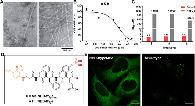
Figure 2.
ALP-instructed assembly of peptides in cell-free conditions and in live cells. (A) Transmission electron microscope (TEM) images of nanostructures formed before and after adding ALP (2 U/mL) to the solution of 1P (200 μM). (B) Cell viability of Saos-2 cells treated with 1P for 0.5 h. (C) IC50 values of 1P (24 h, 48 h, 72 h) against Saos-2 or HepG2 cells. (D) Molecular structures of NBD-ffypeMe2 and NBD-ffye. (E) CLSM images of Saos-2 cells treated with NBD-ffypeMe2 or NBD-ffye (200 μM) for 0.5 h. Scale bars, 10 μm.

