The increasing risk of Rift Valley fever virus (RVFV) infection as a global veterinary and public health threat demands the development of safe and accurate diagnostic tests. The aim of this study was to assess the suitability of a baculovirus expression system to produce recombinant RVFV nucleoprotein (N) for use as serodiagnostic antigen in an indirect enzyme-linked immunosorbent assay (ELISA).
KEYWORDS: ELISA, PRNT, Rift Valley fever virus, diagnostic test, livestock, nucleoprotein
ABSTRACT
The increasing risk of Rift Valley fever virus (RVFV) infection as a global veterinary and public health threat demands the development of safe and accurate diagnostic tests. The aim of this study was to assess the suitability of a baculovirus expression system to produce recombinant RVFV nucleoprotein (N) for use as serodiagnostic antigen in an indirect enzyme-linked immunosorbent assay (ELISA). The ability of the recombinant N antigen to detect RVFV antibody responses was evaluated in ELISA format using antisera from sheep and cattle experimentally infected with two genetically distinct wild-type RVFV strains and sera from indigenous sheep and goat populations exposed to natural RVFV field infection in The Gambia. The recombinant N exhibited specific reactivity with the N-specific monoclonal antibody and various hyperimmune serum samples from ruminants. The indirect ELISA detected N-specific antibody responses in animals with 100% sensitivity compared to the plaque reduction neutralization test (6 to 21 days postinfection) and with 97% and 100% specificity in sheep and cattle, respectively. There was a high level of correlation between the indirect N ELISA and the virus neutralization test for sheep sera (R2 = 0.75; 95% confidence interval [CI] = 0.73 to 0.92) and cattle sera (R2 = 0.80; 95% CI = 0.67 to 0.97); in addition, the N-specific ELISA detected RVFV seroprevalence levels of 26.1% and 54.3% in indigenous sheep and goats, respectively, in The Gambia. The high specificity and correlation with the virus neutralization test support the idea of the feasibility of using the recombinant baculovirus-expressed RVFV N-based indirect ELISA to assess RVFV seroprevalence in livestock in areas of endemicity and nonendemicity.
INTRODUCTION
Rift Valley fever virus (RVFV) is a mosquitoborne zoonotic pathogen that causes Rift Valley fever (RVF) in humans and domestic ruminants. The virus belongs to the family Phenuiviridae and the genus Phlebovirus. In sheep and cattle, the disease is characterized by high mortality in neonates and widespread abortion (1–3). Human disease is often characterized by abrupt onset of high fever, severe headache, myalgia, and conjunctivitis, with a small proportion of patients developing fatal hemorrhagic fever or late complications of encephalitis or ocular disease (4–6). RVFV is a single-stranded negative-sense RNA virus and is composed of three segments, namely, the large (L), medium (M), and small (S) segments. The L segment encodes the L protein, which is the viral RNA-dependent RNA polymerase. The M segment encodes the precursor for the glycoproteins forming the surface subunits Gn and Gc and the nonstructural NSm protein, a 78-kDa protein of unknown function. The S segment encodes the nucleoprotein (N) and nonstructural protein NSs (7–9).
In the United States, RVFV is classified as a tier 2 select agent and is considered a potential tool for bioterrorism or agroterrorism. The wide distribution of competent vectors in different geographic regions globally, coupled with increased international trade and travel, has substantially increased the risk of introduction and spread of RVFV to areas of nonendemicity. Thus, there is an urgent need to develop diagnostic assays that are economically viable, robust, sensitive, and specific for detecting potential RVFV infections in susceptible hosts, especially in areas of nonendemicity. Several diagnostic tests for detection of antibodies to RVFV following natural infection have been developed. These include the hemagglutination inhibition, complement fixation, indirect immunofluorescence, and virus neutralization tests (10). These classical serological methods for antibody detection require antigen, virus, or infected cell production in biocontainment facilities, thus restricting their use outside areas of endemicity. To address these concerns, enzyme-linked immunosorbent assays (ELISAs) designed for high-throughput serological detection of infection were developed (11–14). Some of these assays were based on the of β-propiolactone inactivated or gamma-irradiated whole-virus antigens derived from infected tissue culture or mouse brain and have been validated for diagnosis of RVFV infection in humans and animals (11–14). However, these antigens bind poorly to ELISA plates, thus affecting their suitability for usage in indirect ELISAs (15, 16). Additionally, the use of whole virus as antigen in indirect ELISAs could result in low specificity due to the potential for presenting cross-reactive epitopes and lacks the ability to distinguish infected from vaccinated animals. To overcome these limitations, recombinant RVFV N has been produced using Escherichia coli expression systems and applied as diagnostic antigens in different ELISA formats (12, 17–21). Although these ELISAs exhibited high specificity in some areas of endemicity in Africa, they have been observed to detect cross-reactive antibodies in seronegative North American sheep sera (W. C. Wilson, unpublished; cited previously by Faburay et al. [22]). The reasons for the cross-reactions are unknown; however, possible explanations include cross-reactivity with an unknown closely related agent infecting U.S. sheep populations and potential cross-reaction with antibodies raised against common E. coli microflora infecting North American sheep. The recombinant baculovirus expression system has been used in our laboratory for production of recombinant proteins of a variety of pathogens for subunit vaccine development (23, 24). However, the suitability of the system as a platform for production of recombinant antigens for RVFV serology has not been systematically evaluated. Indeed, we have previously used recombinant baculovirus-expressed RVFV N as a serodiagnostic antigen in-house for experimental purposes (23, 25, 26). In this study, we carried out further comparative analyses of the performance of the indirect N antibody ELISA format given the need for such an assay in countries of RVF nonendemicity. Against the backdrop of promising assay performance with experimental sera, we proceeded to perform preliminary evaluation in an country in which RVF is endemic, namely, The Gambia. Here, we report that the recombinant baculovirus system could provide a suitable alternative platform for production of RVFV N antigen for use in an indirect ELISA for the detection and serosurveillance of RVFV in countries of nonendemicity and endemicity.
MATERIALS AND METHODS
Cloning and recombinant protein expression.
The cloning and creation of a recombinant baculovirus construct expressing RVFV nucleoprotein have been described previously (22). Briefly, the RVFV N gene (based on sequences of RVFV strain ZH548) was amplified from a recombinant plasmid, pUC57N, by high-fidelity PCR using Accuprime Supermix per the manufacturer’s instructions. Following cloning of the PCR product into a pFastBac/CT/TOPO plasmid to create a donor plasmid, pFastBacN, a recombinant bacmid was created via a site-specific transposition step in an E. coli host system, DH10 Bac competent E. coli. Recombinant bacmid was purified using a HiPure plasmid kit (Life Technologies). Recombinant bacmids were used to transfect Spodoptera frugiperda (Sf9) cells using Cellfectin reagent per the manufacturer’s instruction to rescue a recombinant baculovirus. Recombinant baculovirus passage 2 (P2) and subsequent passages were used to express RVFV N in Sf9 cells.
Recombinant protein purification.
Recombinant RVFV N protein was expressed with a C-terminal 6×His-tagged fusion protein that allowed purification by affinity column chromatography using nickel nitrilotriacetic acid (Ni-NTA) His Bind Superflow resins (Millipore Corp., Billerica, MA) as described previously (22). Briefly, recombinant baculovirus-infected Sf9 cells expressing recombinant RVFV N protein were pelleted by centrifugation at 500 × g for 5 min. The pellet was resuspended in Ni-NTA binding buffer (300 mM NaCl, 50 mM Na3PO4 [pH 8.0], and 10 mM imidazole) containing 1× complete protease inhibitor. Insect Popculture reagent (Novagen) was then added at 0.05 volumes from the original culture volume. The lysate was incubated at room temperature for 15 min and the supernatant further clarified by centrifugation at 1,500 × g for 10 min. The clarified lysate was mixed with previously equilibrated Ni-NTA His Bind Superflow resin. Following binding for 1 h at 4°C, the column was washed with 10 column volumes of wash buffer (300 mM NaCl, 50 mM Na3PO4 [pH 8.0], and 20 mM imidazole) and then eluted with elution buffer (300 mM NaCl, 50 mM Na3PO4 [pH 8.0], and 250 mM imidazole). The purified recombinant protein was dialyzed overnight against phosphate-buffered saline (PBS) (pH 7.4). Protein concentrations were measured by bicinchoninic acid (BCA) assay (Thermo Scientific, Rockford, IL) at an absorbance of 562 nm, using bovine serum albumin (BSA; Sigma-Aldrich, St. Louis, MO) as the protein standard. Aliquots of the protein were stored at –80°C.
Coomassie staining and immunoblot analysis.
To assess expression of recombinant RVFV N, approximately 5 μg of purified recombinant N protein was resolved in 12% Bis-Tris polyacrylamide gel with 1× MOPS (morpholinepropanesulfonic acid) running buffer. After fixing for 30 min, the gel was subjected to Coomassie staining per standard protocol (RPI, Mount Prospect, IL) or to immunoblot analysis to detect and/or authenticate specific recombinant protein expression. Briefly, following gel electrophoresis, the protein was transferred by electroblotting onto a polyvinylidene difluoride (PVDF) membrane per standard protocol. The membrane was blocked in PBS (pH 7.4) containing 0.1% Tween 20 and 3% BSA for 1 h at room temperature. The blot was probed with mouse anti-His horseradish peroxidase (HRP) (1:5,000) or mouse anti-RVFV N monoclonal antibody (R3-1D8; 1:2,000) or RVFV antisera obtained from sheep or cattle experimentally infected with either of two wild-type RVFV strains, SA01-1322 (SA01) or Kenya-128-15 (Ken06) (25). Goat anti-mouse HRP antibody conjugate (Santa Cruz Biotechnology) (1:5,000) or protein G-HRP (Abcam, Cambridge, MA) (1:6,000) was used as the secondary detection antibody. Detection of specific reactivity was performed using an ECL (enhanced chemiluminescence) detection system.
Clinical specimens from experimental infections.
A panel of archived antisera obtained from previous studies involving experimental infections of sheep and calves with wild-type RVFV strain Ken06 or SA01 (25, 26) was used to evaluate the diagnostic sensitivity of the ELISA. These sera were collected from animals at different time points postinfection, i.e., at 0 to 21 days postinfection (dpi), at the Kansas State University Biosecurity Research Institute biosafety level 3 (BSL3) facility. Prior to use in a BLS2 setting, the sera had been inactivated by addition of 0.25% Tween 20 at a dilution of 1:10 to the serum and by heating the samples at –60°C in a water bath for 2 h as previously described (26). A panel of sera obtained from naive sheep and calves purchased from a private farm in Kansas, USA, as well as Ehrlichia ruminantium antisera (from sheep) and Schmallenberg virus antisera (from sheep and cattle), was used to evaluate diagnostic specificity.
Sera from natural RVFV infections.
Sera from natural infections were obtained from indigenous sheep of Djallonké breed (n = 46) and West African dwarf goats (n = 46) of both sexes aged 1 to 3 years. The animals were located at sites in Kiang West District in the Lower River Degion of The Gambia, West Africa, and were randomly sampled for initial field evaluation of the assay. All animals were maintained using a traditional husbandry system without insecticide treatment or insect control. Blood samples were collected using Vacutainer tubes without anticoagulant and transported to the laboratory at the West Africa Livestock Innovation Center (WALIC). Serum was separated after 2 to 4 h by centrifugation and stored at –20°C until use.
Detection of specific IgG in clinical specimens by indirect ELISA.
An indirect ELISA format using the recombinant RVFV N as a diagnostic antigen to detect RVFV N-specific IgG antibodies in sera from experimental and natural infections was used. The ELISA was performed as described previously (24), with minor modifications. Briefly, the plates (Nunc MaxiSorp) were coated overnight at 4°C with approximately 150 ng per well of purified recombinant baculovirus-expressed N protein. All washing steps were performed three times with 1% Tween 20–PBS. The antigen-coated plates were blocked with PBS containing 1% skim milk and 0.1% Tween 20 for 15 min at 37°C. After washing was performed, each serum was tested in duplicate, including positive serum (mouse anti-RVFV N monoclonal antibody, R1-ID8) and negative serum (serum from naive sheep and cattle in Kansas, USA). Sera were diluted 1:200 (anti-mouse RVFV N monoclonal at 1:500) and incubated with antigen at 37°C for 1 h. Plates were then incubated with recombinant protein G-HRP (Abcam, Cambridge, MA) (1:50,000). Chromogenic detection, termination of reactions, and measurement of optical density (OD) values were performed as previously described (24). The detection cutoff value was determined by addition of 2 standard deviations to mean OD values from negative-testing sera.
Virus neutralization test.
To validate the performance of the indirect N ELISA, a plaque reduction neutralization test (PRNT) was performed on the sera as described previously (24). Briefly, 2-fold dilutions were made from aliquots of serum from each animal. The stock of MP12 RVFV was diluted to 50 PFU and mixed with an equal volume of diluted serum, and the mixture was incubated at 37°C for 1 h. Each mixture of serum plus RVFV was used to infect confluent monolayers of Vero E6 cells in 12-well plates. After 1 h of adsorption at 37°C and 5% CO2, the mixture was removed, and 1.5 ml of nutrient agarose overlay (1× minimal essential medium [MEM], 4% bovine serum albumin, and 0.9% SeaPlaque agar) was added to the monolayers. After 4 to 5 days of incubation, the cells were fixed with 10% neutral buffered formalin for 3 h prior to removal of the agarose overlay. The monolayer was stained with 0.5% crystal violet–PBS, and plaques were enumerated. The calculated 80% PRNT titers (PRNT80) corresponded to the reciprocal titer of the highest serum dilution, which reduced the number of plaques by 80% or more relative to the virus control results.
Detection of specific IgG in sheep exposed to natural RVFV infection.
An indirect ELISA was used as described above to detect RVFV N-specific IgG antibodies in sera obtained from indigenous sheep and goats exposed to natural RVFV infection in The Gambia. Briefly, each ELISA plate was coated with approximately 150 ng of recombinant RVFV N per well in 100 μl of Dulbecco’s coating buffer (22, 24) and maintained overnight at 4°C. Each serum sample was tested in duplicate at a dilution of 1:200, and each test included duplicate negative-control sera obtained from a naive sheep in Kansas (USA) or from an indigenous sheep that tested strongly negatively (with significantly high OD values) in a competitive ELISA based on the use of recombinant RVFV N protein as the antigen (unpublished data). The experiment included duplicate positive-control sera from sheep experimentally infected with RVFV at 21 dpi as well as indigenous sheep sera that tested strongly positively in a competitive RVFV ELISA based on the use of recombinant RVFV N antigen. Detection was performed with protein G-HRP, which binds with high affinity the Fc region of IgG of sheep. The cutoff point for the indirect ELISA was determined by the addition of 2 standard deviations (SD) to the mean optical density (OD) values of the negative-control sera included in each test run.
Data analysis.
Data on IgG response OD values are presented as means ± standard deviations for each time point postinfection. Plaque reduction neutralization test (PRNT) titers are also presented as mean values per time point postinfection. Correlations between IgG OD values and PRNT titers were examined by the use of Pearson correlation coefficients (GraphPad Prism) at a 95% confidence interval (CI).
RESULTS
Recombinant baculovirus expression and reactivity of RVFV N.
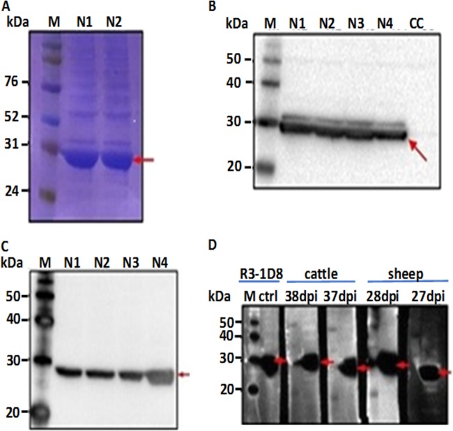
To assess expression of recombinant RVFV N, a fraction of the affinity purified protein was resolved in a polyacrylamide gel and stained with Coomassie blue. An estimated 30-kDa protein corresponding to the expected molecular weight of recombinant RVFV N was detected (Fig. 1A). The purified RVFV N protein was also examined by immunoblot analysis using a mouse anti-His (C-terminal)-HRP monoclonal antibody. Immunoblot analysis demonstrated expression of a recombinant N protein of the expected molecular size of 30 kDa (Fig. 1B). Additional immunoblot analyses were performed with RVFV-specific monoclonal and polyclonal antibodies. Expression of the RVFV N protein was confirmed by positive reactivity with mouse anti-RVFV monoclonal antibody (R3-1D8) (Fig. 1C) and with antiserum from sheep (at 27 and 28 dpi) and cattle (at 37 and 38 dpi) that had been experimentally infected with wild-type RVFV (Fig. 1D). The immunoreactive bands corresponded to the expected molecular weight of recombinant RVFV N.
FIG 1.
Analysis of baculovirus-expressed RVFV N. (A) Coomassie blue stain shows overexpression of the target protein. (B and C) Detection of a recombinant protein of the expected molecular weight by the use of anti-His (C-terminal) monoclonal antibody (B) and mouse anti-RVFV N monoclonal (R3-1D8) antibody (C). (D) Authentication of recombinant RVFV N expression using mouse anti-RVFV N monoclonal (R3-1D8) antibody and convalescent-phase sera from sheep and cattle at different time points postinfection. N1 to N4 represent various recombinant baculovirus clones tested for expression of RVFV N; CC represents uninfected Sf9 cell lysate. M, molecular weight marker. Arrows indicate specific molecular size or a band representing RVFV N protein.
Serology.
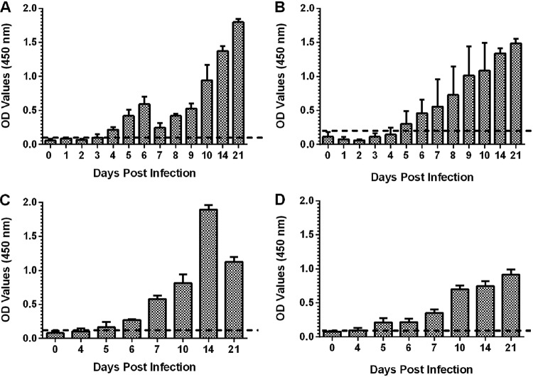
(i) Detection of IgG antibodies in clinical specimens obtained from experimental infections. A panel of antisera collected from sheep and calves experimentally infected with genetically distinct wild-type strains of RVFV was tested to evaluate the performance of RVFV N-specific indirect ELISA in detecting the kinetics of specific IgG antibody responses. Antisera sera from sheep infected with two wild-type RVFV strains (Ken06 and SA01) exhibited time-dependent increases in antibody reactivity as manifested by corresponding increases in IgG OD readings (Fig. 2A and B). The indirect ELISA detected specific IgG antibodies in all sheep (n = 5) infected with RVFV Ken06 as early as 4 dpi, with stronger reactivity observed at later time points postinfection (10 through 21 dpi) (Fig. 2A). Sample mean OD values ranged from 0.159 (4 dpi) to 1.745 (21 dpi), with a positive cutoff threshold value of 0.106. The assay exhibited 100% sensitivity (n = 23) (4 to 21 dpi). A similar time-dependent increase in antibody activity was detected in sheep experimentally infected with the RVFV SA01 strain, with peak antibody activity detected at 21 dpi (Fig. 2B). However, in contrast to the Ken06 group, only one of five animals infected with SA01 tested positive at 4 dpi. The assay detected that 3 of 4 animals tested positive at 5 dpi, and thereafter all samples tested consistently positive from 6 through 21 dpi. Positive mean sample OD values ranged from 0.254 (5 dpi) to 1.532 (21 dpi) with the cutoff threshold value set at 0.240. Overall, for sheep sera, the indirect RVFV N-specific ELISA exhibited 94.7% diagnostic sensitivity (n = 17) in testing sera from 5 to 21 dpi, or 100% diagnostic sensitivity (n = 13) in testing sera starting at day 6 to 21 dpi, and 97% diagnostic specificity using naive sheep sera (n = 32; Table 1). For cattle, the assay exhibited a relatively higher sensitivity of 95.5% in testing sera from 5 to 21 dpi or an equal sensitivity of 100% in testing sera from 6 to 21 dpi. The ELISA also exhibited a comparatively higher specificity of 100% in cattle (Table 1). The naive sheep and cattle sera were obtained from the same animals mentioned above prior to exposure to experimental inoculation. Table 1 summarizes the overall performance of the indirect ELISA in sheep. Importantly, convalescent-phase sera from sheep experimentally infected with Ehrlichia ruminantium (a rickettsial pathogen of ruminants) or Schmallenberg virus (at 21 dpi) (a member of the order Bunyavirales) were not cross-reactive with the RVFV N protein in the indirect ELISA.
FIG 2.
(A and B) Detection of kinetics of anti-RVFV N IgG responses in sheep experimentally infected with one of two wild-type RVFVs, Ken06 (A) or SA01 (B). The antibody profile shows a time-dependent increase in antibody activity. Horizontal dashed lines depict cutoff points as follows: 0.106 (A) and 0.240 (B). (C and D) Detection of kinetics of anti-RVFV N IgG response in calves experimentally infected with one of two wild-type RVFVs, Ken06 (C) or SA01 (D). The antibody profile shows time-dependent increase in antibody activity. Horizontal dashed lines depict cutoff points as follows: 0.138 (A) and 0.095 (B).
TABLE 1.
Sensitivity and specificity of the indirect RVFV N ELISA in detection of specific antibodies in sheep and cattle sera
| Source of sera | % sensitivity |
% specificitya | |
|---|---|---|---|
| 5–21 dpi | 6–21 dpi | ||
| Sheep | 94.7 | 100 | 97 |
| Cattle | 95.5 | 100 | 100 |
Specificity was determined using serum specimens from naive sheep and cattle.
The indirect RVFV N ELISA exhibited kinetics of time-dependent increase in IgG activity in calves similar to the kinetics seen in sheep (Fig. 2C and D). With the exception of one animal from the Ken06 group that showed a borderline positive OD value (0.1375) at 4 dpi (cutoff point = 0.138), the earliest time point of detecting specific IgG responses was 5 dpi (Fig. 2C), with 2 of 3 Ken06-infected animals testing positive. Thereafter, all serum specimens from the Ken06 -infected animals tested positive, with mean sample OD readings ranging from 0.1535 to 1.842. In total, 10 of 11 Ken06 serum samples tested positive from 5 dpi through 21 dpi, indicating a diagnostic sensitivity of 91%, or 100% diagnostic sensitivity for serum samples tested from 6 dpi through 21 dpi (Fig. 2C). Similar detection performance was noted in antisera from calves infected with the SA01 strain. Except for one animal that exhibited an elevated mean OD value (0.147) at 4 dpi (n = 4; cutoff value = 0.0951), the earliest time point at which the ELISA detected RVFV-positive IgG OD readings in the SA01-infected calves was 5 dpi (Fig. 2D). Thereafter, all specimens tested consistently positive until 21 dpi, the study endpoint, with mean sample OD values ranging from 0.193 to 0.866. Overall, the ELISA exhibited 100% diagnostic sensitivity (5 to 21 dpi) (n = 16) and specificity (n = 10) (Table 1). Serum from a calf experimentally infected with Schmallenberg virus (at 21 dpi) tested negative, exhibiting significantly low OD readings (P < 0.0001).
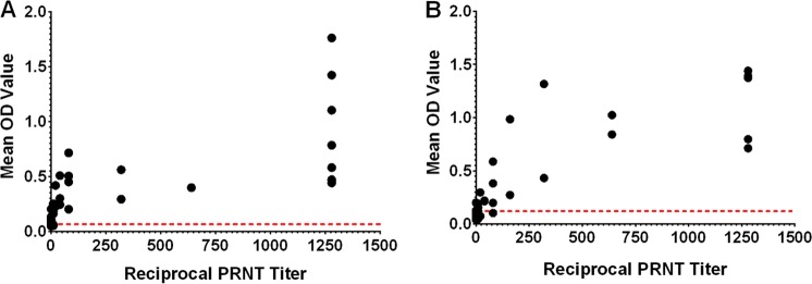
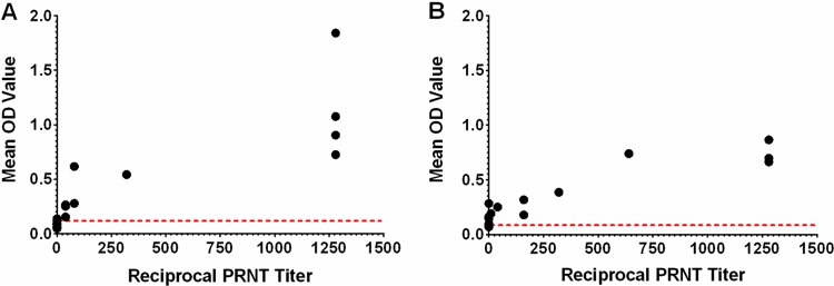
(ii) Correlation between RVFV N ELISA and PRNT. The PRNT is the gold standard for serological confirmation of RVFV infection. Thus, the performance of the indirect RVFV N ELISA was compared with that of the PRNT assay. Data obtained from sheep and cattle serum specimens indicated significant correlation (P < 0.0001) between PRNT titer and ELISA IgG OD readings in the two animal species as determined by Pearson correlation coefficient analysis (Fig. 3; see also Fig. 4). Detection of neutralizing antibody titers was generally associated with positive IgG OD readings in both ruminant species (Table 2; see also Table 3).
FIG 3.
Analysis of correlation between the indirect RVFV N ELISA and PRNT in detecting antibodies to RVFV. Antisera were obtained from sheep experimentally infected with one of two wild-type viruses, Ken06 (A) or SA01 (B). Results show significant agreement between the two assays (P < 0.0001) in detecting antibodies against both viruses. Dashed horizontal lines depict ELISA cutoff points for positive IgG response.
FIG 4.
Analysis of correlation between the indirect RVFV N ELISA and PRNT in detecting antibodies to RVFV. Antisera were obtained from calves experimentally infected with one of two wild-type viruses, Ken06 (A) or SA01 (B). Results show significant agreement between the two assays (P < 0.0001) in detecting antibodies against both viruses. Dashed horizontal lines depict ELISA cutoff points for positive IgG response.
TABLE 2.
Correlation between mean neutralizing antibody titer and indirect N ELISA results in sheep
| Day postinfection |
Ken06 RVFV challenge |
SA01 RVFV challenge |
||||
|---|---|---|---|---|---|---|
| PRNT titer | ELISA OD valuea |
ELISA result |
PRNT titer | ELISA OD valueb |
ELISA result |
|
| 0 | 0 | 0.059 | − | 0 | 0.112 | − |
| 4 | 16 | 0.217 | + | 15 | 0.146 | − |
| 5 | 45 | 0.419 | + | 60 | 0.325 | + |
| 6 | 160 | 0.593 | + | 107 | 0.49 | + |
| 7 | 240 | 0.247 | + | 400 | 0.557 | + |
| 8 | 960 | 0.421 | + | 480 | 0.728 | + |
| 9 | 1,280 | 0.525 | + | 800 | 1.015 | + |
| 10 | >1,280 | 0.944 | + | >1,280 | 1.086 | + |
| 14 | >1,280 | 1.375 | + | >1,280 | 1.339 | + |
| 21 | >1,280 | 1.798 | + | >1,280 | 1.487 | + |
ELISA cutoff value = 0.106.
ELISA cutoff value = 0.240.
TABLE 3.
Correlation between mean neutralizing antibody titer and indirect N ELISA results in cattle
| Day postinfection |
Ken06 RVFV challenge |
SA01 RVFV challenge |
||||
|---|---|---|---|---|---|---|
| PRNT titer | ELISA OD valuea |
ELISA result |
PRNT titer | ELISA OD valueb |
ELISA result |
|
| 0 | 0 | 0.079 | − | 0 | 0.08 | − |
| 4 | 0 | 0.106 | − | 3 | 0.097 | + |
| 5 | 13 | 0.166 | + | 120 | 0.213 | + |
| 6 | 60 | 0.272 | + | 100 | 0.216 | + |
| 7 | 200 | 0.580 | + | 240 | 0.352 | + |
| 10 | 1,280 | 0.815 | + | 960 | 0.703 | + |
| 14 | 1,280 | 1.892 | + | 1,280 | 0.748 | + |
| 21 | 1,280 | 1.127 | + | 1,280 | 0.0917 | + |
ELISA cutoff value = 0.138.
ELISA cutoff value = 0.095.
Specifically, there was a significant correlation between PRNT titer and ELISA IgG OD reading in sheep infected with both Ken06 (R2 = 0.761, 95% CI = 0.73 to 0.92, P < 0.0001) or SA01 (R2 = 0.739, 95% CI = 0.72 to 0.91, P < 0.0001) (Fig. 3). A similar correlation between the two assays was observed with serum specimens from calves infected with either wild-type virus isolate, Ken06 (R2 = 0.756, 95% CI = 0.67 to 0.95, P < 0.0001) or SA01 (R2 = 0.853, 95% CI = 0.81 to 0.97, P < 0.0001) (Fig. 4).
Overall, correlation analysis indicated that the PRNT titers had a positive linear relationship to ELISA IgG OD readings, with high PRNT titers generally correlating with high serum OD readings. Comparison of detection performances at 6 dpi (the earliest time point when most RVFV-infected animals tested as distinctly seropositive) through 21 dpi showed complete agreement between the two assays.
(iii) Detection of RVFV antibodies in sheep exposed to natural field infection. Evaluation of the performance of the indirect RVFV-N protein ELISA in detecting RVFV-specific IgG antibodies in sera of indigenous small ruminants exposed to potential natural field infection was performed in a setting of endemicity in The Gambia, West Africa. The RVFV N ELISA detected seroprevalence rates of 26.1% (12/46) in sheep and 54.3% (25/46) in goats. The ELISA cutoff point for sheep sera was determined at 0.098 and for goat sera at 0.065. The degree of positivity was variable and ranged from weakly positive (with OD values of 0.15 for sheep and 0.068 for goats) to strongly positive (with OD values of 1.017 for sheep and 1.424 for goats). Sheep and goat samples that tested positive in the indirect RVFV N protein ELISA also tested positive in a commercial indirect ELISA (data not shown).
DISCUSSION
The veterinary and public health impact of RVF in sub-Saharan Africa coupled with the threat of spread of the disease to areas of nonendemicity urgently demand the development of robust, economical, high-throughput, sensitive, and accurate diagnostic tests. The recombinant RVFV nucleocapsid protein has been used extensively in various ELISA formats (indirect and competitive ELISAs) as an antigenic target for detection of RVFV infection in countries where RVF is endemic (17, 19–21, 27). The recombinant N antigen has mostly been produced in E. coli, representing a prokaryotic expression system (19, 21, 27). Although the E. coli N-based assays demonstrated satisfactory sensitivity, issues pertaining to specificity with respect to application to sera from North American sheep have been recently reported (cited by Faburay et al. [22]). The baculovirus expression system, representing a eukaryotic protein expression platform, has been extensively used in our laboratory to express recombinant antigens for diagnostic and subunit vaccine development. However, the utility of the system as a platform for production of serodiagnostic antigens for RVFV detection has not been evaluated. This report presents results from the first study to have evaluated the performance of an indirect ELISA based on a recombinant RVFV N produced in the eukaryotic baculovirus expression system to detect antibodies to RVFV infection in target host species. We compared the indirect RVFV N ELISA with the virus neutralization test as the gold standard or reference test for serodiagnosis of RVFV infection. There was high correlation between the two assays in both animal species, with the virus neutralizing antibody titer generally correlating positively with the ELISA OD test result. For example, from 6 through 21 dpi, the ELISA exhibited 100% diagnostic sensitivity in detecting N-specific IgG antibodies in clinical specimens from experimental infections of sheep and cattle while exhibiting 97% and 100% specificity for sera obtained from resident (Kansas, USA) naive sheep and cattle, respectively.
Diagnostic specificity and sensitivity are the most critical parameters defining the utility of a serodiagnostic assay for use in areas of nonendemicity. The level of diagnostic specificity for the baculovirus N-based ELISA in sheep and cattle is acceptable for areas of nonendemicity, especially when used in conjunction with a confirmatory virus neutralization test. This is particularly relevant to North America, where significant serological cross-reactivity in small ruminants has been detected using E. coli-produced RVFV N-based ELISAs. The cause of the cross-reactivity remains unknown; however, it has mostly been attributed, without empirical evidence, to an unidentified antigenically related agent infecting small ruminants in North America. Nonetheless, the possibility that cross-reacting antibodies might be raised against commensal or pathogenic E. coli microflora in ruminants cannot be ruled out. Unlike E. coli, baculovirus does not typically infect mammalian hosts, including ruminant livestock, which makes it a desirable platform for production of recombinant diagnostic antigens, especially for use in environments where antigenic cross-reactivity could be a major concern. Indeed, it has been observed that immunization with pathogenic E. coli O157:H7 induced production of homologous antibodies, which showed cross-reactivity in brucellosis serological tests (28). It is noteworthy, however, that in areas of endemicity in Africa, serological assays based on the use of E. coli-produced recombinant N as the diagnostic antigen have been reported to perform satisfactorily in antigenic cross-reactivity studies (27, 29) and in field studies in cattle (30, 31). There was no demonstrated evidence that other phleboviruses could hamper the serodiagnosis of RVF in areas of endemicity. The data presented in this study indicate that the serological assay based on the recombinant baculovirus-expressed RVFV N as a diagnostic antigen could provide an alternative platform for serodiagnosis of RVFV in susceptible hosts in regions of endemicity and nonendemicity.
In the present study, the performance of the baculovirus-expressed RVFV N protein-based indirect ELISA was comparable to the performance of similar serodiagnostic assays used for detection of antibodies to RVFV in experimentally infected ruminant livestock in Africa (18). For example, the performance of the assay was comparable to that of an indirect N ELISA based on E. coli-expressed recombinant N antigen used in indigenous domestic ruminants (18), exhibiting 100% diagnostic sensitivity in both sheep and cattle (6 to 21 dpi). Indeed, several ELISAs based on recombinant RVFV N antigen produced in E. coli have been evaluated in different animal species for diagnostic sensitivity and specificity (12, 18, 20, 21). These assays yielded diagnostic sensitivities and specificities ranging from 99.4% to 100% and from 98.3% to 100%, respectively, depending on the antibody detection system (anti-species-specific IgG antibody conjugate or protein G conjugate) and/or animal species (domestic and wild ruminants) (11, 18, 20, 21). Using clinical specimens from experimentally infected animals, the baculovirus-expressed indirect RVFV N ELISA could detect specific IgG as early as 4 to 5 days postinfection; this performance was comparable to that of a recombinant E. coli-expressed RVFV N protein-based indirect ELISA reported previously (19). The baculovirus-expressed indirect RVFV N ELISA revealed various levels of sensitivity at the early time points after infection (3 to 5 dpi compared to 6 to 21 dpi). The relatively lower level of sensitivity of the assay at the earliest time points of detectable immunological response (3 to 5 dpi) is similar to that seen with other N-specific ELISAs and may be insignificant if the test is used in prevalence studies and population-based disease surveillance programs.
Preliminary evaluation of the RVFV N-specific ELISA using sera from field-exposed indigenous sheep and goats raised in an endemic setting in The Gambia, West Africa, detected seroprevalences of 26.1% in sheep and 54.3% in goats. These rates are within the range of RVFV seroprevalence estimates reported elsewhere in regions of endemicity in sub-Saharan Africa. In Senegal, West Africa, a serological prevalence of 30% (with variability within households ranging from 0% to 66.7%) was reported among indigenous sheep (32), whereas in Mozambique, in Southern Africa, an overall seroprevalence of 44.2% has been reported among local sheep by the use of a commercial ELISA kit (33). Although a wider study may be required to further corroborate the findings of our preliminary field evaluation, detection of high RVFV seroprevalence in an endemic setting in domestic ruminants that are in close contact with humans signifies a significant risk factor for zoonotic transmission of this pathogen in the local communities. Evidently, in sub-Saharan Africa, outbreaks of RVF have been a frequent occurrence among individuals with a known history of association with infected livestock (3, 34).
In summary, the performance of the indirect RVFV N ELISA, based on the use of the baculovirus-expressed recombinant antigen, correlated well with the PRNT in detecting antibody responses to experimental RVFV infection. Applied to sera from both ruminant species, the ELISA also exhibited high degrees of diagnostic sensitivity and specificity. Although testing a larger sample size will be required in future studies, the degree of sensitivity and specificity exhibited by the assay in this study represents an important attribute for serological detection and surveillance for RVFV introduction in regions of endemicity and nonendemicity such as North America. Furthermore, given that RVFV infection induces lifelong antibody titers in humans (35) and animals (36), the baculovirus-based RVFV N ELISA could be a reliable substitute for the virus neutralization test for investigation of RVFV seroprevalence and disease as well as for risk mapping for humans and livestock in countries of endemicity in sub-Saharan Africa with limited biosafety and biosecurity capabilities.
ACKNOWLEDGMENTS
We thank the staff of Kansas State University Biosecurity Research Institute and Dane Jasperson, Lindsey Reister, Chester McDowell, Tammy Koopman, and Haixia Liu, who contributed to previous studies that produced the archived antisera used in this study. We thank Asia Fernandes, as well as Amadou Keita at the West Africa Livestock Innovation Center (WALIC), for assistance in performing the serology tests.
This work was supported by the Science and Technology Directorate of the United States Department of Homeland Security (DHS) under award instrument number D15PC0027, grants of the Department of Homeland Security Center of Excellence for Emerging and Zoonotic Animal Diseases (CEEZAD), grant no. 2010-ST061-AG0001, USDA Agricultural Services Project no. 5430-050-005-00D, and NBAF Transition Funds from the State of Kansas. The funders had no role in study design, data collection and interpretation, or the decision to submit the work for publication.
REFERENCES
- 1.SIB (Swiss Institute of Bioinformatics). 2018. ViralZone; phlebovirus. https://viralzone.expasy.org/252.
- 2.Blitvich BJ, Beaty BJ, Blair CD, Brault AC, Dobler G, Drebot MA, Haddow AD, Kramer LD, LaBeaud AD, Monath TP, Mossel EC, Plante K, Powers AM, Tesh RB, Turell MJ, Vasilakis N, Weaver SC. 2018. Bunyavirus taxonomy: limitations and misconceptions associated with the current ICTV criteria used for species demarcation. Am J Trop Med Hyg 99:11–16. doi: 10.4269/ajtmh.18-0038. [DOI] [PMC free article] [PubMed] [Google Scholar]
- 3.Faburay B, LaBeaud AD, McVey DS, Wilson WC, Richt JA. 19 September 2017, posting date. Current status of Rift Valley fever vaccine development. Vaccines (Basel) doi: 10.3390/vaccines5030029. [DOI] [PMC free article] [PubMed] [Google Scholar]
- 4.Mansfield KL, Banyard AC, McElhinney L, Johnson N, Horton DL, Hernandez-Triana LM, Fooks AR. 2015. Rift Valley fever virus: a review of diagnosis and vaccination, and implications for emergence in Europe. Vaccine 33:5520–5531. doi: 10.1016/j.vaccine.2015.08.020. [DOI] [PubMed] [Google Scholar]
- 5.Bird BH, Ksiazek TG, Nichol ST, Maclachlan NJ. 2009. Rift Valley fever virus. J Am Vet Med Assoc 234:883–893. doi: 10.2460/javma.234.7.883. [DOI] [PubMed] [Google Scholar]
- 6.Bird BH, McElroy AK. 2016. Rift Valley fever virus: unanswered questions. Antiviral Res 132:274–280. doi: 10.1016/j.antiviral.2016.07.005. [DOI] [PubMed] [Google Scholar]
- 7.Gerrard SR, Nichol ST. 2002. Characterization of the Golgi retention motif of Rift Valley fever virus G(N) glycoprotein. J Virol 76:12200–12210. doi: 10.1128/JVI.76.23.12200-12210.2002. [DOI] [PMC free article] [PubMed] [Google Scholar]
- 8.Gerrard S, Bird B, Albarino C, Nichol S. 2007. The NSm proteins of Rift Valley fever virus are dispensable for maturation, replication and infection. Virology 359:459–465. doi: 10.1016/j.virol.2006.09.035. [DOI] [PMC free article] [PubMed] [Google Scholar]
- 9.Won S, Ikegami T, Peters CJ, Makino S. 2007. NSm protein of Rift Valley fever virus suppresses virus-induced apoptosis. J Virol 81:13335–13345. doi: 10.1128/JVI.01238-07. [DOI] [PMC free article] [PubMed] [Google Scholar]
- 10.Swanepoel R, Struthers JK, Erasmus MJ, Shepherd SP, McGillivray GM, Erasmus BJ, Barnard BJ. 1986. Comparison of techniques for demonstrating antibodies to Rift Valley fever virus. J Hyg (Lond) 97:317–329. doi: 10.1017/s0022172400065414. [DOI] [PMC free article] [PubMed] [Google Scholar]
- 11.Paweska JT, Burt FJ, Anthony F, Smith SJ, Grobbelaar AA, Croft JE, Ksiazek TG, Swanepoel R. 2003. IgG-sandwich and IgM-capture enzyme-linked immunosorbent assay for the detection of antibody to Rift Valley fever virus in domestic ruminants. J Virol Methods 113:103–112. doi: 10.1016/S0166-0934(03)00228-3. [DOI] [PubMed] [Google Scholar]
- 12.Paweska J, Smith S, Wright I, Williams R, Cohen A, Van Dijk A, Grobbelaar A, Croft J, Swanepoel R, Gerdes G. 2003. Indirect enzyme-linked immunosorbent assay for the detection of antibody against Rift Valley fever virus in domestic and wild ruminant sera. Onderstepoort J Vet Res 70:49–64. [PubMed] [Google Scholar]
- 13.Paweska JT, Burt FJ, Swanepoel R. 2005. Validation of IgG-sandwich and IgM-capture ELISA for the detection of antibody to Rift Valley fever virus in humans. J Virol Methods 124:173–181. doi: 10.1016/j.jviromet.2004.11.020. [DOI] [PubMed] [Google Scholar]
- 14.Paweska JT, Mortimer E, Leman PA, Swanepoel R. 2005. An inhibition enzyme-linked immunosorbent assay for the detection of antibody to Rift Valley fever virus in humans, domestic and wild ruminants. J Virol Methods 127:10–18. doi: 10.1016/j.jviromet.2005.02.008. [DOI] [PubMed] [Google Scholar]
- 15.Meegan JM, Yedloutschnig RJ, Peleg BA, Shy J, Peters CJ, Walker JS, Shope RE. 1987. Enzyme-linked immunosorbent assay for detection of antibodies to Rift Valley fever virus in ovine and bovine sera. Am J Vet Res 48:1138–1141. [PubMed] [Google Scholar]
- 16.Niklasson B, Peters CJ, Grandien M, Wood O. 1984. Detection of human immunoglobulins G and M antibodies to Rift Valley fever virus by enzyme-linked immunosorbent assay. J Clin Microbiol 19:225–229. [DOI] [PMC free article] [PubMed] [Google Scholar]
- 17.Cetre-Sossah C, Billecocq A, Lancelot R, Defernez C, Favre J, Bouloy M, Martinez D, Albina E. 2009. Evaluation of a commercial competitive ELISA for the detection of antibodies to Rift Valley fever virus in sera of domestic ruminants in France. Prev Vet Med 90:146–149. doi: 10.1016/j.prevetmed.2009.03.011. [DOI] [PubMed] [Google Scholar]
- 18.Fafetine J, Tijhaar E, Paweska J, Neves L, Hendriks J, Swanepoel R, Coetzer J, Egberink H, Rutten V. 2007. Cloning and expression of Rift Valley fever virus nucleocapsid (N) protein and evaluation of a N-protein based indirect ELISA for the detection of specific IgG and IgM antibodies in domestic ruminants. Vet Microbiol 121:29–38. doi: 10.1016/j.vetmic.2006.11.008. [DOI] [PubMed] [Google Scholar]
- 19.Jansen van Vuren P, Potgieter A, Paweska J, van Dijk A. 2007. Preparation and evaluation of a recombinant Rift Valley fever virus N protein for the detection of IgG and IgM antibodies in humans and animals by indirect ELISA. J Virol Methods 140:106–114. doi: 10.1016/j.jviromet.2006.11.005. [DOI] [PubMed] [Google Scholar]
- 20.Paweska J, Jansen van Vuren P, Swanepoel R. 2007. Validation of an indirect ELISA based on a recombinant nucleocapsid protein of Rift Valley fever virus for the detection of IgG antibody in humans. J Virol Methods 146:119–124. doi: 10.1016/j.jviromet.2007.06.006. [DOI] [PubMed] [Google Scholar]
- 21.Paweska J, van Vuren P, Kemp A, Buss P, Bengis R, Gakuya F, Breiman R, Njenga M, Swanepoel R. 2008. Recombinant nucleocapsid-based ELISA for detection of IgG antibody to Rift Valley fever virus in African buffalo. Vet Microbiol 127:21–28. doi: 10.1016/j.vetmic.2007.07.031. [DOI] [PubMed] [Google Scholar]
- 22.Faburay B, Wilson W, McVey DS, Drolet BS, Weingartl H, Madden D, Young A, Ma W, Richt JA. 2013. Rift Valley fever virus structural and nonstructural proteins: recombinant protein expression and immunoreactivity against antisera from sheep. Vector Borne Zoonotic Dis 13:619–629. doi: 10.1089/vbz.2012.1285. [DOI] [PMC free article] [PubMed] [Google Scholar]
- 23.Faburay B, Wilson WC, Gaudreault NN, Davis AS, Shivanna V, Bawa B, Sunwoo SY, Ma W, Drolet BS, Morozov I, McVey DS, Richt JA. 2016. A recombinant Rift Valley fever virus glycoprotein subunit vaccine confers full protection against Rift Valley fever challenge in sheep. Sci Rep 6:27719. doi: 10.1038/srep27719. [DOI] [PMC free article] [PubMed] [Google Scholar]
- 24.Faburay B, Lebedev M, McVey DS, Wilson W, Morozov I, Young A, Richt JA. 2014. A glycoprotein subunit vaccine elicits a strong Rift Valley fever virus neutralizing antibody response in sheep. Vector Borne Zoonotic Dis 14:746–756. doi: 10.1089/vbz.2014.1650. [DOI] [PMC free article] [PubMed] [Google Scholar]
- 25.Faburay B, Gaudreault NN, Liu Q, Davis AS, Shivanna V, Sunwoo SY, Lang Y, Morozov I, Ruder M, Drolet B, Scott McVey D, Ma W, Wilson W, Richt JA. 2016. Development of a sheep challenge model for Rift Valley fever. Virology 489:128–140. doi: 10.1016/j.virol.2015.12.003. [DOI] [PubMed] [Google Scholar]
- 26.Wilson WC, Davis AS, Gaudreault NN, Faburay B, Trujillo JD, Shivanna V, Sunwoo SY, Balogh A, Endalew A, Ma W, Drolet BS, Ruder MG, Morozov I, McVey DS, Richt JA. 2016. Experimental infection of calves by two genetically-distinct strains of Rift Valley fever virus. Viruses 8:145. doi: 10.3390/v8050145. [DOI] [PMC free article] [PubMed] [Google Scholar]
- 27.Jansen van Vuren P, Paweska JT. 2009. Laboratory safe detection of nucleocapsid protein of Rift Valley fever virus in human and animal specimens by a sandwich ELISA. J Virol Methods 157:15–24. doi: 10.1016/j.jviromet.2008.12.003. [DOI] [PubMed] [Google Scholar]
- 28.Bonfini B, Chiarenza G, Paci V, Sacchini F, Salini R, Vesco G, Villari S, Zilli K, Tittarelli M. 2018. Cross-reactivity in serological tests for brucellosis: a comparison of immune response of Escherichia coli O157:H7 and Yersinia enterocolitica O:9 vs Brucella spp. Vet Ital 54:107–114. doi: 10.12834/VetIt.1176.6539.2. [DOI] [PubMed] [Google Scholar]
- 29.Swanepoel R, Struthers JK, Erasmus MJ, Shepherd SP, McGillivray GM, Shepherd AJ, Hummitzsch DE, Erasmus BJ, Barnard BJ. 1986. Comparative pathogenicity and antigenic cross-reactivity of Rift Valley fever and other African phleboviruses in sheep. J Hyg (Lond) 97:331–346. doi: 10.1017/s0022172400065426. [DOI] [PMC free article] [PubMed] [Google Scholar]
- 30.Davies FG. 1975. Observations on the epidemiology of Rift Valley fever in Kenya. J Hyg (Lond) 75:219–230. doi: 10.1017/s0022172400047252. [DOI] [PMC free article] [PubMed] [Google Scholar]
- 31.Swanepoel R. 1976. Studies on the epidemiology of Rift Valley fever. J S Afr Vet Assoc 47:93–94. [PubMed] [Google Scholar]
- 32.Wilson ML, Chapman LE, Hall DB, Dykstra EA, Ba K, Zeller HG, Traore-Lamizana M, Hervy JP, Linthicum KJ, Peters CJ. 1994. Rift Valley fever in rural northern Senegal: human risk factors and potential vectors. Am J Trop Med Hyg 50:663–675. doi: 10.4269/ajtmh.1994.50.663. [DOI] [PubMed] [Google Scholar]
- 33.Blomstrom AL, Scharin I, Stenberg H, Figueiredo J, Nhambirre O, Abilio A, Berg M, Fafetine J. 2016. Seroprevalence of Rift Valley fever virus in sheep and goats in Zambezia, Mozambique. Infect Ecol Epidemiol 6:31343. doi: 10.3402/iee.v6.31343. [DOI] [PMC free article] [PubMed] [Google Scholar]
- 34.LaBeaud AD, Kazura JW, King CH. 2010. Advances in Rift Valley fever research: insights for disease prevention. Curr Opin Infect Dis 23:403–408. doi: 10.1097/QCO.0b013e32833c3da6. [DOI] [PMC free article] [PubMed] [Google Scholar]
- 35.Findlay GM, Howard EM. 1951. Notes on Rift Valley fever. Arch Gesamte Virusforsch 4:411–423. doi: 10.1007/BF01241162. [DOI] [PubMed] [Google Scholar]
- 36.Barnard BJH. 1979. Rift Valley fever vaccine antibody and immune response in cattle to a live and an inactivated vaccine. J S Afr Vet Assoc 50:155–157. [PubMed] [Google Scholar]


